



Abstract
Background
Lipedema is an adipose disorder associated with multiple impairments. Conservative treatments remain the mainstay of management, yet evidence regarding the effects of physical therapies on clinical, imaging, and body composition outcomes is limited. Radial extracorporeal shock wave therapy (rESWT) has been proposed as a non-invasive therapeutic option, although its impact is not fully established.
Methods
This was a prospective, longitudinal, within-patient study conducted in women with clinically diagnosed lipedema. One lower limb was treated with radial extracorporeal shock wave therapy (rESWT), whereas the contralateral limb served as an internal control. A total of 16 patients were initially assessed, of whom 12 completed the full follow-up and were included in the final analysis. rESWT was applied over six sessions (two sessions per week) using standardized parameters. Clinical outcomes (LEFS, EQ-5D, SF-36 Physical Function, and IPAQ) were assessed at baseline, 6 weeks, and 3 months. Ultrasound and elastography were used to evaluate subcutaneous tissue thickness and stiffness at predefined leg and thigh sites, while segmental bioimpedance analysis assessed body composition and fluid distribution. Longitudinal changes were analyzed using mixed-effects models.
Results
Significant improvements were observed in functional capacity, quality of life, and physical activity levels at both 6 weeks and 3 months compared with baseline (p < .05). In contrast, no statistically significant changes were detected in ultrasound-derived tissue thickness, elastography measurements, or bioimpedance parameters over time, and no significant differences were detected between treated and control limbs within the constraints of the available sample size.
Conclusions
rESWT was associated with meaningful clinical and functional improvements in patients with lipedema, despite the absence of detectable changes in tissue thickness, stiffness, or body composition. These findings suggest that the benefits of rESWT may be mediated through symptom modulation and functional adaptation rather than structural tissue modification, supporting its role as part of conservative, symptom-oriented treatment strategies in lipedema.
Introduction
Lipedema is a chronic, progressive disorder of adipose tissue marked by a disproportionate and symmetrical accumulation of subcutaneous fat, primarily involving the lower extremities and occurring almost exclusively in women.1,2 The condition is frequently associated with pain, tenderness, increased capillary fragility, and a tendency to bruise easily, features that help differentiate lipedema from obesity and lymphedema.3,4 Despite increasing awareness, lipedema continues to be underdiagnosed and frequently misclassified, resulting in delayed or inappropriate management. 5
Beyond cosmetic concerns, lipedema has a substantial impact on physical function, mobility, and health-related quality of life. Patients frequently report exercise intolerance, functional limitations, and reduced participation in daily and social activities.6,7 These impairments are reflected in reduced scores on validated functional and quality-of-life questionnaires, such as the Lower Extremity Functional Scale (LEFS), EQ-5D, and SF-36.8–10 As a result, treatment approaches are progressively directed not only toward changes in tissue characteristics but also toward improving functional status and patient-reported outcomes.
Imaging techniques are playing an increasingly important role in the objective evaluation of lipedema. High-resolution ultrasound enables quantification of subcutaneous tissue thickness and structural alterations, whereas elastography offers complementary information on tissue stiffness and mechanical properties.11–13 Together, these modalities provide non-invasive and reproducible measures that may complement clinical assessment and facilitate treatment monitoring. Nevertheless, the association between imaging-derived parameters and functional or bioimpedance-based outcomes remains insufficiently investigated.
Bioimpedance analysis (BIA) has also been proposed as a useful method for characterizing body composition in lipedema, offering estimates of segmental water content, lean mass, phase angle, and the extracellular-to-total body water ratio.14,15 Phase angle, in particular, has been suggested as an indicator of cellular health and tissue integrity and may reflect changes related to therapeutic interventions. 16 Nevertheless, data combining BIA parameters with imaging findings in lipedema remain limited.
Extracorporeal shock wave therapy (ESWT) has emerged as a potential conservative treatment option in lipedema, based on its reported effects on microcirculation, tissue remodeling, and pain modulation.17–19 While early studies suggest improvements in symptoms and quality of life, data on its effects on tissue characteristics evaluated by ultrasound, elastography, and bioimpedance are still scarce, and longitudinal evidence is not yet available.
Therefore, the aim of this study was to evaluate the clinical, functional, imaging, and bioimpedance changes following ESWT in patients with lipedema, using a within-patient design with the contralateral limb as control. In addition, we explored the associations between changes in imaging-derived parameters and bioimpedance measures, seeking to better understand the relationship between tissue-level changes and systemic or functional outcomes.
Materials and methods
Study design and participants
The study was carried out at the University Hospital Complex of Santiago de Compostela during 2023. Women diagnosed with lipedema according to established clinical criteria were consecutively recruited. Inclusion criteria were: adult women (≥18 years), clinical diagnosis of lipedema affecting the lower limbs, and ability to complete the treatment protocol and follow-up assessments.
Exclusion criteria included pregnancy, active malignancy, uncontrolled systemic disease, previous surgical treatment for lipedema, current participation in other interventional studies, contraindications to shock wave therapy, or inability to complete follow-up evaluations.
This was a prospective, longitudinal, within-patient study conducted in women with clinically diagnosed lipedema. One lower limb was treated with radial extracorporeal shock wave therapy (rESWT), whereas the contralateral limb served as an internal control. An initial cohort of 16 patients was enrolled; 12 patients completed the full treatment protocol and follow-up assessments and were therefore included in the final analysis. Ultrasound and elastography measurements were available for all participants at baseline, 6 weeks, and 3 months. Bioimpedance-derived parameters (segmental water, segmental lean mass, phase angle, and ECW/TBW ratio) were available in all patients at baseline, in 8 patients at 6 weeks, and in 3 patients at 3 months. The reduced availability of 3-months bioimpedance assessments was mainly due to logistical scheduling constraints in the endocrinology department rather than to treatment-related discontinuation. For phase angle, one baseline measurement from each limb was unavailable because of segment-specific acquisition failure during the original assessment; therefore, baseline phase angle analyses included 8 paired observations. Questionnaire-based outcomes (IPAQ, SF-36 Physical Function, EQ-5D VAS, and LEFS) were available at the three time points. This pattern of missing data reflects scheduled follow-up availability and was addressed using mixed-effects models.
Baseline comparisons between the treated and control limbs were performed to evaluate the assumption of bilateral equivalence. No statistically significant baseline differences were observed between limbs in subcutaneous thickness, shear-wave velocity, segmental water, segmental lean mass, phase angle, or ECW/TBW ratio (all p > .05), supporting the use of the contralateral limb as an internal comparator (Supplemental Table 1).
The study was approved by the Research Ethics Committee of Galicia (approval code 2023/004). All participants provided written informed consent prior to inclusion. The study was conducted in accordance with the Declaration of Helsinki.
Intervention and study protocol
All included patients underwent radial extracorporeal shock wave therapy (rESWT) applied to the right lower limb in all cases. The left lower limb served as the internal control. The right limb was selected systematically to standardize the intervention procedure and facilitate reproducibility across participants; no randomization of limb assignment was performed, because most patients showed similar bilateral clinical involvement at baseline. The contralateral limb served as an internal control, allowing a within-subject comparison and minimizing interindividual variability. Treatments were delivered using a BTI Medical shock wave device (BTI Biotechnology Institute, Spain), following internationally accepted therapeutic parameters for musculoskeletal and soft tissue conditions.
The treatment protocol consisted of
• Pressure: 2.5 bar • Frequency: 10 Hz • Total pulses per session: 6.000 • Treatment schedule: 6 sessions in total, administered twice weekly
These parameters are consistent with international recommendations and previously published protocols for radial shock wave therapy in soft tissue disorders. 20
Assessments were performed at three predefined time points: baseline (prior to treatment), 6 weeks, and 3 months after the start of the intervention. Clinical questionnaires, ultrasound and elastography measurements, and bioimpedance analyses were obtained according to the same standardized protocol at each visit. To ensure measurement consistency and minimize inter-observer variability, all assessments were performed by the same professionals throughout the study period. Clinical questionnaires were supervised by the same clinician at all visits, ultrasound and elastography examinations were conducted by the same radiologist, and bioimpedance measurements were performed by the same endocrinologist.
Participants were instructed to maintain their usual conservative management during the study period. No participant initiated new compression therapy, manual lymphatic drainage, or other physical interventions after study entry.
Clinical and functional assessment
Clinical and functional out comes were assessed using validated questionnaires: • • • •
Questionnaires were self-administered under standardized conditions at each visit.
Ultrasound and elastography assessment
High-resolution ultrasound (Canon Aplio i800, Canon Medical Systems, Japan) examinations were performed to measure subcutaneous tissue thickness (in millimeters) at two predefined anatomical sites: the leg (primary site) and the thigh (secondary site). Measurements were obtained bilaterally in both the treated and control limbs.
Ultrasound elastography was used to assess tissue mechanical properties, expressed as shear wave velocity (m/s). Elastography measurements were performed at the same anatomical locations and under the same conditions as thickness measurements.
Subcutaneous tissue thickness and elastographic measurements were obtained at standardized anatomical landmarks: • •
These locations were selected to ensure reproducibility and consistency across follow-up visit. All imaging assessments were conducted by an experienced operator using a standardized acquisition protocol to minimize inter- and intra-observer variability.
Bioimpedance analysis
Segmental bioimpedance analysis (BIA) was assessed using bioimpedance analysis (InBody 770, InBody Co., South Korea) to evaluate body composition parameters of the lower limbs, including: • Segmental water content (L), • Segmental lean mass (kg), • Phase angle (degrees), • Extracellular-to-total body water ratio (ECW/TBW).
Measurements were obtained following standard manufacturer recommendations, with participants in a fasting state and under controlled conditions. Segmental values were analyzed separately for treated and control limbs at each time point.
Statistical analysis
Descriptive statistics are presented as mean ± standard deviation (SD) and median, as appropriate. Changes over time in clinical outcomes were analyzed using mixed-effects models, accounting for repeated measurements within subjects.
For imaging and bioimpedance outcomes, mixed-effects linear models were used to assess the effects of time, limb (treated vs control), and their interaction, with subject included as a random effect. For shear-wave elastography velocity, mixed-effects models including subject as a random intercept were initially fitted using the same structure. However, these models failed to converge because of sparse repeated observations, particularly at 3 months, and the low variability of the elastography measurements. Alternative simplified parameterizations, including models without the interaction term and models with random intercept only, also showed convergence instability and singular covariance estimates. Therefore, ordinary least squares models were used for elastography outcomes as an exploratory approach.
Pairwise comparisons between baseline and follow-up visits were performed, and results are reported as mean changes with 95% confidence intervals (95% CI). Correlations between changes in elastography, ultrasound, and bioimpedance parameters were explored using Spearman’s rank correlation coefficient.
A retrospective post-hoc power analysis was performed for the primary imaging outcome (subcutaneous thickness at the leg level). Based on the observed between-limb difference at baseline, the estimated effect size was small (Cohen’s d = 0.078), corresponding to a statistical power of 5.7% with the available sample size.
Given the exploratory nature of the study and the limited sample size, no formal adjustment for multiple comparisons was applied. Therefore, secondary outcomes and correlation analyses should be interpreted as exploratory and hypothesis-generating.
All statistical tests were two-sided, and a p-value <.05 was considered statistically significant. Statistical analysis was performed using IBM SPSS Statistics™ version 29.0.
Results
Patient-reported functional outcomes
Clinical and patient-reported outcomes showed a progressive improvement over time (Figure 1). Functional capacity (LEFS), physical function (SF-36), perceived health status (EQ-5D VAS), and physical activity levels (IPAQ) increased from baseline to 6 weeks and were maintained or further improved at 3 months. Clinical and patient-reported outcomes over time. Evolution from baseline to 6 weeks and 3 months in functional capacity, health-related quality of life, and physical activity. (A) Lower Extremity Functional Scale (LEFS) score. (B) Health status assessed using the EQ-5D visual analogue scale (VAS). (C) Physical functioning evaluated by the SF-36 Physical Function domain. (D) Physical activity assessed by the International Physical Activity Questionnaire (IPAQ), expressed as MET-minutes per week. Data are presented as mean values with error bars representing standard deviation.
Baseline values did not differ significantly between the treated and control limbs for any ultrasound, elastography, or bioimpedance variable (Supplemental Table 1).
Lower Extremity Functional Scale (LEFS)
Clinical and functional outcomes over time.

Descriptive statistics of lower limb function (Lower Extremity Functional Scale, LEFS), health-related quality of life (EQ-5D visual analogue scale), physical functioning (SF-36 Physical Function subscale), and physical activity level (International Physical Activity Questionnaire, IPAQ MET-min/week) at baseline, 6 weeks, and 3 months after treatment. Data are presented as mean, standard deviation (SD), and median. Sample size (n) reflects available data at each time point.
Changes in clinical and functional outcomes from baseline.
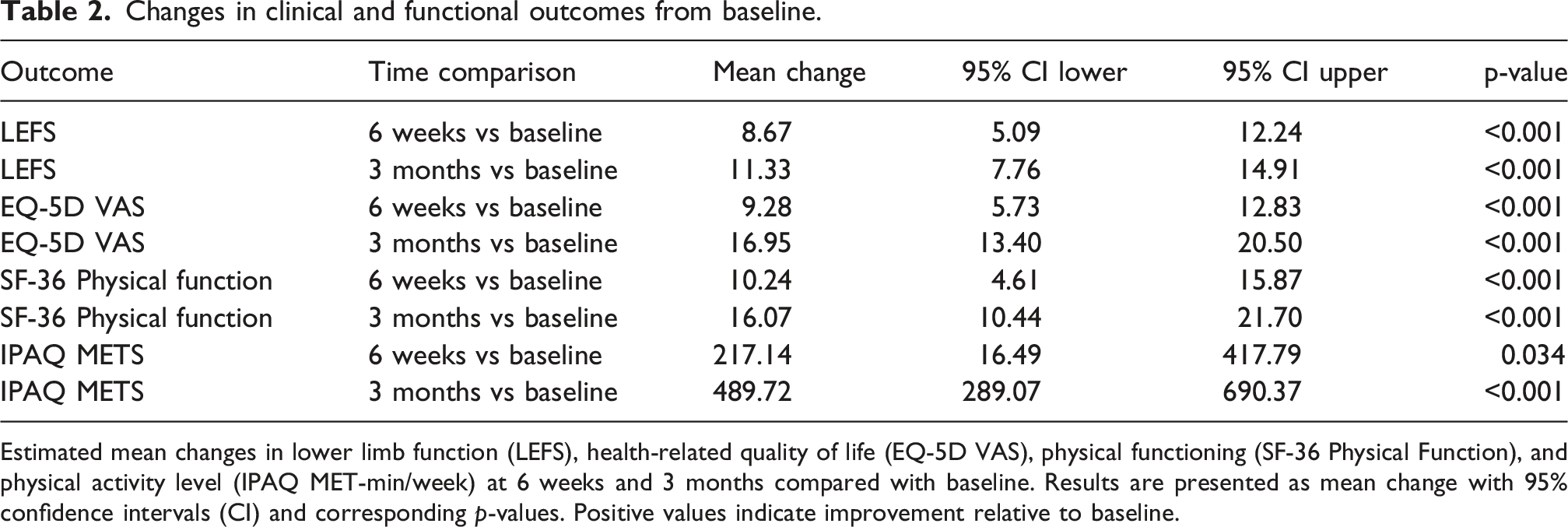
Estimated mean changes in lower limb function (LEFS), health-related quality of life (EQ-5D VAS), physical functioning (SF-36 Physical Function), and physical activity level (IPAQ MET-min/week) at 6 weeks and 3 months compared with baseline. Results are presented as mean change with 95% confidence intervals (CI) and corresponding p-values. Positive values indicate improvement relative to baseline.
Health-related quality of life (EQ-5D VAS)
Baseline EQ-5D VAS was 44.0 (SD 20.3; n = 16). Mean scores increased to 53.9 (SD 17.6; n = 12) at 6 weeks and to 61.6 (SD 21.4; n = 12) at 3 months (Table 1).
The mixed-effects model demonstrated significant improvements in EQ-5D VAS compared with baseline, with a mean increase of 9.3 points at 6 weeks (95% CI 5.7 to 12.8; p < .001) and 16.9 points at 3 months (95% CI 13.4 to 20.5; p < .001) (Table 2).
Physical function (SF-36 physical function)
Mean SF-36 Physical Function score at baseline was 39.3 (SD 18.3; n = 16). Scores increased to 51.2 (SD 17.9; n = 12) at 6 weeks and to 57.0 (SD 21.1; n = 12) at 3 months (Table 1).
In the mixed-effects model, SF-36 Physical Function improved significantly over time, with increases of 10.2 points at 6 weeks (95% CI 4.6 to 15.9; p < .001) and 16.1 points at 3 months (95% CI 10.4 to 21.7; p < .001) compared with baseline (Table 2).
Physical activity (IPAQ METS)
Baseline physical activity measured by IPAQ METS was 742.3 MET-min/week (SD 688.5; n = 16). Mean values increased to 898.3 MET-min/week (SD 778.7; n = 12) at 6 weeks and to 1170.8 MET-min/week (SD 905.0; n = 12) at 3 months (Table 1).
The mixed-effects model showed a significant increase in physical activity over time. Compared with baseline, IPAQ METS increased by 217.1 MET-min/week at 6 weeks (95% CI 16.5 to 417.8; p = .034) and by 489.7 MET-min/week at 3 months (95% CI 289.1 to 690.4; p < .001) (Table 2).
Ultrasound thickness
Longitudinal changes in subcutaneous thickness measured by ultrasound at the thigh and leg are shown in Figure 2. Individual trajectories and group means suggested a modest reduction over time, more evident at the thigh than at the leg. Ultrasound-measured subcutaneous tissue thickness over time. Longitudinal changes in ultrasound-measured subcutaneous tissue thickness (mm) from baseline to 6 weeks and 3 months. Values at 3 months are based on n = 3 and should be interpreted with caution. Left panel: thigh (secondary site). Right panel: leg (primary site). Thin coloured lines represent individual patients, while bold lines with markers indicate group means for the treated and control legs. Error bars represent standard deviation.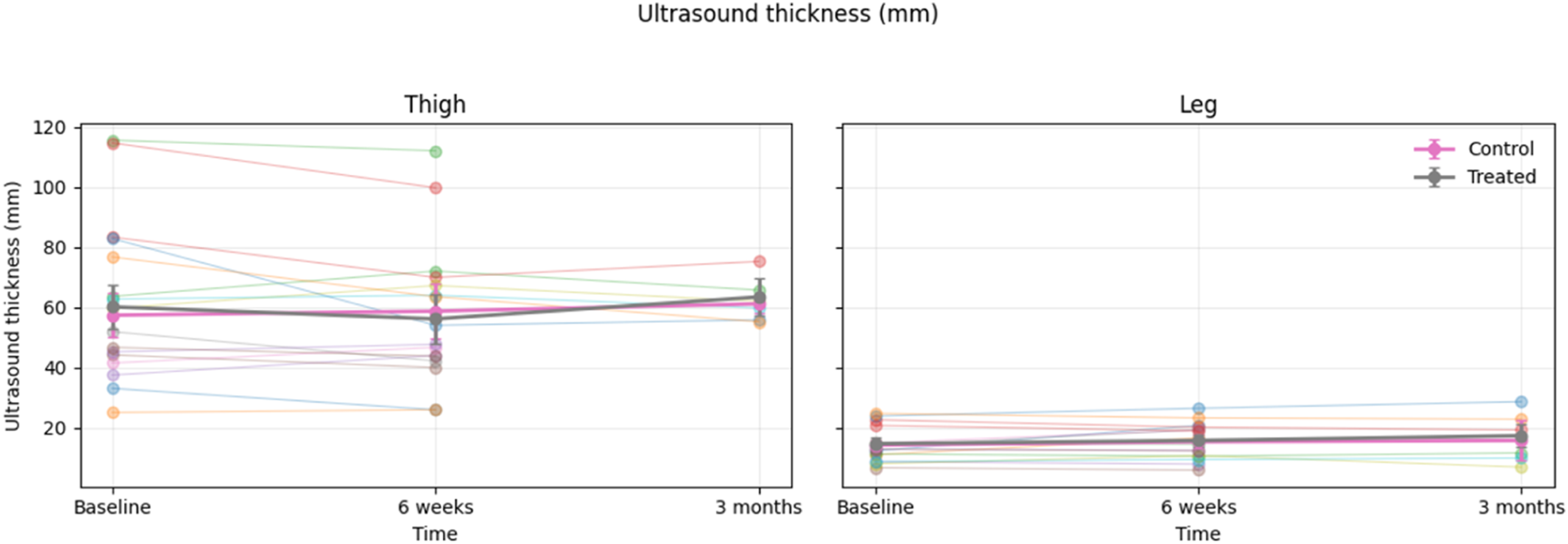
Ultrasound thickness and elastography measurements over time.
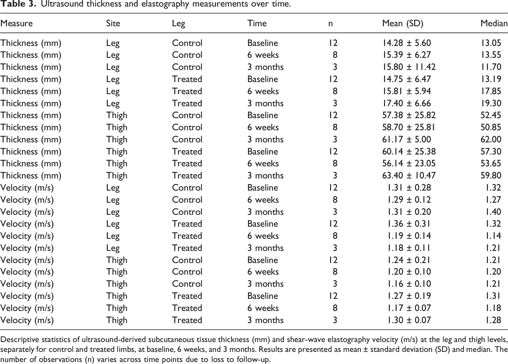
Descriptive statistics of ultrasound-derived subcutaneous tissue thickness (mm) and shear-wave elastography velocity (m/s) at the leg and thigh levels, separately for control and treated limbs, at baseline, 6 weeks, and 3 months. Results are presented as mean ± standard deviation (SD) and median. The number of observations (n) varies across time points due to loss to follow-up.
Shear-wave elastography velocity
Elastography results are presented in Figure 3. Both thigh and leg showed small changes over time, with a general tendency toward lower velocities at follow-up. Elastography shear-wave velocity over time. Longitudinal changes in shear-wave elastography velocity (m/s) from baseline to 6 weeks and 3 months. Values at 3 months are based on n = 3 and should be interpreted with caution. Left panel: thigh (secondary site). Right panel: leg (primary site). Thin coloured lines represent individual patients, while bold lines with markers indicate group means for the treated and control legs. Error bars represent standard deviation.
Exploratory ordinary least squares models did not detect statistically significant effects of time, treatment, or time × treatment interaction at either anatomical site. (Table 3). At the thigh, the estimated change at 3 months compared with baseline was −0.077 m/s (95% CI −0.286 to 0.131; p = .458). At the leg, no meaningful temporal variation was observed (Figures 4 to 6). Segmental bioimpedance parameters over time. Longitudinal changes in segmental bioimpedance-derived variables from baseline to 6 weeks and 3 months. Values at 3 months are based on n = 3 and should be interpreted with caution. (A) Segmental water content (L). (B) Segmental lean mass (kg). (C) Phase angle (°). (D) Extracellular water ratio (ECW/TBW). Thin coloured lines represent individual patients, while bold lines with markers indicate group means for the treated and control legs. Error bars represent standard deviation. Changes in segmental bioimpedance parameters at follow-up. Changes (Δ) in segmental bioimpedance-derived variables from baseline to 6 weeks and from baseline to 3 months, comparing treated and control limbs. (A) Change in segmental water content (L). (B) Change in segmental lean mass (kg). (C) Change in phase angle (°). (D) Change in extracellular water ratio (ECW/TBW). Points represent group means for treated and control legs, with error bars indicating standard deviation. Thin lines correspond to individual patient trajectories. The horizontal dashed line indicates no change from baseline. Association between changes in elastography and phase angle at 6 weeks. Scatter plots showing the relationship between the change in shear-wave elastography velocity and the change in bioimpedance phase angle from baseline to 6 weeks. (A) Leg. (B) Thigh. Spearman correlation coefficients (r), p-values and sample size (N) are shown in each panel. The solid line represents the fitted linear trend, with the shaded area indicating the 95% confidence interval. Each point corresponds to an individual patient.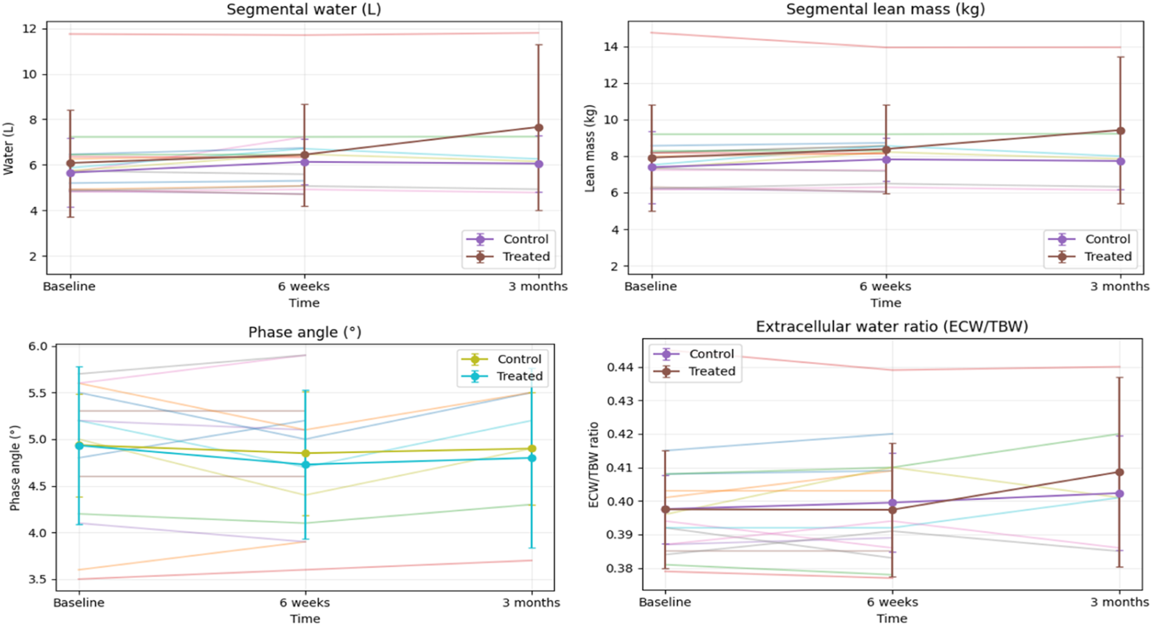


Segmental bioimpedance outcomes
Segmental bioimpedance analysis parameters over time.
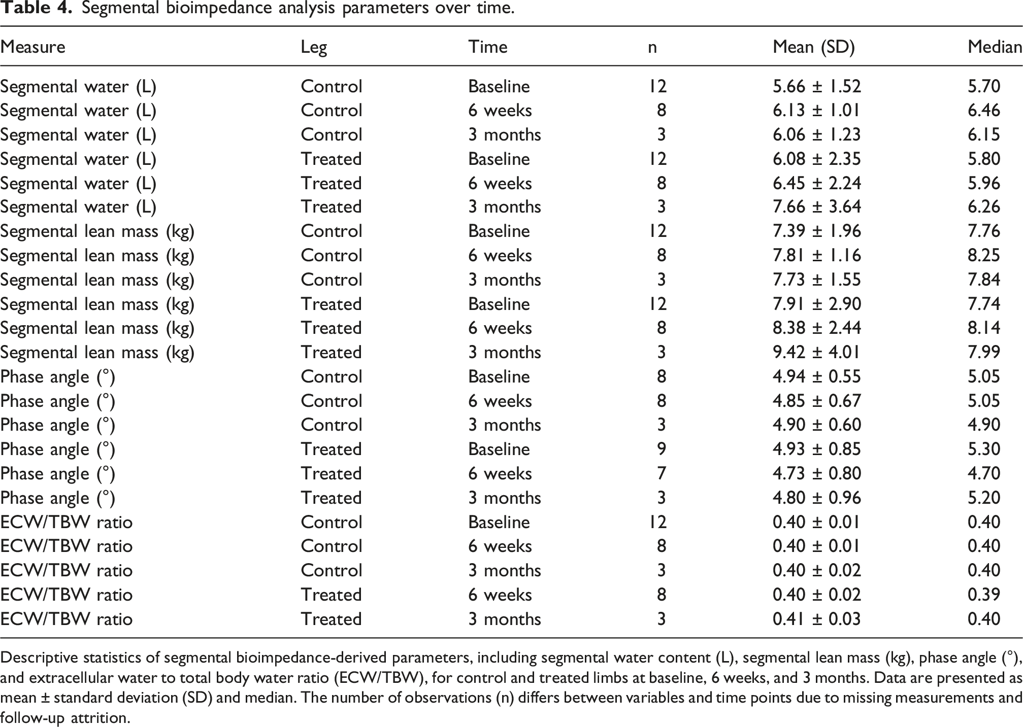
Descriptive statistics of segmental bioimpedance-derived parameters, including segmental water content (L), segmental lean mass (kg), phase angle (°), and extracellular water to total body water ratio (ECW/TBW), for control and treated limbs at baseline, 6 weeks, and 3 months. Data are presented as mean ± standard deviation (SD) and median. The number of observations (n) differs between variables and time points due to missing measurements and follow-up attrition.
Segmental water and lean mass tended to increase slightly over time in the treated limb, while remaining relatively stable in the control limb. Phase angle showed small transient reductions at 6 weeks, with partial recovery at 3 months. ECW/TBW ratio remained stable over time in both limbs.
Mixed-effects models
Longitudinal mixed-effects and regression models for imaging and bioimpedance outcomes.
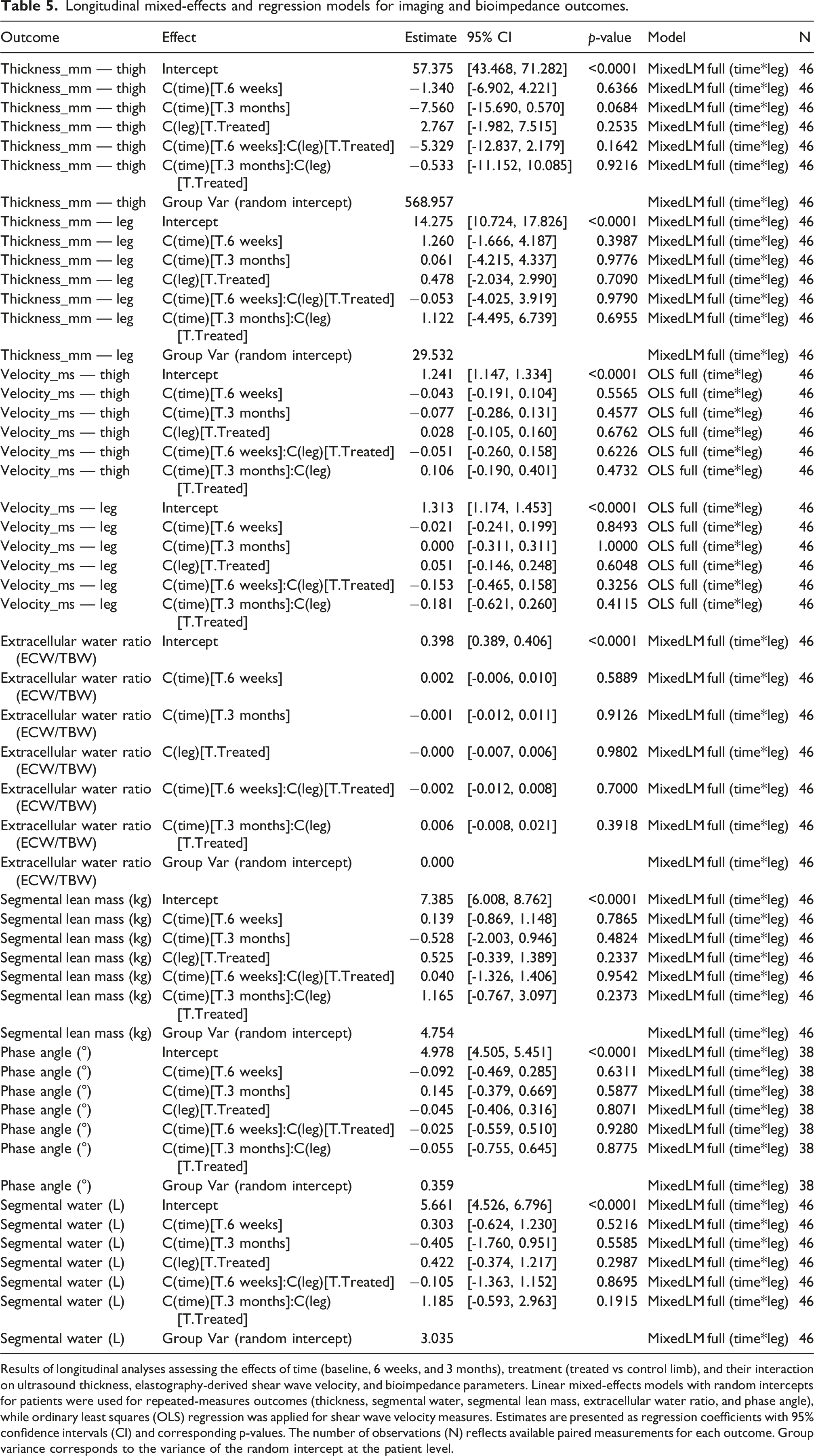
Results of longitudinal analyses assessing the effects of time (baseline, 6 weeks, and 3 months), treatment (treated vs control limb), and their interaction on ultrasound thickness, elastography-derived shear wave velocity, and bioimpedance parameters. Linear mixed-effects models with random intercepts for patients were used for repeated-measures outcomes (thickness, segmental water, segmental lean mass, extracellular water ratio, and phase angle), while ordinary least squares (OLS) regression was applied for shear wave velocity measures. Estimates are presented as regression coefficients with 95% confidence intervals (CI) and corresponding p-values. The number of observations (N) reflects available paired measurements for each outcome. Group variance corresponds to the variance of the random intercept at the patient level.
No statistically significant time-by-leg interactions were detected for ultrasound thickness, elastography shear-wave velocity, segmental water, lean mass, phase angle or ECW/TBW ratio. These findings indicate that changes over time were not significantly different between treated and control limbs within the limits of sample size and follow-up duration.
Associations between imaging and bioimpedance changes
Correlation between early changes in elastography and bioimpedance phase angle.
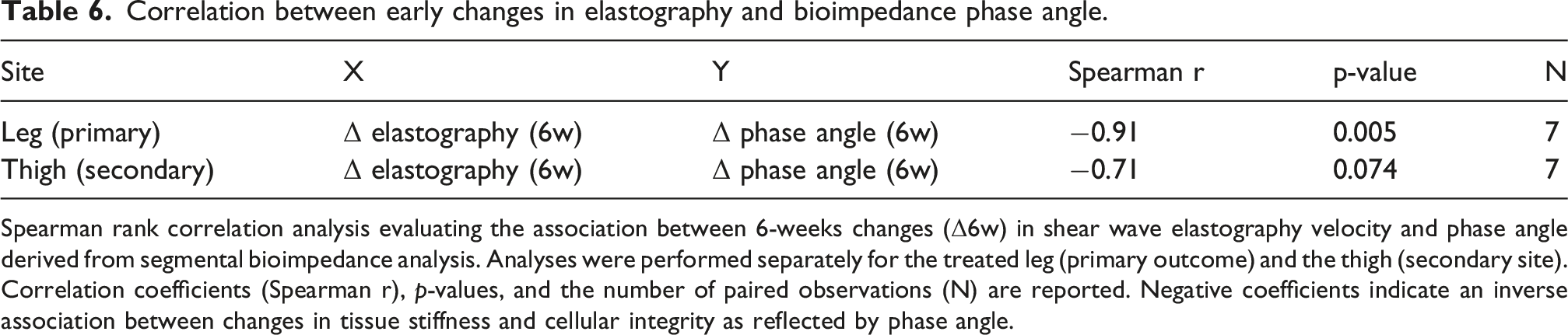
Spearman rank correlation analysis evaluating the association between 6-weeks changes (Δ6w) in shear wave elastography velocity and phase angle derived from segmental bioimpedance analysis. Analyses were performed separately for the treated leg (primary outcome) and the thigh (secondary site). Correlation coefficients (Spearman r), p-values, and the number of paired observations (N) are reported. Negative coefficients indicate an inverse association between changes in tissue stiffness and cellular integrity as reflected by phase angle.
At 3 months, correlation analyses were limited by the small number of available observations and did not yield statistically interpretable results.
Discussion
In this prospective intra-subject study, radial shock wave therapy (rESWT) applied in the right limb (control in left limb) in women with lipedema was associated with a clinically meaningful improvement in functional capacity, health-related quality of life, and physical activity levels over short- and mid-term follow-up. These clinical benefits were accompanied by an absence of statistically significant changes in imaging and bioimpedance parameters, suggesting that functional improvement may precede or occur independently of detectable structural changes within the limits of the available sample.
From a clinical standpoint, patients showed significant improvements in LEFS, EQ-5D VAS, SF-36 Physical Function, and IPAQ scores at both 6 weeks and 3 months compared with baseline. Baseline IPAQ values showed considerable inter-individual variability, which is common in lipedema populations because of differences in baseline mobility, pain severity, and habitual physical activity. Therefore, the IPAQ findings should be interpreted cautiously. Changes in LEFS and EQ-5D surpassed established minimal clinically important difference thresholds for musculoskeletal and chronic conditions, indicating that the observed effects are clinically meaningful rather than merely statistically significant. These findings align with previous studies reporting symptomatic improvements in pain, mobility, and quality of life after conservative or physical treatments in lipedema, even in the absence of substantial objective changes in tissue composition or volume.3,5
Interestingly, imaging outcomes evaluated by ultrasound and elastography did not show statistically significant time × leg interactions in the mixed-effects models. Across follow-up, subcutaneous thickness and shear wave velocity showed minimal change in both the treated and control limbs. This pattern aligns with the chronic, fibrotic nature of lipedematous adipose tissue, which typically exhibits a slow response to conservative treatments. 22 Accordingly, rESWT may primarily produce neuromodulatory, microcirculatory, or anti-inflammatory effects, rather than prompt macroscopic alterations in adipose tissue structure.
A recent prospective study by Bruno and Cilluffo reported that shock wave therapy after liposuction in women with stage II–III lipedema reduced postoperative fibrosis and improved skin elasticity, pain, and patient satisfaction. 23 However, that study evaluated shock wave therapy as an adjunctive postoperative intervention after surgical treatment, whereas the present study investigated rESWT as a conservative intervention in non-operated patients. In addition, the previous study focused primarily on fibrosis-related postoperative changes, while our work combined patient-reported outcomes with ultrasound, elastography, and segmental bioimpedance. Although both studies support a beneficial clinical effect of shock wave therapy in lipedema, our findings suggest that, in the conservative setting, clinically meaningful improvement may occur despite the absence of statistically significant changes in tissue thickness, stiffness, or body composition. This difference may reflect distinct mechanisms according to whether shock wave therapy is applied to postoperative fibrotic tissue or to untreated lipedematous tissue.
A notable exploratory finding was the inverse association between changes in elastography and phase angle at 6 weeks in the treated leg. Phase angle is regarded as a marker of cell membrane integrity and tissue quality, whereas elastography reflects tissue mechanical stiffness. The observed inverse relationship may suggest that early biomechanical adaptations induced by rESWT are accompanied by changes in tissue quality, even in the absence of overt morphological modification. However, because multiple exploratory comparisons were performed without formal adjustment for multiplicity, this association should be interpreted cautiously and requires confirmation in larger studies. The analysis was based on only seven participants with complete paired data, was restricted to the treated limb. Therefore, this finding should be considered purely exploratory and hypothesis-generating and should not be interpreted as evidence of an underlying mechanistic effect. Comparable dissociations between functional improvement and stable imaging findings have been described in other chronic soft tissue disorders treated with shock wave therapy.24,25
Bioimpedance analysis did not reveal statistically significant differences over time between treated and control limbs in segmental water content, lean mass, or the ECW/TBW ratio. This stability is clinically relevant, as it indicates that the intervention did not produce adverse fluid shifts or worsen extracellular fluid accumulation, which is a common concern in the management of lipedema. The consistently stable ECW/TBW ratio around 0.40 across limbs and time points may reflect genuine stability in extracellular fluid distribution. However, because this parameter has a narrow physiological range and limited variability, the absence of detectable change may also partly reflect the restricted sensitivity of segmental bioimpedance to identify subtle fluid shifts in a small sample. Previous reports have noted the limited sensitivity of bioimpedance to detect subtle regional changes in lipedema, particularly in small cohorts, which may partially account for these results. 26
The discrepancy between robust clinical improvement and minimal objective tissue changes reinforces the concept that patient-reported outcomes are critical endpoints in lipedema research. Functional impairment, pain, and reduced quality of life are the core drivers of disability in this condition, often outweighing purely anatomical considerations. 7 Therefore, interventions capable of improving function and perceived health status, even without measurable structural change, may still hold substantial therapeutic value.
A number of limitations need to be considered. While 16 patients were initially recruited, only 12 completed all scheduled follow-up evaluations and were included in the final analysis. This loss to follow-up illustrates the practical difficulties of conducting longitudinal research in chronic diseases and limits statistical power, particularly for imaging-based and correlation analyses at the 3-months time point. In addition, the relatively short follow-up period may not adequately capture delayed structural changes in adipose or connective tissues. Finally, despite the strength of the intra-subject control design, the absence of a sham-treated limb prevents firm attribution of the observed effects solely to rESWT.
Notwithstanding its limitations, this study has important strengths, such as standardized evaluations performed by the same clinicians, a multimodal approach combining clinical, imaging, and bioimpedance measures, and the use of a clearly defined rESWT protocol based on international guidelines. These findings support the integration of rESWT within conservative treatment pathways focused on symptom relief and functional improvement rather than volume reduction. Future studies with larger samples, longer follow-up, and sham-controlled designs are warranted to clarify the mechanisms underlying the observed clinical benefits and to determine whether repeated or prolonged treatment courses may induce measurable structural changes. The limited sample size also reduced statistical power for imaging and bioimpedance endpoints. A retrospective post-hoc power analysis for the primary imaging outcome demonstrated a very small observed effect size (Cohen’s d = 0.078) and an estimated power of only 5.7%, indicating that the study was underpowered to detect small between-limb differences. Therefore, non-significant findings should be interpreted cautiously and should not be considered evidence of absence of treatment effect. In addition, elastography velocity had to be analysed using ordinary least squares models because mixed-effects models did not converge. Consequently, the repeated nature of these measurements may not have been fully captured, and the corresponding standard errors may have been underestimated. Bioimpedance data at 3 months were available in only three participants because of logistical constraints in follow-up scheduling. Therefore, the missing-at-random assumption may not be fully satisfied, and these analyses should be interpreted with caution. In particular, the correlation between elastography and phase angle was based on only seven complete paired observations and was therefore highly vulnerable to the influence of individual data points. Lipedema stage was not systematically recorded, which may limit the interpretation of imaging findings because tissue characteristics may differ across disease stages. Although no new conservative interventions were initiated during follow-up, continuation of usual background management may have contributed in part to the observed clinical changes.
Conclusion
In conclusion, rESWT appears to be a safe and clinically effective adjunctive treatment for women with lipedema, leading to significant improvements in function, quality of life, and physical activity. These benefits occurred despite the absence of statistically significant changes in imaging and bioimpedance findings, underscoring the importance of patient-centered outcomes in this population. As such, rESWT may be considered a useful element within a multimodal conservative treatment approach for lipedema.
Supplemental material
Supplemental Material - Clinical, ultrasound, elastography and bioimpedance changes after radial extracorporeal shock wave therapy in patients with lipedema: A prospective within-patient study
Supplemental Material for Clinical, ultrasound, elastography and bioimpedance changes after radial extracorporeal shock wave therapy in patients with lipedema: A prospective within-patient study by Manuel Novo Rigueiro, Martín Bravo González, Teresa Prado Moraña, Alberto Pena Dubra, Sara Villarroel Comesaña, Pablo Navarro Núñez, Bibiana Villamayor Blanco and Ignacio Novo Veleiro in Phlebology
Supplemental material
Supplemental Material - Clinical, ultrasound, elastography and bioimpedance changes after radial extracorporeal shock wave therapy in patients with lipedema: A prospective within-patient study
Supplemental Material for Clinical, ultrasound, elastography and bioimpedance changes after radial extracorporeal shock wave therapy in patients with lipedema: A prospective within-patient study by Manuel Novo Rigueiro, Martín Bravo González, Teresa Prado Moraña, Alberto Pena Dubra, Sara Villarroel Comesaña, Pablo Navarro Núñez, Bibiana Villamayor Blanco and Ignacio Novo Veleiro in Phlebology
Footnotes

Ethical consideration
This was a prospective, longitudinal, within-patient study conducted in women diagnosed with lipedema. Initially, 16 patients were initially assessed for eligibility. 12 patients completed the predefined follow-up protocol and were therefore included in the final analysis. Patients not included did not complete all scheduled imaging and bioimpedance assessments. This approach ensured consistency and completeness of longitudinal data. The study was carried out at the University Hospital Complex of Santiago de Compostela during 2023. The study was approved by the Research Ethics Committee of Galicia (approval code 2023/004).
Consent to participate
All participants provided written informed consent prior to inclusion. The study was conducted in accordance with the Declaration of Helsinki.
Author Contributions
1Manuel Novo Rigueiro: Conceptualization; Methodology; Investigation; Data curation; Formal analysis; Writing – original draft; Writing – review & editing; Supervision.
2Martín Bravo González: Investigation; Data curation; Writing – review & editing.
3Teresa Prado Moraña: Investigation; Imaging assessment; Data curation; Writing – review & editing.
4Alberto Pena Dubra: Conceptualization; Methodology; Writing – review & editing.
5Sara Villarroel Comesaña: Formal analysis; Statistical analysis; Data interpretation; Writing – review & editing.
6Pablo Navarro Núñez: Investigation; Data collection; Writing – review & editing.
7Bibiana Villamayor Blanco: Conceptualization; Critical revision of the manuscript; Writing – review & editing.
8Ignacio Novo Veleiro: Conceptualization; Supervision; Critical revision of the manuscript; Writing – review & editing.
All authors read and approved the final version of the manuscript and agree to be accountable for all aspects of the work.
Funding
The authors received no financial support for the research, authorship, and/or publication of this article.
Declaration of conflicting interests
The authors declared no potential conflicts of interest with respect to the research, authorship, and/or publication of this article.
Data Availability Statement
The datasets generated and analyzed during the current study are available from the corresponding author on reasonable request.
AI declaration
As non-native English speakers, the authors used artificial intelligence tools exclusively for language editing and readability improvement. All content was generated, reviewed, and approved by the authors, who take full responsibility for the accuracy and integrity of the manuscript.
Supplemental material
Supplemental material for this article is available online.
References
Supplementary Material
Please find the following supplemental material available below.
For Open Access articles published under a Creative Commons License, all supplemental material carries the same license as the article it is associated with.
For non-Open Access articles published, all supplemental material carries a non-exclusive license, and permission requests for re-use of supplemental material or any part of supplemental material shall be sent directly to the copyright owner as specified in the copyright notice associated with the article.
