



Abstract
The recombinant enzyme glutamate oxaloacetate transaminase (rGOT) has emerged as a promising neuroprotective candidate for acute ischaemic stroke. This study examined whether rGOT can attenuate the neurotoxic side effects of recombinant tissue plasminogen activator (rtPA) and enhance the therapeutic benefit of thrombolytic reperfusion. First, primary cortical neurons were exposed to rtPA with or without rGOT under normoxia or oxygen–glucose deprivation. rGOT did not reduce rtPA-induced cytotoxicity, indicating that the pathways underlying rtPA neurotoxicity are mechanistically distinct from those modulated by rGOT. Second, a thromboembolic mouse model was used to assess the interaction between rGOT and rtPA across clinically relevant treatment scenarios. rGOT alone or when administered prior to rtPA had no effect on infarct size or haemorrhagic transformation. However, co-administration of rGOT with rtPA significantly reduced infarct volume at 24 h (p < 0.05), without increasing bleeding risk. These findings demonstrate that rGOT is compatible with thrombolytic therapy and can enhance rtPA efficacy when delivered during reperfusion, supporting its potential use as an adjuvant treatment in acute ischaemic stroke.
Introduction
Reperfusion therapies, including intravenous thrombolysis using recombinant tissue plasminogen activator (rtPA) and, more recently, mechanical endovascular thrombectomy (MET), are currently the most widely accepted reperfusion therapies in clinical practice for the acute phase of ischaemic stroke. Both therapeutic strategies are restricted to patients who present within 4.5–6 h of symptom onset, although MET may be offered up to 24 h in carefully selected cases.1,2
Many pharmacological and non-pharmacological strategies have been developed to counteract the early stages of the ischaemic cascade, a period that typically overlaps with the administration of recanalization therapies. Therefore, rather than evaluating cytoprotective agents as isolated treatments, current recommendations emphasise the need to determine whether new protective candidates can be effectively implemented as adjuncts to reperfusion therapies. This approach is essential not only to identify potential synergistic effects, but also to detect any pharmacodynamic antagonism that could compromise recanalization before progressing to costly clinical trials.3–5 A clear example is the ESCAPE-NA1 trial, which evaluated nerinetide, a promising neuroprotective peptide targeting the PSD-95/N-methyl-D-aspartate (NMDA) receptor (NMDAR) to limit excitotoxicity. Although thrombolytic efficacy remained intact, nerinetide lost therapeutic benefit specifically in rtPA-treated patients due to plasmin-mediated cleavage of its C-terminal sequence.5,6
The recombinant form of the circulating enzyme glutamate oxaloacetate transaminase (rGOT) has emerged as a promising neuroprotective candidate for the acute phase of stroke, owing to its ability to neuronal protection. 7 In previous studies, rGOT protection was first demonstrated in the transient middle cerebral artery occlusion (tMCAO) model, which recreates the clinical scenario of arterial recanalization after mechanical thrombectomy. 8 In this model, due to the short circulating half-life of the drug in blood (~2 h), four consecutive doses of 1 mg/kg rGOT administered during the first 8 h were established as the most protective protocol. 7 In the same study, according to the importance of testing neuroprotection in combination with reperfusion therapies, the effect of rGOT on pharmacological thrombolysis was explored. In vitro analysis revealed that rGOT did not alter rtPA-mediated fibrinolytic activity in a dose-dependent manner, and rtPA did not compromise rGOT enzymatic activity. The absence of pharmacological interactions between these drugs was later confirmed in vivo in both healthy and ischaemic rodents undergoing surgery involving embolic middle cerebral artery occlusion with blood clots. The administration of rGOT was carried out in two distinct clinical scenarios; the first corresponded to a prehospital setting, in which rGOT was administered after stroke suspicion and before thrombolytic therapy. The second represented an in-hospital scenario, where rGOT and rtPA were administered simultaneously. In both cases, the pharmacological activities of both drugs were not affected. 7
Despite its proven fibrinolytic efficacy, rtPA can exert detrimental effects after stroke, including an increased risk of haemorrhagic transformation and the induction of NMDAR-mediated neurotoxicity.9,10 Whether the neuroprotective actions of rGOT can mitigate these well-described rtPA-induced toxic effects or enhance the therapeutic benefit of thrombolysis, however, remains insufficiently explored.
To address this question, we adopted a two-step experimental approach. First, primary mouse cortical neurons were exposed to rtPA, with or without rGOT, under both normoxic and oxygen–glucose deprivation (OGD) conditions to determine whether the rGOT enzyme is capable of modulating rtPA-induced cytotoxicity. Second, we employed a cerebral in in situ thromboembolic mouse stroke model, widely accepted for evaluating recanalization dynamics comparable to those observed in patient,11–16 to assess whether rGOT can enhance the benefits of thrombolytic recanalization when administered in combination with rtPA.
Methods
Ethics statement
Male Swiss mice (Harlan Laboratories) weighing 25–30 g were used for in vivo assays. Mice were kept in separate rooms under controlled temperature (22 °C ± 1 °C) and humidity (60% ± 5%) with a 12/12-h light/dark cycle for a week prior to surgery and up to 14 days after surgery. The animals had access to food and water ad libitum. All procedures were performed under anaesthesia induced by inhalation of 5% sevoflurane in a nitrous oxide/oxygen mixture (70/30). The rectal temperature was monitored and maintained at 37 °C ± 0.5 °C using a feedback-controlled heating system. At the end of the procedure, the mice were sacrificed under deep anaesthesia (8% sevoflurane). The experimental protocol was approved by the Local Animal Experimental Committee (Health Research Institute of Santiago de Compostela (IDIS) Animal Care Committee) and the animal experiments were conducted under the procedure number 15011/2024/006 and according to the Spanish and European Union rules (86/609/CEE, 2003/65/CE, 2010/63/EU, RD 1201/2005 and RD 53/2013). The studies followed the Animal Research: Reporting In Vivo Experiments (ARRIVE) guidelines.
Primary cortical neuron cultures
Primary neuronal cells were cultured according to a previous protocol. 9 Cortices from embryonic day 15 Swiss mice were dissociated with 0.02% trypsin for 15 min at 37 °C, triturated and plated at 2 × 10⁵ cells/cm2 on poly‑D‑lysine‑coated plates in Neurobasal medium supplemented with 2% B‑27, 2 mM L‑glutamine and 1% penicillin‑streptomycin. Cultures matured for 8–9 days at 37 °C in 5% CO2. Neuronal purity was determined by β‑III‑tubulin (IC1195A; R&D Systems, Minneapolis, USA) verified by flow cytometry (FACSAria II) after fixation/permeabilization and concentration 104 cells/µL. The analysis was performed using a FACSDiva software (BD Biosciences, Denmark). Unlabelled cells were used as negative controls.
Toxicity and viability assays
Cell viability was determined via 3-(4,5-dimethylthiazol-2-yl)-2,5-diphenyl-2H-52 tetrazolium bromide (MTT; Sigma–Aldrich, EE. UU) precipitation. Toxicity based on cell death was evaluated by measuring the leakage of lactate dehydrogenase (LDH) into the medium using a commercial kit (CyQUANT LDH Cytotoxicity Assay Kit; Gibco–Invitrogen, OR, USA), according to manufacturer’s protocol.
The experiments were performed under normoxic and OGD conditions. For OGD, cultures were transferred to glucose‑free medium and placed in an anaerobic chamber continuously gassed with 95% N2/5% CO2 at 37 °C for 60 min; normoxic controls remained in complete medium. Immediately after OGD or normoxia, cells received: (1) vehicle (phosphate buffered saline (PBS)), (2) 100 µg/mL rtPA (Actilyse, Boehringer, Germany), (3) 5 µg/mL rGOT, (4) 10 µg/mL rGOT, (5) 100 µg/mL rtPA + 5 µg/mL rGOT and (6) 100 µg/mL rtPA + 10 µg/mL rGOT.
rGOT was administered in four doses of 5 or 10 µg/mL. The first dose was administered at the end of normoxia or OGD and 2, 5 and 8 h later. The groups that were not treated with rGOT received the same volume of PBS as a control. This four-dose rGOT regimen corresponds to the dosing sequence that we have previously demonstrated to be effective in vivo. 7 rtPA dose represents the concentration in the blood of mice treated 10 mg/kg of rtPA and that showed neuronal in vitro toxicity. 9 rGOT was produced and supplied by BiotechPharma UAB (Vilnius, Lithuania) using E. coli as host cells. See Perez-Mato et al. 7 for protein synthesis procedure.
GOT measurement in blood
GOT levels in blood were measured before the surgery and 8 h after the first treatment administration. The blood was extracted from the jugular vein of mice and the GOT and Reflotron system (Reflotron® plus; Roche, Switzerland) was used to measure GOT levels. The analysis was conducted following the manufacturer’s instructions. In general, 32 µL of blood were added to reactive stripes for GOT (10745120202; Roche).
Thromboembolic stroke model
Thromboembolic stroke model was induced by injection if thrombin in the middle cerebral artery (MCA) of mice as originally described by Orset et al. 12 in Swiss male mice with minimal modifications adapted to our conditions. 17 Briefly, mice were placed in a stereotaxic frame, the skin between the right ear and eye was cut, the temporal muscle was retracted and the temporal and parietal bones exposed. A small craniotomy was performed over the artery bifurcation, the meninges were cut using a 25 G needle (BD Microlance, Italy) and the MCA was exposed. A micropipette (tip size: 20–40 µm), made with haematologic glass capillaries (World Precision Instruments, FL, USA) using a puller (Sutter Instruments, CA, USA), was pneumatically filled with 1.5 µL of 1.5 U/µL thrombin (murine thrombin 0.05 mg MIIA; Stago-BNL, Belgium). The micropipette was placed in a micromanipulator and 1 µL of thrombin solution was injected into the lumen of the artery bifurcation to induce the formation of a clot. The micropipette was removed 15 min later, when the clot was stabilised.
Cerebral blood flow (CBF) was monitored with a Periflux 5000 laser Doppler perfusion monitor (Perimed AB, Sweden) by placing the probe (Perimed AB, Sweden) in the parietal territory of the MCA. Basal CBF and throughout the experiment was measured. The occlusion was considered successful when CBF decreased more than 60%. After the treatment administration the reperfusion was considered when it recovered more than 20% of the occlusion. Magnetic resonance imaging (MRI) was performed 24 h, 7 and 14 days after the surgery to study infarct volumes and the presence of haemorrhages.
Animal experimental groups and treatments administration
Treatments were randomised to simulate five clinically relevant scenarios within the following groups: (1) vehicle, representing a stroke condition without reperfusion or protective treatment; (2) 10 mg/kg rtPA infusion, modelling thrombolytic therapy during acute phase; (3) four boluses of 1 mg/kg rGOT administered over 8 h starting 30 min after ischaemia and 2, 5 and 8 h after this first administration, representing neuroprotection without reperfusion; (4) four boluses of 1 mg/kg rGOT over 8 h starting 20 min before rtPA (and 10 min after ischaemia; 10 mg/kg; rGOTpreR + rtPA), simulating pre-hospital neuroprotection followed by thrombolysis; (5) four boluses of 1 mg/kg rGOT over 8 h simultaneously with rtPA infusion (10 mg/kg; rGOTpostR + rtPA), modelling concurrent administration of both therapies in a hospital setting.
For rGOT, we have previously demonstrated that four continuous intravenous doses of rGOT (1 mg/kg) administered within the first 8 h after stroke onset was requited to induce significant protection effect, 7 therefore similar protocol was selected to test the effect in combination with rtPA. In the case of group rGOTpreR + rtPA the first administration was performed 10 min after the onset of the ischaemia (pre-reperfusion) and 2, 5 and 8 h after. In the group rGOTpostR + rtPA, rGOT was administered 30 min after the ischaemia, in combination with rtPA infusion (post-reperfusion), and 2, 5 and 8 h after.
Doses of rtPA was selected based on our previous study to evaluate arterial recanalization on the thrombolytic drug on the same ischaemic model. 9 Note that, since mice are less sensitive to thrombolytic therapies than humans, these animals require doses 10-fold higher than the established clinical dose (0.9 mg/kg), as we have recently reported. 9 rtPA was administered intravenously, 10% as bolus and 90% as infusion during 30 min using a micro-pump (4.5 µL/min). Due to rtPA requires a visual infusion administration through jugular vein, the researchers were not blinded to the treatments.
Magnetic resonance imaging
MRI was performed at 24 h, 7 and 14 days after ischaemia induction to evaluate infarct size and the presence of haemorrhages, following the same imaging protocol previously used for rtPA analysis in this thromboembolic ischaemic model. 9 MRI studies were conducted on a 9.4 T horizontal bore magnet (BrukerBioSpin) with 12 cm wide actively shielded gradient coils (440 mT/m). Radiofrequency transmission was achieved using a birdcage volume resonator and the signal was detected using a two-element arrayed surface coil (RAPID Biomedical), positioned over the head of the animal, which was fixed with a tooth bar, earplugs and adhesive tape. The respiratory frequency and body temperature were monitored during the experiments. The transmission and reception coils are actively decoupled from each other. Gradient echo pilot scans were performed at the beginning of each imaging session to accurately position the animals inside the magnetic bore. The progression of ischaemic lesions and infarct volumes was determined from T2-maps calculated from T2-weighted images. Ischaemic lesions were identified by counting the pixels with apparent T2-map values above the threshold in the ipsilateral brain hemisphere. In the ipsilateral ischaemic hemisphere, hyperintensity on the T2-map determined the analysis of the ischaemic damage (T2 map values >60 ms). T2-weighted images were acquired using a multi-slice multi-echo (MSME) sequence with a 11 ms echo time (TE), 2.8 s repetition time (TR), 12 echoes with 11 ms echo spacing, FA of 180°, two averages, 50 KHz spectral bandwidth (SW), 16 slices of 0.5 mm, 19.2 × 19.2 mm2 field of view (FOV) with saturation bands to suppress signal outside this FOV, and a matrix size of 256 × 256 (isotropic in-plane resolution of 75 × 75 μm/pixel) and implemented without fat suppression option. The acquisition time was 23 min.
The presence of brain haemorrhage was evaluated using T2*-weighted imaging. T2*-weighted images were acquired using a multi-gradient-echo sequence (MGE) with a 5 ms TE, 1.2 s TR, eight echoes with 4.5 ms echo spacing, 100 KHz spectral bandwidth, FA of 20°, 16 slices of 0.55 mm, two averages, 19.2 × 19.2 mm2 FOV with saturation bands to suppress signal outside this FOV and a matrix size of 256 × 256 (isotropic in-plane resolution of 75 × 75 μm/pixel) and implemented with fat suppression option. The acquisition time was 10 min.
Images were processed using ImageJ (Rasband WS; National Institutes of Health, Bethesda, MD, USA, http://rsb.info.nih.gov/ij/) on an independent computer workstation. Lesion segmentation was performed manually using ImageJ software, following the same criteria consistently applied in our previous studies.9,17 The entire segmentation and quantification process was carried out by a researcher blinded to the treatment groups, ensuring objectivity and minimizing the risk of bias in infarct volume assessment.
Statistical analysis
Data are presented as mean and standard deviation (SD; mean ± SD). Data was first examined to assess distribution using Shapiro–Wilk normality test. Non‑parametric data were compared using Kruskal–Wallis followed by Dunn’s multiple comparison. One-way analysis of variance (ANOVA) followed by Tukey’s multiple comparison test was performed to determine significant differences in parametric data. Significance was set at p < 0.05. Statistical analysis and graphs were generated using GraphPad Prism 10.0.
The sample size for the present study was determined a priori based on variability estimates obtained from our previous studies using the same thromboembolic ischaemia model and identical outcome measures.9,17,18 Infarct volume at 24 h, assessed by MRI, was defined as the primary endpoint for the power calculation, as this variable is the most sensitive and reproducible readout of reperfusion-related effects in this model. Based on these prior datasets, the expected standard deviation of infarct volume ranged between 20% and 25% of the mean. Using these variance estimates, sample size calculations were performed assuming a two-sided significance level (α) of 0.05 and a statistical power of 80% (β = 0.20). Under these assumptions, the calculated group size was sufficient to detect an effect size corresponding to a 20%–30% reduction in infarct volume between experimental groups, which is consistent with the magnitude of effect reported for rtPA-mediated reperfusion and adjunctive neuroprotective strategies in this model. In addition, the calculated sample size incorporated an expected loss of animals based on historical experience with this thromboembolic model, accounting for animals excluded due to surgical complications, absence of stable occlusion or mortality prior to imaging. Researchers were blinded to data analysis.
Results
rtPA toxicity analysis on neuronal culture in combination with rGOT
To assess whether rGOT can counteract the known neurotoxic effects of rtPA, we first analysed their interaction in primary cortical neurons exposed to normoxia or OGD, represented in the Figure 1(a). The different groups were treated 1 h after normoxia or OGD with vehicle, rtPA or rGOT and 2, 5 and 8 h after the first treatment with vehicle or rGOT. Before performing the experiments, flow cytometry analysis confirmed a purity of 90.4% primary cortical neuron cultures (Figure 1(b)).
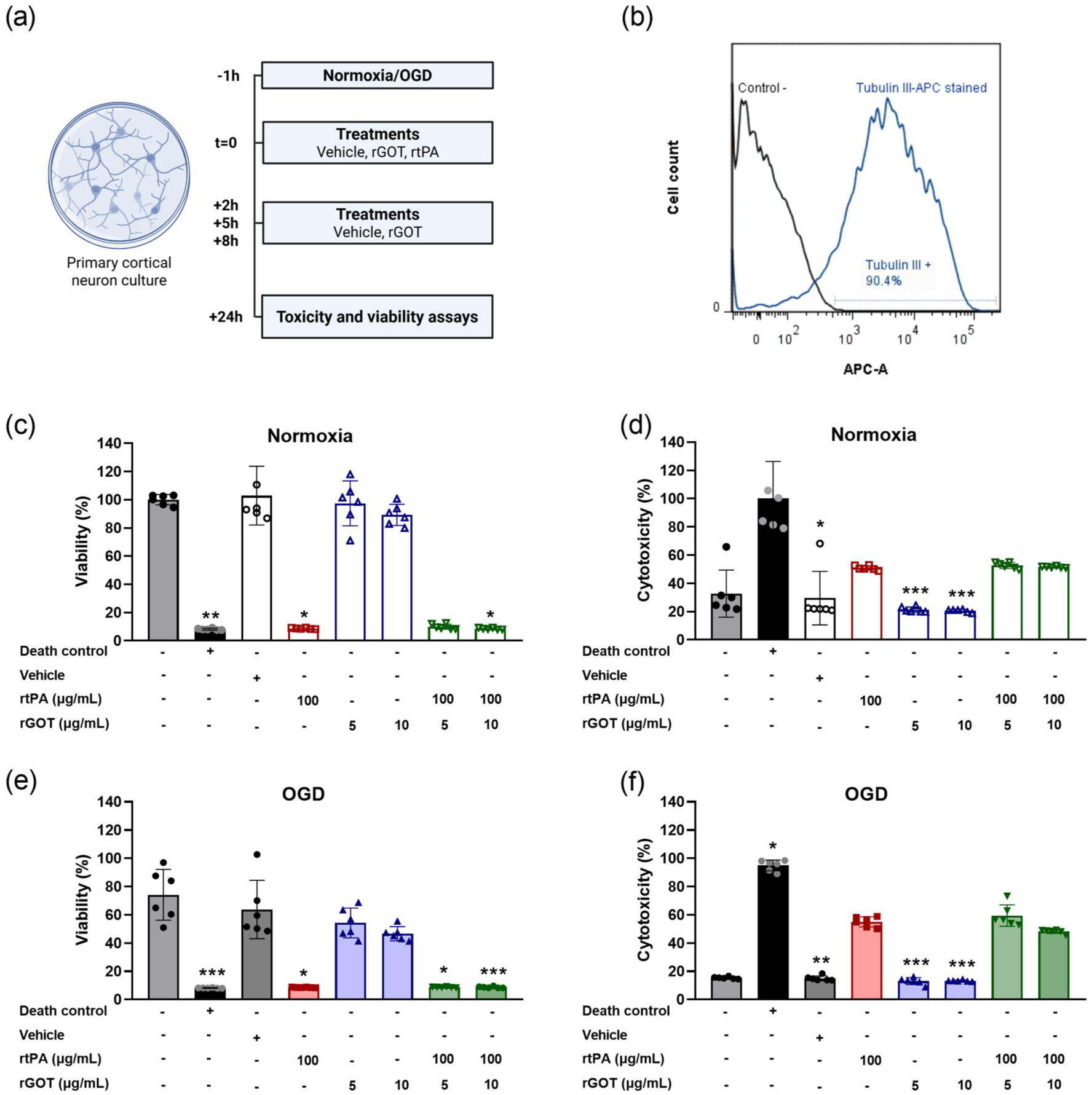
(a) Evaluation of the effect of the rGOT and rtPA combination on primary neuron cultures after 1 h of normoxia or OGD. BioRender (https://biorender.com/), (b) cell phenotype analysis based on flow cytometry. Unlabelled cells were used as a negative control (black line). Blue line corresponds to positive cells for the sample incubated with beta-III tubulin APC antibody. Neuronal viability and cytotoxicity were assessed under (c, d) normoxia and (e, f) OGD conditions. Data are expressed as percentage (%) relative to the normoxic viability control (for viability assays) and to the normoxic cell death control group (for toxicity assays). Results are shown as mean ± SD (n = 6 independent cultures). Statistical analysis was performed using the Kruskal–Wallis test followed by Dunn’s multiple comparisons: *p < 0.05, **p < 0.01 and ***p < 0.001 compared with the control group in the viability assay and with the death control in the toxicity assay.
Under normoxic conditions 100 µg/mL rtPA reduced neuronal viability from 100% ± 20% (vehicle) to 8.7% ± 0.5% (p < 0.01, compared to the viability group) and increased LDH release from 29.5% ± 19.0% (p < 0.05, compared to the death control) to 50.9% ± 1.6% of maximal lysis. rGOT at 5 µg/mL (97.4% ± 15.9% viability; 21.4% ± 1.9% LDH release) or 10 µg/mL (89.2% ± 7.5%; 20.6% ± 1.1%) did not differ from vehicle. Co-administration of rGOT with rtPA failed to rescue neuronal toxicity caused by the thrombolytic drug (viability 10.0% ± 1.8% and 8.6% ± 0.6%; toxicity 52.8% ± 2.2% and 51.6% ± 0.6% for 5 and 10 µg/mL, (Figure 1(c) and (d), respectively). Similar pattern was observed under 1 h of OGD. The vehicle group viability after OGD was 63.8% ± 20.7% with 15.0% ± 1.8% in toxicity assays. rtPA further depressed viability to 8.6% ± 0.2% (p < 0.05, compared to the viability control) and raised toxicity to 55.0% ± 3.6%. Addition of 5 or 10 µg/mL rGOT to 100 µg/mL rtPA did not alter these values in viability (8.8% ± 0.3% and 8.7% ± 0.6%, respectively) and toxicity (59.4% ± 7.6% and 48.2% ± 1.4%, respectively) assays (Figure 1(e) and (f)).
Effect of rGOT and rtPA recanalization in ischaemic thromboembolic model
A total of 73 mice were used (Figure S1 represents the number of included and excluded animals). The interaction between rtPA and rGOT was evaluated in five groups in an ischaemic thromboembolic model (represented in the Figure 2(a)): (1) vehicle (saline), (2) rtPA (10 mg/kg), (3) rGOT (four doses of 1 mg/kg over 8 h), (4) rGOT (four doses of 1 mg/kg over 8 h) starting the administration 20 min before rtPA (10 mg/kg; rGOTpreR + rtPA) and (5) rGOT (four doses of 1 mg/kg over 8 h) starting the administration simultaneously with rtPA (10 mg/kg; rGOTpostR + rtPA). These groups allowed assessment of individual and combined effects of rtPA and rGOT on reperfusion, infarct size and haemorrhagic transformation.
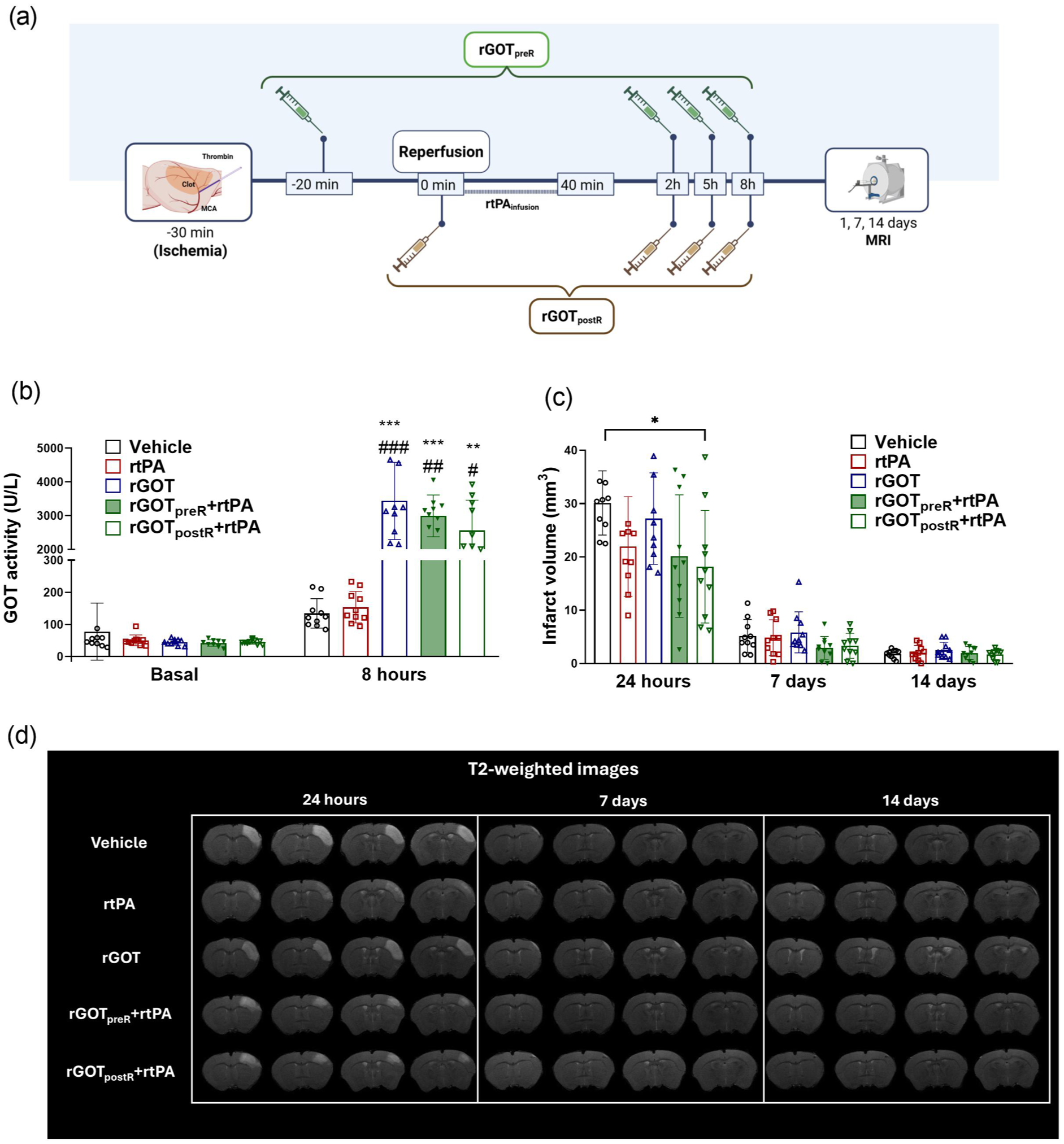
Therapeutic effect of the rtPA and rGOT combination in an animal model of thromboembolic ischaemia: (a) schematic representation of the experimental design. BioRender (https://biorender.com/), (b) GOT blood levels before surgery and 8 h after the first treatment administration. All data are presented as mean ± SD (n = 10). Statistical analysis was performed using the Kruskal–Wallis test followed by Dunn’s multiple comparisons: **p < 0.01 and ***p < 0.001 compared to the vehicle group; #p < 0.05, ##p < 0.01 and ###p < 0.001 compared to the rtPA group, (c) infarct volume measured at 24 h, 7 and 14 days post-ischaemia. All data are presented as mean ± SD (n = 10). Statistical analysis was performed using one-way ANOVA followed by Tukey’s multiple comparison test: *p < 0.05 and (d) representative T2-weighted images of ischaemic lesions.
GOT activity in blood increased after administration of four boluses of rGOT (0, 2, 5 and 8 h) in all treated animals. Baseline GOT activity levels before rGOT administration were: GOT group, 45 ± 9 U/L; rGOTpreR + rtPA, 42 ± 10 U/L; and rGOTpostR + rtPA, 46 ± 7 U/L. Eight hours after the first bolus, GOT activity rise to 3436 ± 1145, 2993 ± 619 and 2559 ± 900 U/L, respectively (Figure 2(b)).
In line with our previous studies, 7 infarct volumes determined via MRI T2-weighted images revealed that the group treated with the vehicle had an average infarct volume of 30.1 ± 6.0 mm3 at 24 h, 5.2 ± 3.1 mm3 at 7 days and 1.8 ± 0.8 mm3 at 14 days. At 24 h, a reduction of infarct volume, but not significant, were observed in the groups treated with rtPA (21.9 ± 9.3 mm3) or rGOTpreR + rtPA (20.1 ± 11.5 mm3). Similarly, at 7 days (rtPA: 4.8 ± 3.4 mm3; rGOT: 5.8 ± 3.8 mm3; rGOTpreR + rtPA: 2.9 ± 2.1 mm3) and 14 days (rtPA: 2.0 ± 1.4 mm3; rGOT: 2.5 ± 1.5 mm3; rGOTpreR + rtPA: 2.0 ± 1.2 mm3), none of these treatments showed significant differences compared with the vehicle. The only group that exhibited a significant infarct volume reduction was rGOTpostR + rtPA, where rGOT was administered simultaneously with rtPA, resulting in a volume of 18.2 ± 10.6 mm3 at 24 h (p < 0.05 compared to the vehicle group; Figure 2(c) and (d)).
As in previous results, the analysis of haemorrhagic transformations was performed using T2*-weighted images. 9 These sequences revealed an overall increase in hyposignal detection at 7 and 14 days after ischaemia in comparison with 24 h images. However, there were no significant differences in haemorrhagic transformation volume among treatment groups, indicating that rGOT, alone or in combination with rtPA, did not exacerbate bleeding risk (Figure 3(a) and (b)).
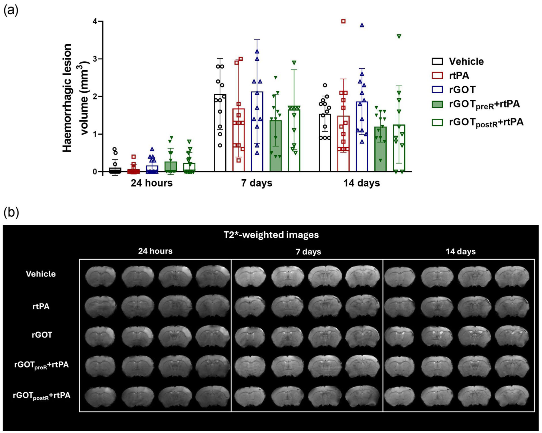
Evaluation of haemorrhagic transformations: (a) haemorrhagic volume measured at 24 h, 7 and 14 days post-ischaemia. All data are presented as mean ± SD (n = 10). Statistical analysis was performed using one-way ANOVA followed by Tukey’s multiple comparison test and (b) representative T2*-weighted images of ischaemic lesions.
Discussion
The combination of thrombolysis and neuroprotection has long been considered a promising approach for the treatment of acute ischaemic stroke. Several strategies have been explored to extend the thrombolytic time window (e.g. atorvastatin), 19 to enhance the benefits of rtPA by reducing ischaemic lesion size (e.g. uric acid), 20 or mostly to reduce thrombolysis-associated toxicity (e.g. 7β-oestradiol or memantine).21,22
rtPA is a glycoprotein belonging to the serine protease family that, in addition to converting plasminogen into plasmin to induce clot lysis, is associated with neurotoxic side effects mediated by the activation of glutamate NMDAR, 23 as we have also recently confirmed in primary neuronal culture. 9 Thus, some glutamate antagonists such as memantine 22 or selected NMDAR antibody (named Glunomab) 23 have been used as neuroprotective to use in combination with rtPA after stroke. 22
The therapeutic mechanisms proposed to underlie the protective effects of rGOT are primarily linked to its ability to modulate glutamate metabolism and lower circulating glutamate levels, thereby limiting the pathological glutamate increase that occurs in the brain parenchyma after stroke. Additional neuroprotective actions have been associated with the induction of cytoprotective autophagy and the reduction of neuronal apoptosis. 7 Based on these mechanisms, we hypothesised that rGOT might also mitigate the neuronal toxicity induced by rtPA. Our current in vitro analysis on neuronal culture demonstrates that rGOT alone does not counteract the neurotoxicity induced by rtPA, either under normoxic or oxygen glucose-deprived conditions. This result suggests that, unlike other neuroprotectants such as memantine that directly act on NMDA receptor–mediated excitotoxicity, 22 rGOT does not interact with receptor-level pathways and therefore cannot prevent rtPA-induced toxicity.
Additionally, we examined whether treatment with rGOT could enhance the therapeutic benefits of rtPA in a thromboembolic model of brain ischaemia. In a previous study, 7 we investigated the pharmacological interaction between rGOT and rtPA using an embolic middle cerebral artery occlusion model induced by homologous blood clots. However, that model did not allow a conclusive assessment of potential additive benefits, as it reproduces the human thromboembolic mechanism but is characterised by high variability, elevated mortality and inconsistent infarct sizes, which significantly limits its usefulness for evaluating therapeutic efficacy. 24 In contrast, the thromboembolic stroke model employed in the present study is widely recognised as suitable for preclinical evaluation of rtPA.11–16 This model generates fibrin-rich clots in situ, providing better control over the location and timing of occlusion, improved reproducibility and higher animal survival rates. These features make it particularly advantageous for assessing combination therapies such as rGOT and rtPA in a more reliable and clinically relevant manner.11,12
In vivo analysis in the thromboembolic stroke model revealed that, although rGOT and rtPA showed a trend in the reduction of infarct volume at 24 h, only the co-administration of rGOT with rtPA (rGOTpostR + rtPA) produced a significant reduction in infarct volume at 24 h. Neither rGOT alone nor rGOT administered 20 min before rtPA (rGOTpreR + rtPA) had any significant effect on lesion size or haemorrhagic risk. The main differences between groups were observed at 24 h, whereas infarct volumes converged by 7 and 14 days. This evolution is typical of this thromboembolic model, where small-to-moderate cortical infarcts rapidly lose MRI contrast as edema resolves making later measurements less reliable for detecting treatment effects. For this reason, 24 h is the optimal timepoint for evaluating therapeutic efficacy in this model. The absence of long-term differences therefore reflects the natural temporal evolution of cortical infarcts in this model rather than a lack of sustained biological effect. Importantly, this pattern has been consistently reported in previous studies employing similar methodologies, reinforcing that the acute phase provides the most sensitive window for capturing treatment-related changes.9,17,18
From a clinical perspective, these results indicate that co-administration of the current rGOT with rtPA in the hospital setting represents the most realistic and effective therapeutic scenario, supporting its implementation as an adjuvant therapy closely linked to the timing of reperfusion. These observations are in line with the principle that neuroprotective agents can only reach the salvageable ischaemic penumbra once adequate recanalization has been achieved.3,4,25
Importantly, our findings also highlight the relevance of pharmacokinetic constraints. rGOT displays a relatively short circulating half-life, which may limit the efficacy of pre-rtPA administration. In this context, it is plausible that the initial dose of rGOT loses a substantial part of its biological activity before rtPA-induced recanalization is achieved. Consequently, the three subsequent doses administered after rtPA may be insufficient to sustain adequate enzyme blood levels during the critical window of reperfusion required to trigger neuroprotection. In this regard, the development of more stable rGOT variants 26 with an extended circulating half-life could represent an important therapeutic advance, potentially allowing pre-rtPA administration to remain effective until recanalization occurs and thereby broadening the clinical applicability of this strategy.
Overall, detecting an additive or synergistic effect of a neuroprotective agent when combined with rtPA remains challenging. The substantial benefit of rtPA alone made it difficult to demonstrate a clear additional advantage from the neuroprotective intervention, especially in optimised clinical settings with early thrombolysis as previously observed in trials with citicoline. 25 Citicoline or CDP-choline, is a drug that combines neurovascular protection and repair effects. It has been used to treat acute ischaemic stroke with excellent safety profile. In 2002, a formal meta-analysis of trials of CDP-choline in acute and subacute stroke suggested a beneficial and substantial treatment effect, with absolute reductions of 10%–12% in the rates of long-term death and disability. 27 However, a new trial (ICTUS trial) on citicoline did not demonstrate effectiveness in the treatment of moderate to severe acute ischaemic stroke. 28 Posterior analyses reported that the inclusion of patients treated very early with rtPA in a multidisciplinary stroke unit could mask the protective benefits of citicoline. 29 In this line, the efficacy and use of MET have increased significantly in recent years, particularly in stroke cases where thrombi are in accessible in large vessel occlusion (LVO). Laboratory and clinical imaging studies have shown that early after LVO, the core of ischaemia is still reduced, whereas the surrounding infarcted tissue corresponds to a salvageable penumbra, an ideal condition for protection analyses. Therefore, patients with LVO awaiting thrombectomy appear to be excellent candidates for trials, as the first step in testing the protective effects of new agents in combination with reperfusion therapies.3,30
In conclusion, rGOT does not appear to be an effective adjunct therapy for reducing the direct neurotoxicity associated with thrombolysis. However, its neuroprotective benefit becomes evident when administered in combination with rtPA at the time of arterial reperfusion, suggesting that rGOT requires restored cerebral blood flow to reach the ischaemic tissue and exert its therapeutic effects. The demonstrated safety profile and absence of pharmacological interactions of concomitant rGOT and rtPA administration further support the feasibility of this combination approach. Therefore, our results advocate for the use of rGOT as a complementary treatment to enhance the efficacy of rtPA when delivered immediately before or during reperfusion in acute ischaemic stroke.
Supplemental Material
sj-docx-1-jcb-10.1177_0271678X261438565 – Supplemental material for Protective recombinant glutamate oxaloacetate transaminase increases the benefits of alteplase recanalization therapy in experimental ischaemic stroke
Supplemental material, sj-docx-1-jcb-10.1177_0271678X261438565 for Protective recombinant glutamate oxaloacetate transaminase increases the benefits of alteplase recanalization therapy in experimental ischaemic stroke by Clara Correa-Paz, Esteban López-Arias, María Pérez-Mato, Antonio Dopico-López, Antonio Cañizo-Outeiriño, Lara Pérez-Gayol, Lucía del Pozo-Filíu, Manuel Rodríguez-Yáñez and Francisco Campos in Journal of Cerebral Blood Flow & Metabolism
Footnotes
Acknowledgements
The authors acknowledge the use of ChatGPT (GPT-5, OpenAI) for assistance in language editing and text revision. The authors reviewed and verified all generated content for accuracy.
Author contributions
CC-P, MP-M and LP-G performed the in vitro experiments. CC-P, EL-A, LP-G and LdP-F performed experiments in animal models. CC-P, EL-A and AD-L developed MRI analysis. MR-Y, AC-O reviewed the manuscript and participated in the clinical interpretation of the results, under the final supervision of FC. CC-P and FC designed and supervised the study.
Funding
The authors disclosed receipt of the following financial support for the research, authorship, and/or publication of this article: Instituto de Salud Carlos III_ICIII, grant numbers RICORS–ICITUS RD24/0009/0017, RD24/0009/0009 and PI24/0065. Fundacion Mutua Madrileña. CC-P acknowledges the support of the Instituto de Salud Carlos III (ISCIII) by research grant Sara Borrell (CD23/00005). AC-O acknowledges the support of the Instituto de Salud Carlos III (ISCIII) by research grant Río Hortega (CM24/00031).
Declaration of conflicting interests
The authors declared no potential conflicts of interest with respect to the research, authorship, and/or publication of this article.

Supplemental material
Supplemental material for this article is available online.
References
Supplementary Material
Please find the following supplemental material available below.
For Open Access articles published under a Creative Commons License, all supplemental material carries the same license as the article it is associated with.
For non-Open Access articles published, all supplemental material carries a non-exclusive license, and permission requests for re-use of supplemental material or any part of supplemental material shall be sent directly to the copyright owner as specified in the copyright notice associated with the article.
