



Abstract
Background
Surgical debridement is often required to treat spinal infections. Successful surgery requires accurate localization of the active infections, however, current imaging technique still requires surgeons’ experience to narrow the surgical fields to achieve less invasive procedures.
Purpose
To investigate the use of F-fluorodeoxyglucose (FDG) positron emission tomography/computed tomography (PET/CT) for successful surgical planning.
Material and Methods
Nine patients with suspected spinal infection underwent magnetic resonance imaging (MRI) and FDG-PET/CT before surgery to locate active foci of infections. The spinal structures were divided into seven compartments at each intervertebral disc level for a total of 315 compartments investigated. The same classification system was used to design operating fields for histological correlation.
Results
FDG-PET/CT diagnosed fewer compartments as active infection (34 compartments, 10.8%) than MRI (62 compartments, 19.7%, P = 0.002). Surgical exploration was performed in 49 compartments, and demonstrated active infection in 25 compartments. The sensitivity / specificity of FDG-PET/CT was 100% / 79%, respectively, which was superior to those of MRI, 76% / 42%. Foci of active infection showed hypermetabolic activity with a SUVmax of 7.1 ± 2.6 (range, 3.0–12.7). Receiver operating characteristic (ROC) analysis indicated an optimal threshold for active spinal infection at a SUVmax of 4.2, corresponding to a sensitivity of 90.3% and specificity of 91.2%.
Conclusion
FDG-PET/CT demonstrated limited areas of abnormality allowing accurate delineation, and is thus useful to narrow the surgical fields. Since overall diagnostic accuracy of FDG-PET/CT was superior to that of MRI, FDG-PET/CT is a useful technique to narrow the surgical field for successful less invasive surgery.
Keywords
Introduction
Surgical debridement is often required to treat antibiotic-resistant spinal infection; however, extensive bone and joint removal may result in biomechanical instability of the spine, leading to loss in locomotive function. Recently introduced minimally invasive surgery is a promising technique, allowing pinpoint surgical debridement for treatment of spinal infection (1).
The success of minimally invasive surgery depends on presurgical planning to accurately localize active infection. Unfortunately, conventional imaging modalities such as plain radiography, computed tomography (CT), and magnetic resonance imaging (MRI) are not accurate enough to demonstrate active regions in chronic or in acute infection because a spine with active infection often shows anatomical changes representing a remodeling and / or degenerative process, which is hard to separate from coexisting active infection with conventional imaging techniques. We hypothesized that assessment of glucose metabolism might demonstrate active infection more specifically because an active inflammatory process is generally associated with highly metabolic tissues (2–6).
The purpose of this study was to evaluate the ability of F-18 fluorodeoxyglucose (FDG) positron emission tomography/computed tomography (PET/CT) to help target the surgical field by localizing active infection. We prospectively investigated the diagnostic accuracy of FDG-PET/CT in detecting regional foci of active infection and correlated with surgical findings. We also compared the accuracy with that of Gd-enhanced MRI, the current standard imaging modality.
Material and Methods
Study design
Candidates for surgical treatment of spinal infection underwent whole-body FDG-PET/CT to locate active spinal infection in addition to the standard presurgical evaluation using an established imaging protocol including Gd-enhanced MRI. The interval between the two imaging examinations was scheduled within a month, and surgical intervention was scheduled as early as possible after stabilizing the patients’ general medical condition. MRI and FDG-PET/CT were reviewed to localize active infection independently by the radiologists. The orthopedic surgeons determined surgical fields based on the radiological results and other clinical findings. The imaging results were correlated with surgical findings, histological analyses, and bacterial cultures.
Patients
The study included nine patients, who were clinically suspected of resistant spinal infection (8 men, 1 woman), with an average age of 65 years (range, 40–84 years) at the time of surgery. Resistant spinal infection was suspected when prior surgery or aggressive antibiotic treatment did not show an effective treatment response. The patients showed one or more clinical symptoms including fever, pain, or skin change. The imaging studies and treatments were performed with the approval of the human subjects and radiation safety committees at the Hokkaido University, Sapporo, Japan (#011-0076). Written informed consent was obtained from all patients.
Imaging procedures
MRI was performed with a 1.5 Tesla MRI units (Magnetom VISION (n = 2), Magnetom AVANTO (n = 5), and Magnetom Symphony (n = 2) (Siemens Medical Systems, Erlangen, Germany). Transverse and sagittal MR examinations of the target spine were performed with a phased-array coil. Images were obtained with T1-weighted (T1W) and fat-saturated T2-weighted (T2W) fast spin-echo (FSE) pulse sequences. The contrast-enhanced study used a gadolinium-based contrast agent (Magnevist; Bayer HealthCare, Osaka, Japan) at a dose of 0.1 mmol/kg of body weight. Fat suppression was also applied. Infection was considered to be present on MRI if: (i) T1W sequences showed confluent decreased signal intensity of the vertebral bone marrow and intervertebral disc space together with absence of a discernible margin between the disc and the adjacent vertebral marrow; (ii) fat-saturated T2W sequences documented an increased signal intensity of the vertebral marrow adjacent to the involved disc and of the disc itself; and (iii) significant contrast enhancement could be seen around the discovertebral junction and in the paravertebral soft tissues. Diagnosis of abscess formation was suggested when fluid collection circumscribed by contrast enhancement was detected in the bone marrow, disc space, or paravertebral soft tissues (7). In the present study, computed tomography (CT) was used in selected circumstances to complement MRI for the detection of calcification and gas to suggest degeneration. The CT was not considered as an independent factor to differentiate active infection from degeneration (8,9).
FDG-PET/CT was performed in three-dimensional mode (3 min per bed position) using a Biograph 64 TruePoint with TrueV (Siemens Japan, Tokyo, Japan). The emission sequence was initiated 60 min after completion of intravenous injections of 400 MBq of F-18 FDG. PET images were reconstructed using a point spread function-based iterative algorithm (TrueX, Siemens) with two iterations per 21 subsets, a matrix size of 168 × 168, a voxel size of 4.1 × 4.1 × 2.0 mm3, and a Gaussian filter of 4.0 mm at full-width half-maximum. Attenuation and scatter corrections were performed using a CT-based method. According to the standardized protocol used at our institution, CT scans were performed using the following imaging parameters: tube voltage, 120 kV; effective mAs, 50 mA; collimation, 24 × 1.2 mm; rotation speed, 0.5 s; pitch, 1.2; matrix, 512 × 512; field of view, 70 cm; slice thickness, 2.0 mm (1.5 mm increment). Intravenous contrast agent was not administered. The resulting PET and CT scans were coregistered using the standard software tool of the system software. Semi-quantitative analysis was performed using SUVmax values. Images were visually interpreted as pathological, if suspected regions showed focal FDG uptake higher than the normal vertebral bodies, muscles, other soft tissues, and blood pool activities around the lesion.
Surgical and histological assessment
The surgical procedure was determined as follows: (i) when the infection was localized at the anterior vertebral column (intervertebral disc spaces and adjacent endplates of vertebral bodies), we chose posterolateral spinal endoscopic debridement (ED) under local anesthesia; (ii) when the infection was localized at the anterior and posterior column (facet joint and paravertebral muscle) simultaneously, we chose intervertebral disc space debridement by endoscopic surgery or trans-spinal canal approach in addition to posterior open debridement (PD) under general anesthesia; (iii) when the spine showed segmental instability, we chose reconstruction surgery with posterolateral fusion (PLF) or a posterior lumbar inter-body fusion (PLIF) with posterior instrumentation in addition to debridement. Surgical findings for active infection were defined as pus formed from yellow and liquefaction tissue except for hard scar tissue observed during surgery. Histological assessment was done using surgical specimens to investigate infiltration of inflammatory cells (neutrophils, lymphocytes, macrophages, etc.) to suggest active infection (10).
Image analysis and histological correlation
One musculoskeletal radiologist and one nuclear radiologist independently read MRI and PET/CT images, respectively. Their image interpretation focused on identifying sites of active infection. The musculoskeletal radiologist was allowed to refer to presurgical clinical information and other imaging results, but was blinded to the results of FDG-PET/CT. Similarly, the nuclear radiologist was blinded to the results of the MRI examinations. The imaging results were summarized in the seven compartments based on the surgical field design at each intervertebral disc level (Fig. 1) as follows: intervertebral disc (#1 right and #2 left); posterior elements of vertebrae involving facet joints and erector muscles of the spine (#3 right and #4 left); iliopsoas muscle (#5 right and #6 left); and spinal canal (#7). All compartments of five disc levels from L2 to S1 (in the case of a lumbar lesion) or from T10–L2 (in the case of a thoracolumbar lesion) were assessed, for a total of 35 regional compartments per patient. The same region classification system was used to design operating fields and surgical procedures to allow comparison of the imaging results with surgical and histological analyses. Effort was made to explore as many compartments as reasonably indicated and to obtain multiple surgical specimens from the compartments for which imaging results (either MRI or FDG-PET/CT) indicated active infection.
Sagittal (a) and axial (b) planes of X-ray CT of a patient are shown. The spinal column and its surrounding structures were divided into seven compartments as follows (c): intervertebral disc (#1 right and #2 left); posterior elements of vertebrae involving facet joints and erector muscles of the spine (#3 right and #4 left side); iliopsoas muscle (#5 right and #6 left); and spinal canal (#7).
Using the histological results as a gold standard, the sensitivity, specificity, positive predictive value (PPV), and negative predictive value (NPV) were calculated as parameters of the diagnostic accuracy of imaging examinations. In addition, semi-quantitative analysis of FDG-PET/CT was done using the SUVmax value as a parameter of regional glucose metabolism.
Statistical analyses
All data were expressed as means ± standard deviations (SD). Differences in diagnostic accuracy between MRI and FDG-PET/CT were investigated using the Chi-square test. Receiver operating characteristics (ROC) analysis was performed to estimate an optimal threshold value of SUVmax for active spinal infection. P values less than 0.05 were considered to denote statistical significance.
Results
Patients’ characteristics
The final study population included nine patients (8 men, 1 woman) with suspected spinal infection, who had prior history of aggressive antibiotic treatment (7/9) and / or surgery (2/9). Mean blood glucose level at the time of FDG injection was 121 ± 25 mg/dl in the range of 80–171 mg/dl. Median interval between the FDG-PET/CT and MRI was 4 days. Median interval between the FDG-PET/CT and surgery was 8 days. One of the patients had a longer interval between the imaging and surgery (239 days) because of the chronic nature of the infection and poor overall medical condition due to diabetes. Surgical exploration was performed in 49 (out of 315) regional compartments, allowing imaging correlation. The remaining compartments (266/315) were not surgically explored because they did not present with signs or symptoms to suggest active spinal infection.
Imaging results
In general, the two imaging modalities showed agreement in the identification of the center of active spinal infection, but MRI tended to show broader areas of non-specific signal changes without significant Gd-enhancement in the adjacent structures that were not metabolically active with FDG-PET/CT. By contrast, FDG-PET/CT demonstrated limited areas of abnormal metabolic activities immediately adjacent to the center of spinal infection (Fig. 2). Regionally, MRI showed abnormal signal changes in 62/315 compartments (19.7%), with median six compartments per patient. FDG-PET/CT showed significantly fewer compartments with abnormal metabolic activity, including 34/315 compartments (10.8%) (P = 0.002), with a median of three compartments per patient (Fig. 3).
MRI (a) and FDG-PET/CT (b) images of a representative patient (case #1). Abnormal MRI signals were seen in L3/4 and L4/5 discs, vertebral bodies of L3–5, and posterior elements of L2–S1 with adjacent soft tissue structures (a). FDG-PET/CT demonstrated abnormal FDG uptake in the relatively limited structures at the level of L3/4 disc and posterior vertebral elements from L2–L5 (b). MRI signal changes were particularly prominent in the posterior compartments and adjacent soft tissues. Surgical findings were positive for active infection at L3/4 (anterior and posterior) and L4/5 (posterior elements). Among 315 compartments investigated, FDG-PET/CT diagnosed fewer compartments as active infection than MRI (P = 0.002).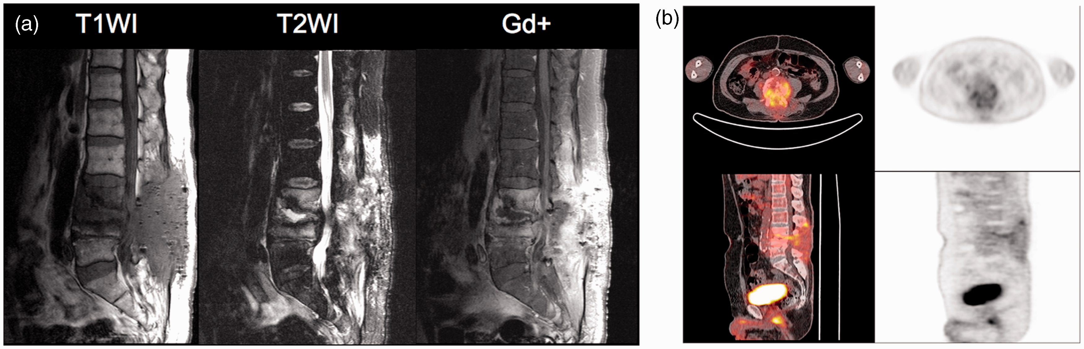

Clinical correlation
Surgical exploration demonstrated pus formed from yellow and liquefaction tissue in eight of nine patients. Bacterial culture was positive in seven of nine patients, including one with Mycobacterium avium complex (MAC) (Table 1). Among 49 compartments with surgical exploration, there were 25 compartments with active infection demonstrated by histological examinations and bacterial cultures. Fig. 4 summarizes the diagnostic accuracy of MRI and FDG-PET/CT in the 49 compartments with surgical exploration. Overall diagnostic accuracy with FDG-PET/CT (89.8%) was superior to that of MRI (59.2%). In particular, FDG-PET/CT was 100% sensitive in identifying active spinal infection, although it had five regions of interest (ROIs) of false positives due to degeneration, resulting in a positive predictive value of 83.3% (Fig. 4).
Comparison of diagnostic accuracy for MRI, FDG-PET/CT(visual analysis) and FDG-PET/CT (SUVmax >4.2) in the 49 compartments with surgical correlation. Anterior compartments include intervertebral disc (#1 right and #2 left) and iliopsoas muscle (#5 right and #6 left); posterior compartments include posterior elements of vertebrae involving facet joints and erector muscles of the spine (#3 right and #4 left side) and spinal canal (#7). NPV, negative predictive value; PPV, positive predictive value. Summary of imaging and surgical findings. AD, anterior debridement from posterior approach; ED, endoscopic debridement; MAC, Mycobacterium avium complex; MRSA, methicillin-resistant Staphylococcus aureus; MSSA, methicillin-sensitive Staphylococcus aureus; P. aeruginosa, Pseudomonas aeruginosa; PD, posterior open debridement; PLF, posterolateral fusion; PLIF, posterior lumbar interbody fusion.

Semi-quantitative analyses
With the surgical correlation, all true positive foci were hyper-metabolic, showing a mean SUVmax of 7.1 ± 2.6, in the range of 3.0–12.7. ROC analysis indicated an optimal threshold for active spinal infection at an SUVmax of 4.2, corresponding to a sensitivity of 90.3% and specificity of 91.2% (Fig. 5). The area under the curve (AUC) was 0.963. Applying this threshold improved the positive predictive value of FDG-PET/CT from 83.3 to 92% (23/25), with a particular improvement of specificity from 79.2 to 91.7% (22/24) (Fig. 4).
ROC curve. An optimal threshold for active spinal infection was estimated at an SUVmax of 4.2, corresponding to a sensitivity of 90.3% and specificity of 91.2%. The area under the curve (AUC) was 0.963.
Discussion
The results of the present study indicated that FDG-PET/CT is superior in detecting active infection compared to MRI. MRI tended to show more compartments with abnormal signal, which did not necessarily correspond to histologically proven active infection. In contrast, FDG-PET/CT demonstrated relatively limited areas of abnormal metabolic activity, although it still overestimated active infection due to degenerative inflammation.
Our results indicate a superior diagnostic accuracy of FDG-PET/CT to MRI. FDG-PET/CT was expected to show higher specificity at the cost of lower sensitivity, because small foci of active infection might suffer from partial volume effect. However, visual investigation of FDG-PET/CT was 100% sensitive at identifying active spinal infection, although it had five ROIs with false positives, resulting in a PPV of 83.3%. The results of the present study indicated that non-specific hyper-metabolic activity associated with degenerative inflammation still resulted in over-diagnosis of active foci of infection with FDG-PET/CT. Sensitivity of MRI was lower than we expected. This is probably because we tried to avoid false positives by employing strict criteria, which focused on specific findings. In general, MRI is sensitive (11) but includes large areas of non-specific signal changes due to degeneration. The strict criteria reduced false positives, but might result in more discordance between MRI findings and histopathological findings. In the present study, MRI tended to under-diagnose infections located in the anterior compartments (low in negative predictive value = 0.33), while it tended to over-diagnose infections in the posterior compartments (low in positive predictive value = 0.45) (Fig. 4). This is probably because degenerative endplates showing Modic type 1 changes (12) tend to mask MRI signal changes associated with active infection in the anterior compartments. In addition, degenerative processes in the posterior compartments were difficult to separate from active infection, because signal changes in MRI can represent the consequences of edema and artifacts (5), as well as hypervascular scar tissue (13).
Quantitative imaging is another advantage of FDG-PET/CT. In the present study, semi-quantitative analyses using SUVmax further improved diagnostic accuracy. The cut-off value at SUVmax = 4.2 appears reasonable compared with the previously reported SUV of 7.5 ± 3.8 in patients with spondylodiscitis (5,14). Semi-quantitative analyses using SUVmax values also provide objective indication of metabolic activity and may thus be used as a marker of therapeutic effect (15). The SUVmax has been established as a metabolic marker of clinical FDG-PET/CT, and it is relatively independent from the partial volume effect in the absence of noise. In the real imaging situation, however, the use of SUVmax is affected by noise and the reconstruction algorithm, smoothing method, and pixel size (16). Therefore, the cut-off value of SUVmax should be optimized in each imaging system, and should not be used in a cross-platform environment. For quantitative analyses, blood glucose level should be controlled to avoid possible altered biodistribution of FDG (17). Extremely high blood glucose level may result in higher background activities, although its influence is relatively limited to the activity of inflammatory lesions (18). In the present study, mean blood glucose level was controlled between 80 and 171 mg/dl, which would not induce altered biodistribution of FDG.
The results of the present study were in agreement with the current consensus supporting the clinical utility of metabolic imaging using FDG to diagnose skeletal infection. Previous reports have demonstrated the excellent diagnostic accuracy of FDG-PET, in the range of 85–100%, which exceeds that of MRI (4,6). FDG-PET was particularly useful in diagnosing active infection in the central skeleton within the active bone marrow (19) and involvement of paravertebral soft tissues (5). The major methodological difference from the previously published reports is the utilization of a PET/CT system for metabolic imaging, which allows for accurate anatomical correlation. Taking advantage of the anatomical information from CT, we analyzed regional metabolic activity at each intervertebral disc level. On the other hand, PET/CT technology may suffer disadvantage due to relatively intensive radiation exposure. In addition to the 400 MBq of FDG causing approximate radiation exposure dose of 10 mSv, it utilized X-ray CT for anatomical localization, which gave additional 6 mSv for CT portion in our standard imaging protocol. A total exposure of 16 mSv is similar to the typical full-dose CT examination for chest imaging (20). This amount will not cause significant biological effect, because spinal infection generally occurs in aged population. However, anatomical correlation can be substituted by MRI in the future, which will reduce radiation exposure and allow one-stop shopping examination to localize active spinal infection (21).
As far as we know, this is the first study to investigate “regional” metabolic activity in the subdivisions of the lumbar spine, and to compare the results with anatomical information obtained by MRI in the corresponding compartment. Utilization of PET/CT enabled detailed assessment of regional metabolic activity, which is important for designing surgical fields for minimally invasive, pinpoint treatments.
The clinical implications of this study include the utility of metabolic imaging to narrow the surgical margins. In general, surgical intervention should be minimized to prevent excessive debridement causing local instability of the spine. Targeting active infection using FDG-PET/CT is considered to be useful to narrow the surgical margins for the minimally invasive surgical technique. FDG-PET/CT can also be used to evaluate the therapeutic effects after the surgery. In the present study population, the surgery could not completely control two of nine patients, who thus required second surgery. The follow-up PET/CT demonstrated the uncontrolled compartments in a part of active foci on the initial PET/CT, which were treated by the second surgery.
The present study has several limitations. First, there was no independent reference for the localization of active infection. Surgical fields should be established according to the institutional standard procedure, which employs MRI as an important reference method. In addition, the procedure requires orthopedic surgeons to narrow the surgical fields to minimize invasive procedures. This process was done based on the surgeons’ experience and all the clinical information available including the results of MRI and FDG-PET/CT to achieve the best results for the patients. Although this might introduce selection bias for the operating fields, the clinical situation would not allow performing surgical procedures on all the regions presenting with MRI signal changes. Second, variations in the stages of infection cannot be controlled in the present study although it may affect FDG uptake. All patients included in the present study had a relatively long history of conservative treatment. Since acute infection tends to show higher metabolic activity (2), the results of the present study may not be applicable in patients presenting with acute spinal infection. Third, different types of infecting organisms may induce different metabolic activities. Although Staphylococcus aureus causes most of the hematogenous spread of spinal infection as in the present study population, tuberculosis and Pseudomonas aeruginosa may also cause spinal infection (22). The present study did not investigate the organism dependence of the metabolic activity due to the limited number of patients. Further research with a larger sample size should stratify patients according to infection stage and causative organism.
In conclusion, metabolic assessment using FDG-PET/CT allowed accurate diagnosis of active infection, and thus is a useful technique to delineate the surgical field for successful minimally invasive surgery.
Footnotes
Conflict of interest
None declared.
Funding
This research received no specific grant from any funding agency in the public, commercial, or not-for-profit sectors.