



Abstract
Background
Magnetic resonance imaging (MRI) is the diagnostic modality of choice in defining soft tissue compromise of the spinal canal.
Purpose
To evaluate the reliability of postoperative MRI in the determination of level and side of lumbar spinal decompression surgery, investigated by two reviewers, in different levels of training and specialization.
Material and Methods
Postoperative MR images of 86 patients who underwent spinal decompression (single level, n = 70; multilevel, n = 16; revision decompression, n = 9) were reviewed independently by an experienced musculoskeletal radiologist and a fourth-year orthopedic surgery resident. The level (single or multiple) and side of previous surgical decompression were determined and compared to the surgical notes. We examined factors that may have influenced the reliability, including demographics, type of surgical decompression, use of a drain, and time interval from surgery to MRI.
Results
Significantly fewer levels were correctly determined by the resident (77/86 cases, 89.5%) compared with the radiologist (84/86 cases, 97.7%) (P = 0.014). The resident interpreted significantly more MR images incorrectly in cases where a drain was used (n = 8; P < 0.001). Re-decompression cases were interpreted incorrectly significantly more often by both the radiologist (n = 2, P = 0.032) and the resident (n = 4, P = 0.014).
Conclusion
Determination of the level and side operated on in previous lumbar spinal decompression surgery on MRI has a high reliability, especially when performed by a musculoskeletal radiologist. However, this reliability is decreased in cases involving surgical drainage and same-level revision surgery.
Introduction
Magnetic resonance imaging (MRI) is the diagnostic modality of choice in defining soft tissue compromise of the spinal canal by disc herniation, degenerative facet joint enlargement, hypertrophy of the ligamentum flavum, and in the evaluation of the postoperative spine (1,2). Surgical decompression of the lumbar spine is most often performed for the treatment of symptomatic radiculopathy or neurogenic claudication. The most common surgical approach is via a posterior midline incision. Various lumbar spinal decompression techniques are used, including fenestration, laminotomy, or uni- or bi-lateral laminectomy sometimes combined with partial facetectomy. Postsurgical changes involving the osseous structures are best appreciated on a computed tomography (CT) scan. However, for most other purposes, MRI is superior as it allows for a better appreciation of the relationship between the osseous structures and the adjacent soft tissue spinal cord and exiting nerve roots (3). Therefore, MRI is routinely used if surgical decompression results in unsatisfactory symptom resolution (4).
Expected postoperative changes may vary depending on the type and extent of surgery and the time since the operation (5). In the case of re-decompression surgery, it is mandatory to know on which level and side scar tissue should be expected to enable precise preoperative planning. In daily clinical practice, surgical reports of previous decompressions are not always available, particularly if the procedure was performed more than 10 years previously. To the best of our knowledge, it is not known how reliable MRI interpretation is in determining the level and side of previously conducted spinal decompression.
The purpose of this investigation was, therefore, twofold: (i) to investigate the reliability of determination of the level and side on which lumbar spinal decompression surgery had previously been conducted on postoperative MR scans by two reviewers, at different levels of training and specialization; and (ii) examine patient-specific, surgical technique, and imaging factors that may influence the accuracy of this determination.
We hypothesized that MRI would be reasonably reliable for determination of the level and side of previous lumbar decompression surgery, but that its reliability would be influenced by the experience of the reader and by patient and surgical factors.
Material and Methods
After approval by the Ethical Committee (Cantonal Ethical Committee number: 2015-0087), 89 patients who underwent MR scanning of the lumbar spine after lumbar decompression surgery between January 2010 and December 2013 were retrospectively identified. Inclusion criteria were: single- or multi-level decompression surgery for symptomatic lumbar disc herniation and/or stenosis at our institution, after which a new MR examination was performed. Participants provided written informed consent. Demographic data (sex, age, body mass index [BMI]) and surgical technique (use of a microscope, fenestration, laminotomy, laminectomy, partial facetectomy, extraforaminal decompression, dural lesion, postoperative use of a drain, re-decompression surgery, level and side of prior decompression) were extracted from the electronic patient charts.
MR examinations were in our institution performed using a 1.5 T scanner (Magnetom Avanto Fit, Siemens Healthcare, Erlangen, Germany) or a 3 T scanner (Magnetom Skyra Fit, Siemens Healthcare, Erlangen, Germany), depending on scanner availability. Standard imaging protocol of the lumbar spine included two sagittal sequences (T1-weighted [T1W] and T2-weighted [T2W] turbo spin-echo) and a transverse T2W sequence. Both sagittal and axial MR sequences were examined for the presence of postsurgical changes. Two investigators, a fellowship-trained musculoskeletal radiologist (FMB) and a fourth-year orthopedic surgery resident (FW) read all MR examinations independently, and were blinded to the levels and side of the surgical decompression that had been performed. The investigators rated each MR examination for the level(s) of the performed decompression (L1/2, L2/3, L3/4, L4/5, L5/S1) and the side of decompression (left, right, bilateral). The MR examination was performed in a different institution in eight patients (9.3%), but the scans were interpreted by the same two readers. In total, a 1.5 T MR scanner was used in 65 cases and a 3 T scanner was used in 21 cases. Contrast media was applied in seven MR examinations (8.1%). Contrast media was used to differentiate recurrent disc herniations (no enhancement) and postsurgical fibrosis (enhancement) or postsurgical seromas (no enhancement) from infections (enhancement).
Three patients were excluded from the study: two because of incomplete surgical notes and one due to a lumbosacral transitional anomaly that could introduce a bias in level determination.
Statistical analysis
Statistical analyses were performed using the statistical software, SPSS 14.0 K (SPSS Inc., Chicago, IL, USA). Descriptive values such as mean, standard deviation, median, and extremes were calculated. The Wilcoxon signed rank test was used to compare differences between the two investigators’ findings. The Mann–Whitney test was used to compare the reliability of determining the correct decompression level and for the comparison of surgical techniques, patient-specific and imaging factors.
Statistical significance was defined as P < 0.05.
Results
Levels operated on in previous lumbar spinal decompression surgery.

Sixteen patients underwent multi-level decompression surgery.
Cases in which at least one reader incorrectly identified the side and/or level of previous spinal decompression surgery.

The letters represent the initials of each patient.
Interpretation from a fourth-year orthopedic surgery resident.
Interpretation from an experienced fellowship-trained musculoskeletal radiologist.
Significantly more MR examinations (n = 9) were read incorrectly by the resident when a drain was used at the time of surgery (P < 0.001). MR examinations from patients that had already undergone a revision decompression surgery (n = 9) prior to imaging were read incorrectly significantly more often than those from patients that had not undergone revision surgery by both the radiologist (n = 2, P = 0.032) and the resident (n = 4, P = 0.014).
None of the following were predictive factors for incorrect identification of the decompression level and side: BMI, sex and age of the patient, time interval between decompression surgery and MR examination, microscope use during decompression surgery, surgical decompression technique (fenestration, laminotomy, laminectomy, partial facetectomy, extraforaminal decompression), repair of a dural lesion at the time of surgery, performance of MR examination performed at a different institution, or the use of intravenous contrast media (Figs. 1 and 2).
(a) Axial and (b) sagittal T2W sequence of a 64-year-old man 43 weeks after unilateral left-sided laminotomy at the level of L4/5. The lamina defect can be easily identified. (a) Axial and (b) sagittal T2W sequence of a 70-year-old woman 10 years after unilateral right-sided laminotomy at the level of L5/S1. This MR scan was performed 18 days after bilateral laminotomy at the level of L2/3 and L3/4. Both reviewers failed to identify the prior unilateral right-sided laminotomy at the L5/S1 level.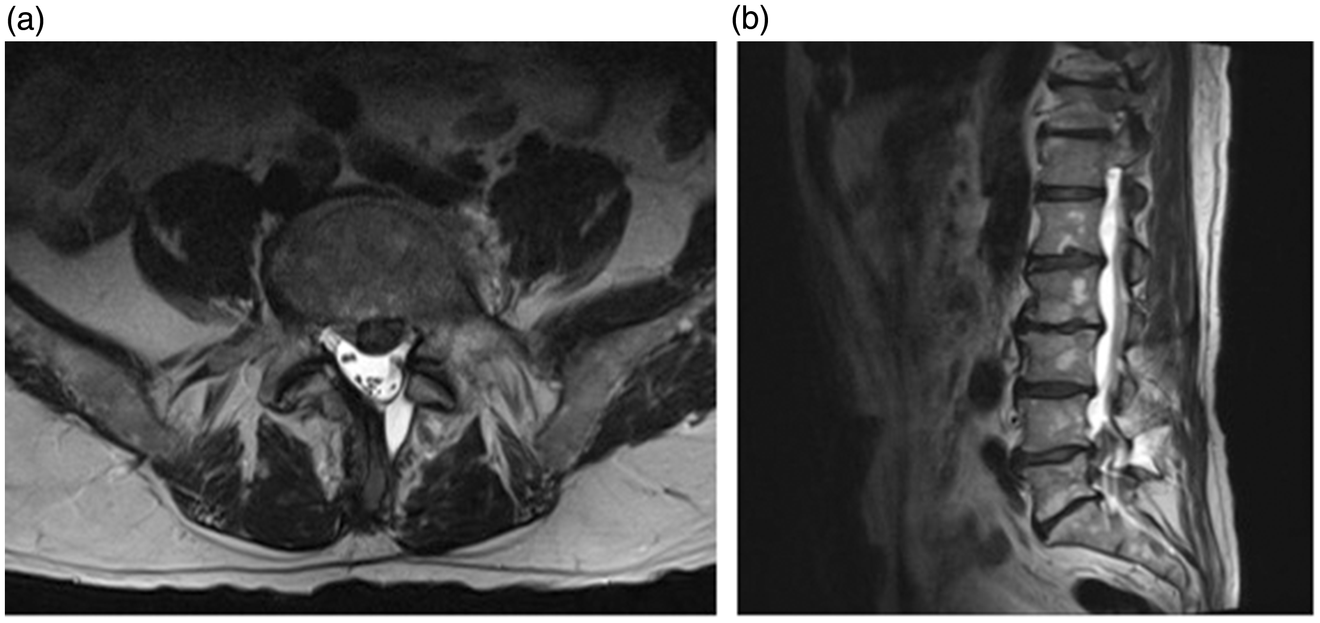

In every case of lumbar decompression surgery, a fluoroscopy was performed intraoperatively to determine the spinal level before the decompression was performed to avoid wrong level surgery. In this study population we did not observe a wrong level surgery. We observed two postoperative hematomas that required surgery for hematoma evacuation and three superficial wound infections that were treated with oral antibiotics in the study collective. The incorrect decompression level was rated wrong by both investigators in one of the cases of postoperative superficial wound infection.
Discussion
To the best of our knowledge this is the first study to evaluate the reliability of postoperative MR examinations in determining the level and side of previous lumbar spinal decompression surgery. Overall, interpretation of MRI was highly reliable in determining the level and side involved in previous lumbar spinal decompression surgery; the fourth-year orthopedic surgery resident could identify the correct level and side in 89.5% of cases on postoperative MRI, while the fellowship-trained radiologist had a higher accuracy of 97.7%. Factors including the use of a surgical drainage or revision surgery decreased the reliability of using MRI imaging to identify the correct level of previous spinal decompression.
This new information is of relevant clinical value: postoperative imaging is needed in cases where preoperative symptoms are not resolved and in cases where new symptoms have developed (6). When surgical revision is being considered, knowledge of the exact level and side of previous decompression surgeries is important for accurate surgical planning and for providing information to the patient regarding potential complications (e.g. dural tear). Previous surgical reports are not always available to the surgeon or the radiologist, and they must then rely on MRI to identify the level and type of previous spine surgery.
Postoperative changes occur within the bones and soft tissues and these vary depending on the type and extent of surgery and the time since the operation (5). The interval between surgery and MRI had no effect on the precision of correct level and side identification in our study. Postoperative changes can be observed on MR images in the area of the paraspinal muscles, facet joints, lamina, epidural space, spinal nerve roots, and the intervertebral disc (7). Axial MR sequences following surgical decompression using a posterior midline incision may demonstrate muscle enhancement or disruption of the margins of the paraspinal muscles, as well as edema of the adjacent soft tissues, fluid collection, and hematoma formation at the levels of surgical dissection in the immediate postoperative period (3,5–8). We observed that MR examinations of revision decompression cases were misinterpreted significantly more often by both investigators compared with cases of primary surgery; this might be due to less pronounced soft tissue changes in older decompression zones. Enhancement of the paraspinal musculature in MRI is marked at 3 weeks and decreases at 6 months after lumbar decompression (9). The incidence of paraspinal soft tissue enhancement decreases from 100% in the immediate postoperative stage to 18% 6 months after surgery (10); there is also often asymmetry in the paraspinal muscle fat planes posteriorly (5). In the current study, the use of a drain at the time of surgery was a risk factor for MR scan misinterpretation by the resident; this might be because additional paraspinal soft tissue trauma was misinterpreted to indicate the surgical approach by the less experienced MR reviewer.
The value of contrast enhancement in improving diagnostic accuracy is controversial (1). Contrast agents shorten T1 relaxation times, and therefore increase the signal on T1W sequences wherever the contrast agent is accumulated (mainly in vessels and hyperemic tissue) (2). Contrast medium administered intravenously helps to differentiate between epidural fibrosis and recurrent disc herniation; epidural fibrosis is enhanced whereas disc hernias are not (1,11–13). In the current study, the use of contrast-enhanced MRI did not influence the accuracy of determining the level and side operated on previously; however, this might be a result of the limited number (n = 7) of contrast-enhanced MR examinations in our patient population.
In conclusion, the determination on postoperative MR images of the level and side operated on during previous lumbar spinal decompression surgery, has a high reliability, especially when performed by a fellowship-trained musculoskeletal radiologist. Revision decompression and use of a surgical drain decreased the ability of the readers to correctly identify the level of previous surgery on MRI.
Footnotes
Acknowledgements
We thank Professor Burkhardt Seifert from the Division of Biostatistics, Institute of Social and Preventive Medicine, for statistical support.
Declaration of Conflicting Interests
The author(s) declared no potential conflicts of interest with respect to the research, authorship, and/or publication of this article.
Funding
The author(s) received no financial support for the research, authorship, and/or publication of this article.