



Abstract
Background
In humans, the left testis generally hangs lower than the right and the right is larger than the left. Magnetic resonance imaging (MRI) represents an important adjuvant modality in the investigation of testicular diseases.
Purpose
To assess if normal testes asymmetry is related to apparent diffusion coefficient (ADC) and magnetization transfer ratio (MTR).
Material and Methods
The normal testes from 106 men were included. Testicular volume (TV) was calculated by using the ellipsoid formula: length × width × height × 0.52. Diffusion-weighted imaging was performed using a SS EPI diffusion pulse sequence and b-values of 0 and 900 s/mm−2. Magnetization transfer imaging was obtained using a 3D GRE sequence both with and without an off-resonance radiofrequency pulse. MTR maps were obtained by the following formula: (SIo–SIm) / (SIo) × 100%, where SIo and SIm represent the signal intensity in the baseline image and that in the corresponding image acquired with an off-resonance MT pulse, respectively. The mean and standard deviation of testicular volume (TV), ADC, and MTR of both testes was calculated and compared using a paired sample t-test.
Results
The mean TV (mL) was greater (P = 0.006) for the right testis (16.77 ± 4.84) compared to the left (15.97 ± 4.45). ADC of the right testis (1.09 ± 0.12 × 10 − 3 mm2 s−1) was not different (P = 0.064) from that of the left testis (1.07 ± 0.12 × 10⁻³ mm2 s−1). Differences (P = 0.032) were observed between MTR of the right (46.6 ± 2.1%) and left testis (46.0 ± 2.2%).
Conclusion
The reported differences in paired testes size was confirmed, introducing a possible relationship with structural and functional asymmetry of normal testes, based on MTR.
Keywords
Introduction
The asymmetry of the human scrotum is an interesting phenomenon, for which many explanations have been proposed (1,2). The right testis is usually bigger than the left and the left testis has a lower position. For centuries, Greek sculptors pursued the idea of perfection in art studying the smallest details in order to create the most realistic replicas of the human body. The Greeks correctly portrayed the right testis in a higher position, but incorrectly portrayed the left testis as larger, using a simple mechanical theory, the left testis being thought to be lower because it was larger and therefore, more subject to the pull of gravity (3,4). Although little is known, there are also reports describing functional asymmetry of the normal testes (5,6).
The relation of genital organ asymmetry in men with sexual and reproductive health issues renders this phenomenon important. Testicular carcinoma may reflect lateral body asymmetry, where the larger testis that is the right may be more likely to be affected by cancer than the smaller (2,7–9). Differences in testes size have also been related to cognitive skills (2,10). Kimura et al. reported that men with a larger right testis were found to perform relatively better on certain spatial tasks, the so-called “masculine” tasks when compared to men with a larger left testis performing better on “feminine” tasks (10).
Although ultrasonography (11) represents the primary imaging modality in the investigation of scrotal diseases, magnetic resonance imaging (MRI) has emerged as an important supplemental diagnostic tool (12–19). MRI is increasingly being used as both a problem-solving technique in patients with inconclusive sonographic findings and as a primary imaging modality in the evaluation of a variety of scrotal diseases (12–19). Recently, functional MRI techniques, including diffusion-weighted imaging (DWI), dynamic contrast-enhanced MRI, and MR spectroscopy have added important diagnostic information in the interpretation of testicular diseases (20–27). Among them, DWI due to its relatively short acquisition time, can easily be added to routine MR protocols. These images provide both qualitative and quantitative data, the latter by means of apparent diffusion coefficient (ADC), useful in the interpretation of a variety of testicular diseases, including both malignant and non-malignant (20–23).
Magnetization transfer imaging (MTI) assesses the interactions of water protons with different macromolecular environments (28–30). Protons in tissue are divided into “free” water protons producing the MR signals detected on conventional sequences and “restricted” protons, bounded to proteins and macromolecules. Exchange and cross-relaxation of magnetization between these two groups result to the magnetization transfer effect. The MT phenomenon is quantified by magnetization transfer ratio (MTR) (28,29). In a recent preliminary report, we studied the MTR of normal testis and its efficacy in the characterization of various testicular lesions (30).
To better understand normal testes asymmetry, in this retrospective study we investigated possible differences of ADC and MTR between right and left testis.
Material and Methods
Study population
This single-center retrospective study included 212 normal testes from 106 men. The age range was 20–84 years and the mean age was 44 years. The participants were referred to the Urology Department from May 2009 to May 2015, with a variety of clinical indications: vague scrotal pain (n = 77, with a history of recently treated acute epididymitis in one case, chronic epididymitis in four patients, chronic prostatitis in five cases, bilateral testicular torsion in two cases, hemospermia in one patient, and prostatic carcinoma in one case); recent scrotal trauma (n = 3); recent penile trauma (n = 1); recent penile enlargement (n = 1); signs of epididymitis (n = 6); painless scrotal enlargement and/or scrotal pain (n = 5); benign sonographic findings (n = 8, including five cases of epididymal cysts, one case of spermatocele and two patients with testis heterogeneity); follow-up after surgery (n = 2, with a history of undescended testis in one case and retroperitoneal germ cell neoplasm in another patient); spermatic cord abscess (n = 1); partial priapism (n = 1); and silicone-induced penile lipogranuloma (n = 1). The records included clinical and sonographic follow-up examinations. Participants having abnormalities of any kind in the scrotum such as varicocele, significant hydrocele, and pathologic conditions of the testis or epididymis were excluded from the analysis.
This study was approved by the institutional review board and written informed consent was obtained from all patients.
MRI protocol
All MRΙ examinations were performed on a 1.5-T scanner (Philips Medical Systems, Cleveland, OH, USA) using a circular surface coil. The participants were examined in supine position, by placing a towel beneath the testes, to keep them at a similar distance from the coil, and the penis draped on the anterior abdominal wall. Axial spin-echo T1-weighted (T1W) images (TR/TE, 500–650/13–15 ms) and fast spin-echo T2-weighted (T2W) images (TR/TE, 4000/100–120 ms) in the three orthogonal planes were used for data analysis.
DWI was performed in 100 men (age range, 20–84 years; mean age, 43 years) from May 2009 to May 2015. DW images were obtained in the transverse plane, during quiet breathing, using a single shot, multi-slice, spin-echo planar sequence and b-values of 0 and 900 s/mm−2. The following parameters were used: TR, 3900 ms; TE, 115 ms; matrix, 180 × 256 mm; field of view (FOV), 240 × 270 mm; number of signals averaged, 1; motion-probing gradient (MPG), 3; and, bandwidth, 1, 5774 kHz/pixel. The total acquisition time was 29 s. No parallel imaging was used.
MTI was performed in 79 men (age range, 20–84 years; mean age, 42 years) from February 2013 to May 2015. MT sequences were obtained in the same plane using a three-dimensional (3D) gradient-echo sequence both with and without an off-resonance binomial prepulse to saturate the broad resonance of immobile macromolecular protons. The following parameters were used: TR, 29 ms; TE, 8 ms; FOV, 190 × 151 × 56 mm; matrix, 256 × 130 mm; voxel size, 0.74 × 1.16 × 4.00 mm; flip angle, 6°; number of signals averaged, 2; and WFS (Water-Fat-Shift, pix)/bandwith (Hz), 6.207/35.0. The scan duration was 150 s. The orientation and location of the transverse slices was identical for all sequences. All images were of 3–4 mm slice thickness with a 0.5-mm intersection gap.
MRI data interpretation
Two radiologists (AN and ACT) in consensus reviewed the MRI data. After the maximum length, width, and height of the testes were measured in the mid-sagittal and mid-transverse T2W images, as shown in Fig. 1, testicular volume (TV) was calculated with the formula for an ellipsoid: length × width × height × 0.52 (31). Coronal T2W images were used as reference to identify the mid-sagittal and mid-transverse planes.
Normal MRI examination of the scrotum in a 31-year old man referred for vague scrotal pain. (a) Sagittal and (b) transverse T2W images. The lines show measurement of length and width in the sagittal plane and height in the transverse plane. (c) Coronal T2W image depicts normal paired testes. The right testis appears larger than the left and the left testis is in lower position compared to the right. Small right hydrocele (arrowhead, normal finding). (d) Transverse ADC map (b = 900 s/mm−2) demonstrates normal testes hypointensity. The ADC of the right and left testis (circles) is 0.88 × 10⁻³ mm2 s−1 and 0.85 × 10⁻³ mm2 s−1, respectively. Axial 3D gradient-echo MR image before (e) and after (f) application of the MT prepulse. The MTR (in percent) of the right and left testis is 49.6 and 47.9, respectively.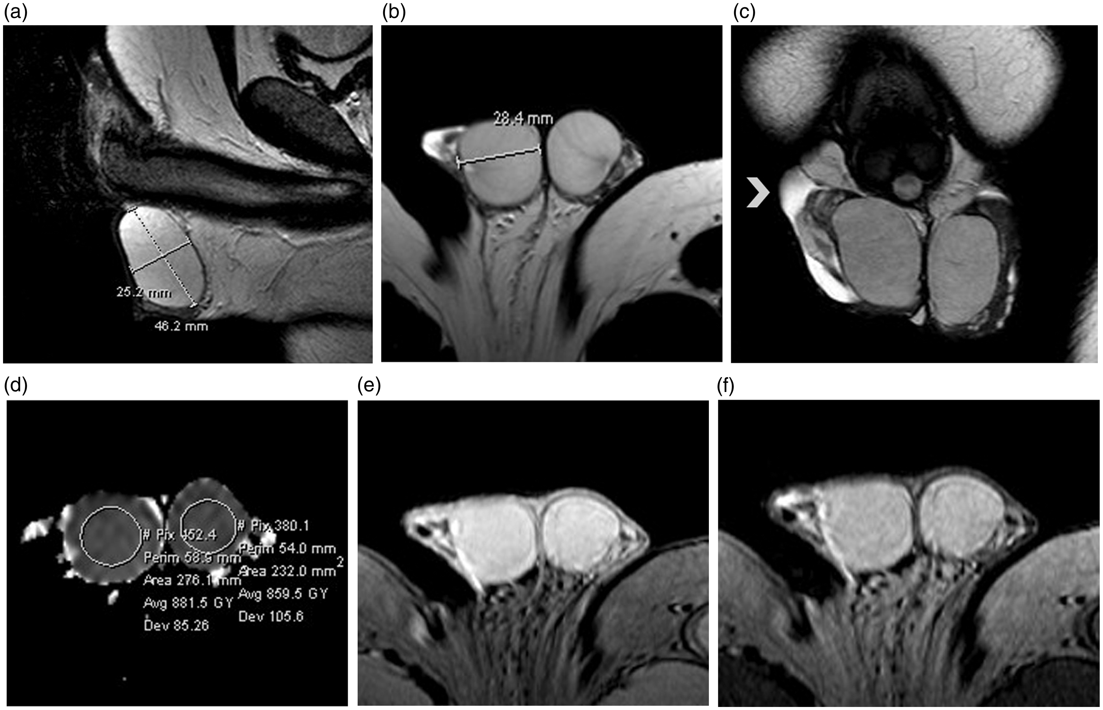
MTR measurements were performed by the following formula: (SIo–SIm) / (SIo) × 100%, where SIm refers to signal intensities after the application of the saturation pulse and SIo refers to signal intensities with the MT pulse turned off. Circular regions of interest (ROIs) as large as possible were placed in the middle of the testis, including the majority of testicular parenchyma. Special care was taken to avoid partial-volume effects and subtraction artifacts. ROIs were placed on the ADC maps and both on images before and after the application of the radio-frequency pulse, with reference to the corresponding T1W and T2W images. Three different ROIs were used for each testis and the measurements were averaged.
Statistical analysis
Statistical analysis was performed using IBM SPSS version 20.0 (IBM Inc., Armonk, NY, USA). The normality of distribution of parameters was assessed by a Kolmogorov–Smirnov test. Mean values of TV, ADC, and MTR for each testis were expressed as mean ± standard deviation. TV, ADC, and MTR of the right and left testis were compared using a paired sample t-test. A P value of <0.05 was considered significant.
Results
Fifteen cases were excluded from data analysis due to the presence of: varicocele (n = 6), significant hydrocele (n = 5), large spermatocele, with pressure-effects on the ipsilateral testis (n = 1), ectopic testis (n = 1), acute epididymoorchitis (n = 1), and testicular hematoma (n = 1). Malposition of the testes was the reason to exclude four more participants from calculation of TV. Also excluded were eight cases with artifacts on the ADC maps and four cases with artifacts on the MTR maps. Testes were characterized as “normal” based on the signal intensity on conventional T1W/T2W images and DW/MT images and/or the absence of abnormal testicular lesions found during subsequent clinical and/or sonographic follow-up studies.
The data followed a normal distribution as evaluated using the Kolmogorov–Smirnov test. The TV was measured and compared in 87 men. The mean TV (mL) of the right testis was 16.77 ± 4.84 and of the left testis 15.97 ± 4.45 (Figs. 1 and 2). Significant differences (P = 0.006) between the two sides were found. The ADC of 154 normal testes from 77 men and the MTR of 120 testes from 60 men were included in the analysis. The mean ± standard deviation of ADC (× 10−3 mm2 s−1) of the right and left testis was 1.09 ±0.12 and 1.07 ± 0.12, respectively (Figs. 1 and 2). No differences in ADC between the right and left testis were observed (P = 0.064). The mean ± standard deviation of MTR (in percent) of the right and left testis was 46.6 ± 2.1 and 46.0 ± 2.2, respectively (Figs. 1 and 2). Significant differences in MTR between the right and left testis were observed (P = 0.032). Table 1 provides numbers of normal testes, mean TV, ADC, MTR, and standard deviations for the right and left testis.
(a) Coronal T2W image depicts normal paired testes in a 58-year-old man. The TV of the right and left testis is 14.9 mL and 12.4 mL, respectively. (b) Transverse ADC map (b = 900 s/mm−2). The ADC of the right and left testis is 1.05 × 10⁻³ mm2 s−1 and 1.11 × 10⁻³ mm2 s−1, respectively. Axial 3D gradient-echo images acquired without (c) and with (d) the application of the MT pulse. The MTR of the right and left testis is 44.2% and 42.0%, respectively. Number of testes, mean ± standard deviations of TV, ADC, and MTR for the right and left testis.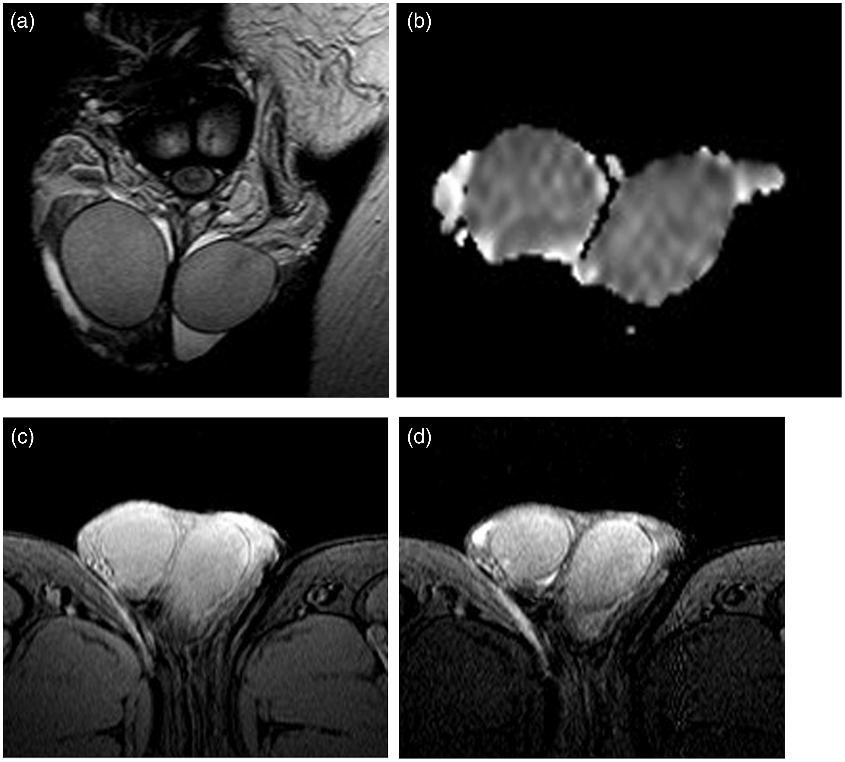

Discussion
In humans, normal testes are actually never quite the same, the right testis is usually larger than the left and the left testis has a lower position in the scrotum (1,2). Many theories have tried to explain scrotal asymmetry in humans. Some have attributed it to the more well-developed and greater flexion of muscles on the right side of the lower abdomen, pulling up the spermatic cord and the ipsilateral testis in a higher position and others to the earlier development of the right testis on human fetuses when compared to the left testis (1,2). Another interesting explanation refers to the venous drainage of the testes, where the right spermatic vein joins the inferior vena cava at an acute angle and the left joins the left renal vein at right angles. The above result in an increase of the resistance to the venous return from the left side, a consequent vascular stasis in the tributaries of the left spermatic vein, and therefore a lower position of the left testis (1).
There are also reports indicating functional asymmetry of the testes, with the right testis showing a greater response either to luteinizing hormone (LH) deficiency treatment or to hemicastration than the left gonad (5,6). Besides its lower functional capacity, the left testis is more vulnerable than the right, and in humans, there is a greater incidence of cryptorchidism and varicocele on the left side (5,6). Thermal asymmetry of the human scrotum was also reported by a French study (32).
Recently published reports have addressed the role of DWI in the interpretation of testicular diseases, including the detection and localization of impalpable testes, the early diagnosis of testicular torsion, the detection of testicular fibrosis in patients with varicocele, the differentiation between normal testis, benign testicular lesions and testicular carcinomas, and the preoperative characterization of the histologic type of testicular germ cell neoplasms (20–23). DW sequences are designed to detect alterations in thermally induced random molecules within tissues. The degree of water diffusion in biologic tissues is inversely related to tissue cellularity and the integrity of cellular membranes. The testis is an example of normal tissue causing restriction of free molecular diffusion and this is correlated to the structural complexity of this organ (33). In a previous study, we reported an ADC of 1.11 ± 0.18 × 10⁻³ mm2 s−1 for normal testis (20). This is in accordance with the results of the present study reporting an ADC of 1.09 ± 0.12 × 10⁻³ mm2 s−1 and 1.07 ± 0.12 × 10⁻³ mm2 s−1 for right and left testis, respectively, with no differences between paired testes.
Magnetization transfer (MT) techniques provide tissue contrast which mainly depends on the concentration of macromolecules and a way of examining tissue structural components that are normally not seen with conventional MR sequences (28,29). The MT phenomenon is quantified by MTR. Tissues with macromolecules transfer magnetization more efficiently and appear hypointense on MT images, with high MTR (28,29). In a retrospective study of 147 normal testes, we found a mean MTR of 46.2 ± 2.5% (30). This is in accordance with our present results, reporting an MTR of 46.6 ± 2.1% and 46.0 ± 2.2% for the right and left testis, respectively. The presence of a number of macromolecules implicated in the secretory activity of the testis, including peptide hormones, as LH, follicle-stimulating hormone (FSH), and steroid hormones, as testosterone and estradiol might justify the high MTR of normal testis. Another factor responsible for the MT effects is the rough endoplasmic reticulum (RER), consisting also of macromolecules, which is important for the synthesis and secretion of hormones. RER plays an important role in the synthesis of testosterone (30,33). Normal testis also contains large amounts of collagen, found in the testicular tunicae, the peritubular tissue, the lamina propria, and the basement membrane of the seminiferous tubules, reflecting of an environment with a high density of macromolecules (30,33).
Our study showed normal paired testes asymmetry, with the left testis smaller than the right, with lower MTR. Whether these observations are correlated with structural and functional differences related to the macromolecular content of paired testes remains to be defined. Johnson et al., in a study of 132 testes obtained at autopsy within 24 h of sudden death, reported left testes smaller than the right, the changes including both the total testis weight and the weight of the testicular parenchyma (34). Specifically, total testicular weight and testicular parenchyma weight were reported about 10% lower on the left than on the right side. In the same report, the values of daily sperm production (DSP)/testis were positively correlated with both the total testis weight and the weight of testicular parenchyma and found 13% lower on the left side (34). Therefore, the smaller testis produces fewer spermatozoa. Another contributing factor to the impairment of spermatogenesis on the left side might be related to the possible differences in blood flow through paired testes, related to vascular stasis on the left side, meaning diminished levels of gonadotrophic stimulation and reduced access to metabolic substrates. Johnson et al. also assessed the testicular tunic weight, a predominantly collageneous structure and found that it was about 8% lower for the left than for the right testis, providing another possible explanation for the lower MTR on the left side (34). Collagen content has been reported as an important determinant of relaxation, with large molecular size and presence of extensive intramolecular and intermolecular cross-linking being responsible for MTI effects (30,35,36).
Testes asymmetry becomes important partly because of its association with a predilection for the right side for testicular cancer. Stone et al., in conducting a survey of 1116 cases of testicular neoplasms in Australia, reported a ratio of right to left-sided tumors of 54:46, suggesting an etiological connection between maldescent and laterality of germ cell neoplasms (9). In a large study involving over 250,000 cancer patients, a significant difference in cancer incidence by laterality was reported for all sites studied, including the breasts, lungs, kidneys, testes, and ovaries (8). Specifically, testicular cancer was found to be commoner in the right testis when compared with the left and a similar mass difference was also reported, with the right testis being larger, suggesting that tissue mass is an important contributor to asymmetry in testicular cancer incidence. Patients with left testicular carcinoma also showed significantly better survival rates than those with contralateral disease (8).
There are several limitations in the current study. This is a retrospective study of a small number of so-called “normal” testes based on the MRI findings and/or the absence of any testicular lesion on subsequent clinical and/or sonographic follow-up. The lack of histologic and laboratory data is a potential criticism. The above cannot exclude the presence of a “non-visible” testicular pathology and this fact might have influenced our results, especially when considering the small differences observed in MTR. No history about handedness in our study population was available, to investigate whether genital asymmetry in men, evaluated by MRI may vary as a function of handedness.
In conclusion, genital asymmetry defined by differences in paired testes size might also include a structural and functional asymmetry, based on differences of MTR between right and left testis. The study of the clinical significance of asymmetries in structures that are bilaterally symmetrical otherwise, including the testes is of great importance. Future work should indicate whether genital asymmetry is due to a specific mechanism, which has not yet been defined and if it is related to spermatogenesis and/or the incidence of testicular carcinomas.
Footnotes
Declaration of Conflicting Interests
The author(s) declared no potential conflicts of interest with respect to the research, authorship, and/or publication of this article.
Funding
The author(s) received no financial support for the research, authorship, and/or publication of this article.