Everyday challenges in radiology 62nd Nordic Congress of Radiology & 23rd Nordic Congress of Radiography Program Overview Thursday June 29th
Friday June 30th
Saturday July 1st
Session 1 – Thursday, June 29th, 12:30–14:00
Session 2 – Friday, June 30th, 12:30–13:40
Session 3 – Friday, June 30th, 14:30–16:00
Scientific Sessions – Poster Presentations – Program Overview
Scientific Session 1 – Abstracts Thursday, June 29th, 12:30–14:00 Topics: Technical Advances, Radiation Protection and Various Chair: H.M.Thorisson
S1P1
Distribution of Citations Received by Scientific Papers Published in the Imaging Literature from 2001 to 2010: Decreasing Inequality and Polarization
S.J. Yoon, D.Y. Yoon, H.J. Lee, J.Y. Moon, S.J. Hong, S.R. Baek, K.J. Lim, Y.K. Cho, Y.L. Seo, E.J. Yun and C.S. Choi
Kangdong Seong-Sim Hospital, Hallym University College of Medicine, Seoul, South-Korea
Introduction: To assess the distribution of citations received by scientific papers published in the imaging literature between 2001 and 2010.
Methods: We extract the number of citations of all articles and reviews for five years from publication using the Scopus citation database in imaging journals between 2001 and 2010. We assessed the quantitative analysis of article and review citations from each journal and each year, including the number, proportion, and annual number of citations of the most- (with ≥20 citations) and least-cited (with ≤ 3 citations) papers, most-cited/least-cited paper ratio, 75/25 percentile citation ratio, 90/10 percentile citation ratio, Gini-coefficient, and Kolkata-index.
Results: Our analysis of 124,332 articles and 13,575 reviews from 121 journals showed that the proportion of most-cited articles (from 19.6% to 27.1%) and reviews (from 19.1% to 37.2%) increased from 2001 to 2010, whereas the proportion of least-cited articles (from 32.3% to 23.0%) and reviews (from 31.9% to 15.8%) declined over the same period. The annual number of citations of most-cited articles/reviews reached a peak in the fourth year after publication, whereas that of least-cited articles/reviews reached a peak in the second/first year after publication and later decreased. The 75/25 and 90/10 percentile ratios of articles/reviews declined from 41.2 to 27.1/from 47.4 to 22.9 and from 1,781.7 to 187.5/from 5,788.0 to 100.7, respectively. The Gini-coefficient and Kolkata-index declined from 0.70 to 0.62 and from 0.75 to 0.68, respectively.
Conclusions: Inequality and polarization of citations consistently decreased in the imaging literature from 2001 to 2010.
S1P2
Stenosis of the superficial femoral artery evaluated in-vivo with vector concentration – a novel ultrasound vector velocity derived flow parameter for measurement of flow complexity
K.L. Hansen1, P.M. Hansen1, C. Ewertsen1, L. Lonn1, J.A. Jensen2 and M.B. Nielsen1
1Rigshospitalet, Copenhagen, Denmark
2Technical University of Denmark, Kgs. Lyngby, Denmark
Introduction: Stenosis of the superficial femoral artery induces complex blood flow with increased velocities. Doppler ultrasound is used to assess stenosis degree by measurement of peak systolic velocity estimation. However, Doppler ultrasound only estimates one-dimensional and angle-dependent velocity estimates. To circumvent these limitations, the ultrasound vector velocity method Transverse Oscillation has been proposed, which provides quantitative, real-time, angle-independent blood flow estimates. The novel parameter vector concentration obtained with Transverse Oscillation is a calculation of the vector angle diversity within a region of interest, and therefore, a measure of flow complexity.
Methods: Transverse Oscillation implemented on a commercial scanner (UltraView 800, BK Medical, Herlev, Denmark) using a linear transducer (8670, BK Medical, Herlev, Denmark) was used on 11 patients with chronic limb ischemia scheduled for endovascular therapy of the lower extremities. Vector concentration was estimated in the 11 patients on a total of 16 stenosis of the superficial femoral artery, and compared with the corresponding stenosis degree percentage obtained with digital subtraction angiography.
Results: The mean vector concentration obtained with Transverse Oscillation was 66.7 (21.2) and the mean stenosis degree percentage obtained with digital subtraction angiography was 33.6 (27.8). The correlation between the two techniques was significant for assessment of stenosis in the superficial femoral artery (R = 0.93; p < 0.001).
Conclusions: The study showed that flow changes in the superficial femoral artery induced by stenosis can be quantified with the novel parameter vector concentration. Transverse Oscillation may become a useful non-invasive diagnostic tool for assessment of atherosclerosis and peripheral arterial disease.
S1P3
How to optimize a CT-liver protocol to patient size? A phantom study on a GE VCT LightSpeed
C H. Kristiansen
University College of Southeast-Norway, Campus Drammen, Drammen, Norway
Introduction: The purpose of this study was to examine noise index (NI) for different patient-sizes with focus on skinny patients, and the extent to which NI affect image quality and radiation dose by the CT-liver protocol at Oslo university hospital(OUS), Rikshospitalet.
Methods: A phantom study was performed with three different extension rings that were placed outside Catphan@600 phantom. Scan was performed with NI ranged from 10 to 30, with and without SmartmA, and with different extension rings. Image quality was assessed objectively and subjectively by looking at the influence of NI have on standard deviation (SD), contrast to noise ratio (CNR) and low contrast resolution (LCR). The radiation dose was compared and evaluated in accordance with national and international guidelines.
Results: Image quality was affected by reduced SD, for extension ring-1 by 27%, extentionring-2 by 10% and extentionring-3 by 6%. There was increased CNR for extentionring-1 by 67%, extentionring-2 by 20%, and decrease on CNR for extension ring-3 by 27%. There was increased LCR with minimum visible diameter of 1%-”supra-slice”-objects for extension ring-1 with 25%, and extentionring-2 by 33%. The dose increased by disabling SmartmA respectively by 12% for extension ring-1, and 13% for extension ring-2. For extension ring-1 with SmartmA the dose was directly below the reference guidelines for abdomen nationally and internationally, while without SmartmA they were right above. For extension ring-2 and extension ring-3 were all the values above the reference guidelines.
Conclusions: The results indicated that deactivation of SmartmA have a positive influence of image quality, but provides a slightly higher dose.
S1P4
Single- and dual energy QCT around acetabular cups in total hip arthroplasty using 3-dimensional segmentation
B.M. Mussmann, P.A. Andersen, T.T. Torfing and S.O. Overgaard
Odense University Hospital, Odense, Denmark
Introduction: Bone density measurements around hip implants are challenged by artifacts and the complex anatomy of the acetabulum. We developed 3D segmentation software and used dual energy CT to reduce artifacts. The between-scan agreement and reliability of the software was tested and bone mineral density (BMD) measurements in single- and dual energy CT (SECT and DECT) were compared.
Methods: 24 patients with total hip arthroplasty were scanned and rescanned using SECT and virtual monochromatic DECT images. 3D-ROIs were defined slice-by-slice and BMD was calculated adjacent to the acetabular cup.
Results: Mean BMD for SECT was 411 mg/ccm with a between scan difference of 21 mg/ccm, p = <0.0001 in the uncemented cup. DECT showed a mean BMD of 153 mg/ccm with a difference of 10 mg/ccm, p < 0.0001. Around the cemented cup the mean BMD for SECT was 523 mg/ccm with a between-scan difference of 14 mg/ccm, p = 0.25 and 186 mg/ccm in DECT with a difference of 6 mg/ccm, p = 0.15. ICC was >0.95 with more narrow limits of agreement in DECT compared with SECT. CTDI was 25% higher with DECT and subjective image quality was better in SECT.
Conclusions: Equally reliable BMD measurements adjacent to acetabular cemented and uncemented cups can be performed using the segmentation software. The absolute between-scan agreement was better in DECT. Virtual monochromatic DECT with fast kVp switching may be beneficial in estimating BMD adjacent to metal implants, but radiation dose and image quality should be taken into consideration. BMD cannot be measured interchangeably with SECT and DECT.
Picture 1: https://www.eventure-online.com/parthen-uploads/154/RADIO/img1_346040_2O7Ve2Cl4k.jpg
Caption 1: Axial (top left) and coronal (bottom left) SECT images with color coded ROI segments and corresponding histograms (right).
S1P5
Kidney perfusion imaging in an isolated ex-vivo pork model using Magnetic Resonance Imaging and Magnetic Particle Imaging
I. Molwitz, J. Salamon, C. Jung, T. Mummert, T. Knopp, G. Adam, H. Ittrich and M.G. Kaul
Universitätsklinikum Hamburg – Eppendorf, Hamburg, Germany
Introduction: The aim of this study was to image and to differentiate arterial and venous blood vessels as well as tissue perfusion using the Magnetic Particle Imaging (MPI) technique in an isolated pork kidney model.
Methods: Slaughterhouse pigs were nephrectomized, the kidneys (n = 16) cannulated, rinsed with NaCl (8°C) and stored on ice. Bimodal fiducial markers filled with MRI and MPI suitable contrast agent were attached to the organ. Imaging was performed using a 7T-MRI (Bruker Biospin) to produce anatomic T2w-images and to characterize the perfusion by a T1w-dynamic contrast enhanced sequence including an injection of Gd-DTPA (Dotarem) via perfusion pump (World Precision Instruments). Afterwards MPI with a preclinical scanner (Bruker Biospin and Philips Medical Systems) was performed dynamically while injecting superparamagnetic iron oxide nanoparticles (MM4 (Micromod), Resovist (Bayer-Schering)) with the same pump at different flow rates. MR- and MP-images were co-registered using the fiducials’ signals and fused using a developed framework.
Results: MPI of kidney vessel and tissue perfusion under ex-vivo conditions using high doses of contrast media (8,4 mg Fe) is feasible with higher temporal resolution than in MRI. Arterial and venous MPI signals can be differentiated by signal to time curves in defined regions of interest. Spatial resolution in MPI decreases with the increase of the field of view’s dimensions which can be compensated for by anatomically matching MP- and MR images.
Conclusions: MPI for blood vessel imaging and perfusion speed measuring in ex-vivo kidneys is feasible. MPI might thus be applicable for measuring a stenosis’ remaining blood flow.
S1P6
Prevention of pulmonary embolism in patients with severe trauma: Role of IVC filters
T.T. Tran1, B. Goss2, H. Bjarnason2, N.O. Skaga3, D. Jenkins2, S. Roy4, P.M. Lauritzen1 and N.E. Kløw1
1University of Oslo, Oslo, Norway
2Mayo Clinic, Rochester, Minnesota, USA
3Oslo University Hospital, Oslo, Norway
4Akershus University Hospital, Norway
Introduction: Prophylactic use of inferior vena cava filters (IVC filters) in patients with severe trauma is a common practice in certain countries. However it remains uncertain whether IVC filters are of benefit.
Methods: Analysis of data collected retrospectively, January 2008 to December 2013, from two participating hospitals, H1 and H2, both university clinics accredited level 1 trauma centers. In H1, IVC filters were commonly used in patients with severe trauma, while in H2, prophylactic placement of IVC filters was not considered. The inclusion criteria were: age >15, injury severity scores (ISS)>15, abbreviated injury score (AIS) >2, survival for more than 24 hours after admission, and for H1, placement of IVC filter. After applying those criteria, 973 patients were included in the study. The primary outcome measure was incidence of in-hospital CT-verified pulmonary embolism (PE). Secondary outcomes included in-hospital mortality and incidence of deep vein thrombosis (LE-DVT).
Results: Use of vena cava filters was not associated with a lower incidence of PE (OR = 1.22, 95% CI [0.39,3.82], p = 0.74), but with a significantly lower mortality (HR = 0.59, 95% CI [0.36, 0.98], p = 0.04). The incidence of LE-DVT was much higher in patients in whom a filter was placed (OR > 10, p < 0.001).
Conclusions: Placement of IVC filters was not associated with lower incidence of PE but a higher incidence of LE-DVT in patients with IVC filters. Patients in H1 had lower mortality rate. This contradictory constellation of findings further underscores the urgent need for a prospective trial of IVC filters in the context of severe trauma.
S1P7
Radiation protection is a convincing argument for the use of diagnostic CT in SPECT/CT of the extremities
T. Bach-Gansmo
Oslo University Hospital, Oslo, Norway
Introduction: The aim was to demonstrate the rationale for using CT, with regards to radiation safety issues, when using CT as a counterpart of intrinsic low resolution SPECT of the extremities.
Methods: The effective radiation dose and the local dose to skin and bone surface of the patients examined with SPECT/CT of the extremities were analysed. Ten patients were examined on a 6 slice Zymiba T6 (Siemens) CT SPECT/CT system with diagnostic CT capabilities. The CT protocol was classified as low dose, using the CT with restraint as to the tube current time product and radiation dose, or diagnostic, when using CT protocols as used in the ordinary orthopaedic radiological practice at our institution. The effective dose and the local dose to skin and bone surface for both diagnostic and low dose CT were compared.
Results: The additional radiation dose when using both low dose CT and diagnostic CT were low, and relatively insignificant compared to the dose from the radiopharmaceutical.
Conclusions: The additional radiation burden when performing diagnostic CT was limited. The incremental radiological value of diagnostic CT could not be determined, but exemplified. The radiological use of diagnostic CT is defined by an optimal ratio diagnostic quality/radiation dose, which gives reason to believe that it is superior to low-dose CT. It may thus be used as standard also when performing SPECT/CT of the extremities, in adults.
S1P8
Mammography quality control practices in five European countries
E. Metsälä1, A. Henner2, N. Richli Meystre3, C. Reis4, J. Pires Jorge3, B. Strøm5 and T. Kukkes6
1Metropolia University of Applied Sciences, Helsinki, Finland
2Oulu University of Applied Sciences (OUAS), Oulu, Finland
3Haute École de Santé Vaud, University of Applied Sciences and Arts Western, LAUSANNE, Switzerland
4Escola Superior de Tecnologia da Saúde de Lisboa/Instituto Politécnico de Lis, Lisboa, Portugal
5F) Western Norway University of Applied Sciences (HVL), Bergen, Norway
6Tartu Health Care College (THCC), Tartu, Estonia
Introduction: Mammography is used for early detection of breast cancer before symptoms (screening mammography) and for diagnosing the patients with symptoms (clinical mammography). Image quality is extremely important in mammography in order to ensure the diagnostic value of the mammograms. Purpose of this study is to describe and compare mammography quality control practices in five European countries.
Methods: Data was collected via a questionnaire study among clinical radiographers in Estonia, Finland, Norway, Switzerland and Portugal, participating in the EBreast project on education and training in early detection of breast cancer for health care professionals, funded by the Erasmus+ Programme. Descriptive statistics was performed, using a Chi2-test for categorical results.
Results: Results were provided by data from 254/677 questionnaires (response rate 38%). Most (96%) units taking part in this study informed doing routinely QC tests, following a written protocol adapted to the specific requirements of a local or national QA programme. Technical QC tests had been implemented in 99% of the participant mammography units. Also, clinical QC was implemented in 87% of the units. There were some statistical differences in the frequency of quality control tests performed by radiographers by country (Table 1).
Conclusions: Although QC practices seemed to be implemented and performed at a good level in the participating countries, differences were found regarding the type of tests and frequency. Further emphasis on implementation of the European QC guidelines is needed in order to ensure equally high mammography IQ in all the countries.
Picture 1: https://www.eventure-online.com/parthen-uploads/154/RADIO/img1_347612_PPakNklpdi.png
Caption 1: Table 1. Differences in performing QC test between five European countries Chi2s.
S1P9
Variability in the assessment of carotid artery plaques with ultrasound; the contribution of acquisition and image analysis
G. Bjornsdottir1,2, S. Sigurdsson1, R. Sturlaugsdottir1, A. Gudmundsdottir1, B. Thorsson1, T. Aspelund1,2 and V. Gudnason1,2
1The Icelandic Heart Association
2University of Iceland
Introduction: Ultrasound imaging can be used to quantitatively evaluate the size and composition of carotid artery plaques. The purpose of this study was to estimate the variability in measurements of plaque and the contribution of image analysis and acquisition separately.
Method: Study sample was from a longitudinal population based study on traditional and emerging risk factors for coronary artery disease. For the assessment of variability in image analysis, two observers analyzed carotid artery ultrasound images of 10 subjects (mean age 59.7 ± 5.7) with known plaques twice with one week apart. The intra-and inter-observer variability was calculated for both observers.
For the assessment of variability in acquisition, twenty different subjects were imaged twice by the same sonographer in two sequential imaging sessions with a pause in-between where the subjects were allowed to stand up before repeated imaging. The images were analyzed by the same observer. Statistical analysis involved calculation of correlation (Spearman’s with 95% confidence interval (95%CI)) and coefficient of variation (%COV).
Results: For observer1 the intra variability for area was 0.96(95%CI: 0.85 to 0.99) and 9.82%. GSM was 0.90(95%CI: 0.76 to 0.96) and 6.84% respectively. For observer2: area was 0.96(95%CI: 0.83 to 1.00) and 16.03%. GSM was 0.88(95%CI: 0.65 to 0.97) and 6.97% respectively. Inter-observer variability for area was 0.91(95%CI: 0.72 to 0.99), 18.20%. GSM was 0.82(95%CI: 0.52 to 0.97) and 7.45% respectively.
Acquisition variability for plaque area was 0.95%(95%CI 0.81 to 0.99) and 12.40%. GSM 0.84 (95%CI 0.62 to 0.96).
Conclusions: Ultrasound can be used to consistently assess changes in plaque size and composition over time.
Scientific Session 2 – Abstracts Friday, June 30th, 12:50–14:00 Topics: Paediatric Radiology, Neuroradiology, Radiation Protection Chair: H. Bjarnason
S2P1
Lack of Utility of Head CT in Concussive Head Injury Amongst Non-Geriatric Patients
T. M. Smith1 and D. Milzman2
1Hartford Hospital and Georgetown University School of Medicine, Hartford, USA
2Georgetown University School of Medicine, Washington, DC, USA
Introduction: There have been a number of publications detailing high-yield criteria for CT imaging of the brain following trauma. Overuse of Head CT has resulted from the efforts of practitioners eager to avoid missed injury with little regard for evidence-based practice outcomes. This study evaluates current metrics in CT ordering and reports on the accuracy of head CT in concussive injury.
Methods: Prospectively collected data on all Head CT performed in ED patients at a level-one trauma center was reviewed for three years. All patients with diagnosed concussive injury, GCS > 13 on arrival, LOC < 5 min, and no distracting injury were included.
Results: 1,198 patients met inclusion criteria. The accuracy of head CT in patients <65 years with GCS 14 and low to moderate mechanism with concussive injury was 0.7%, compared to the finding of TBI in concussion patients >65 years increased at 10.5%; p < 0.01. The overall mortality in pts <65 years was 0.0% and in pts >65 years was 1.9% for all cause 30 day mortality. MRI was performed in 3.1% of patients with a 30% increase in TBI findings not seen on CT but only 1/36 of these patients had a neurosurgically amenable lesion.
Conclusions: Head CT in concussive injury in patients <65 years has little utility. In patients >65 years, 11% of concussion patients had TBI findings with 62% of these patients being on medication which increased risk of bleeding. These results show that non-geriatric use of head CT in concussion patients is a low yield procedure and is not warranted.
S2P2
An evaluation of: radiographic image quality; clinical practice; guidelines, in portable chest x-ray of the neonatal patient
C.P. Pedersen1, M.H. Hardy2 and C.P. Blankholm3
1Aarhus University Hospital, Aarhus N, Denmark
2University of Bradford, Bradford, United Kingdom
3Aarhus University Hospital, CORIR, Aarhus N, Denmark
Introduction: Chest radiography is the most common examination in neonatal paediatrics. Neonates are highly sensitive to radiation exposure. The risk of developing radiation induced cancer is 2–3 times higher in neonates than in adults. No published studies on the impact of radiographic practice on dose and image quality have been identified. We wish to evaluate radiographic practice, identify essential components of an educational intervention for clinical implementation of best practice and recommend revision to practice guidelines.
Methods: Retrospective evaluation of 100 neonatal chest radiographs randomly selected in 2014. Inclusion: Radiographs taken anterior-posterior, supine; examination completed within 30 days of birth. Exclusion: Radiographs undertaken for tube position; actual collimation not visible; combined chest/abdomen examinations. Image evaluation measures were systematically applied to each image and the appropriate statistical test was undertaken to test for statistical significance.
Results: Fifty-three percent of the radiographs were considered straight. Thirty-nine percent were rotated in the lower part of thorax where as 32% were rotated in the upper part. The difference was statistically significant (p < 0.001). Upper thorax rotation was significantly associated with head position (p < 0.001) but arm position had no apparent influence on rotation of the upper thorax (p = 0.148). There was a statistical significant difference between actual and acceptable exposure limits in the cranio-caudally direction (acceptable defined according to the European Commission guidelines)(P = 0.0016), with unnecessary exposure to abdominal structures (20%) and cranial structures (30%).
Conclusions: Image quality depends on patient positioning, and application of appropriate radiographic technique is an essential component of radiographic practice. Further education and guideline revision is required.
S2P3
Are radiographers prepared to meet children with special needs, when seen for an examination?
B. Björkman, I. Gimbler-Berglund, M. Faresjö, K. Enskär and K. Huus
Jönköping University, Jönköping, Sweden
Introduction: Anxiety is often experienced by children undergoing health care procedures, and children with autism spectrum disorders (ADS) experience more anxiety than typically developed children. A prerequisite for obtaining an optimum procedure is firstly based on the health care provider’s knowledge about children with ASD, but may also depend on the use of guidelines. Two previous national surveys showed, that none radiology or paediatric departments and a minority of anaesthesiology departments throughout Sweden use specific guidelines when seeing children with ASD. Following, the purpose was to develop guidelines to use when caring for and preparing children with ASD in those settings.
Methods: A modified Delphi method was used, including 19 experts identified from the two aforementioned surveys. The questions considered in the process, proceeded from previous research and the results from the surveys. The experts’ responses regarding the importance of each item, were analysed and scrutinized between each round.
Results: The Delphi process resulted in guidelines consisting of 15 items and a checklist with 16 aspects. The items cover the areas: planning and involving parents, features in the environment, use of time, communication, the health care professionals. The checklist covers the child’s pattern of communication, anxiety, sensory stimuli, special interests and likes/dislikes.
Conclusions: To obtain an optimum caring encounter when a child with ASD is seen in the preoperative and radiology setting, a meticulous planning is important and the environment should be adjusted for the needs of the child. To accomplish this, guidelines need to be in place and be followed.
S2P4
MR myelography in brachial plexus birth injury (BPBI) regarding to primary reconstructive nerve surgery
T.H. Pöyhiä1, P.M. Grahn2, A.J. Sommarhem2 and A.Y. Nietosvaara2
1HUS Medical Imaging Center,University of Helsinki, Helsinki Univ.Centr.Hospital, Helsinki, Finland
2Children`s Hospital, Helsinki University Central Hospital, Helsinki, Finland
Introduction: Brachial plexus birth injury (BPBI) remains as a significant neonatal problem. Long term outcome can be improved by surgery if injury and root avulsion are adequately diagnosed. MR myelography has been suggested as a beneficial technique for this purpose. However, since there are only few large scale prospective studies with advanced sequences, we have studied the use of MR myelography in preoperative assessment of BPBI patients.
Methods: Between 2007 and 2015 cervical MRI (1.5 T) was performed in 34 patients during first months of life in consideration of primary surgical repair of BPBI. Heavily T2 weighted 0.5 mm slices allowed the evaluation of possible root avulsions. The roots were graded for no avulsion, thinned root, partial avulsion (either anterior or posterior root avulsed), total avulsion (both anterior and posterior roots avulsed). Also pseudomeningoceles were assessed. MRI and surgical findings were compared.
Results: 12 of the studied patients had total avulsion(s) in MRI. Reparative nerve surgery operation was performed to 10 of these patients (2 denied). Additional 6 patients had partial avulsion only, which seemed to have little clinical significance and 4 patients presented thinning of a rootlet. Pseudomeningocele (PMC) accompanied all the total root avulsions, 75% of partial avulsions and 2 patients had PMC with intact roots also. MRI findings were concurrent with the clinical findings in all total avulsion cases that had adequate surgical exposure for comparison.
Conclusions: MR myelography detected root avulsion(s) in addition to clinical symptoms are good indicators for brachial plexus surgery in BPBI.
Picture 1: https://www.eventure-online.com/parthen-uploads/154/RADIO/img1_341690_pWoeDrnpen.jpg
Caption 1: Cor BFFE 0.5 mm image in 3-month-old girl with left BPBI. Right:Intact ventral roots (arrows). Left:No C7-Th1 roots, pseudomeningoceles(*).
S2P5
Association of aortic stiffness with cerebral perfusion in a large population based study
S. Sigurdsson1, L. Forsberg1, G.F. Mitchell2 and V. Gudnason1
1The Icelandic Heart Association, Kopavogur, Iceland
2Cardiovascular Engineering, Inc., Norwood, MA 02062, USA
Introduction: Aortic Stiffness increases with age and is associated with increased risk for structural and functional brain changes. High ambient flow and low impedance with consequent excessive pressure and flow pulsatility are thought to result in higher risk of cerebral microvascular damage. Scarce evidence exists on if aortic stiffness is associated with cerebral perfusion. The purpose of this study was to examine whether cerebral perfusion measured with arterial spin labelling (ASL) was directly associated with aortic stiffness.
Methods: The sample consists of 297 men and women from a community-based cohort (mean-age 63 ± 4 yeas). Imaging with 3D pseudo-continuous-ASL was followed by anatomical MRI sequences for brain tissue segmentation. Quantitative cerebral blood-flow maps were generated for the whole brain and template driven brain regions. Arterial tonometry was obtained from the brachial, radial, femoral and carotid arteries using a custom transducer. Common carotid artery images and flows were assessed with ultrasound. The arterial tonometry and ultrasound data were used to derive the carotid–femoral pulse wave-velocity (CFPWV).
Results: Study participants were divided into two groups by median CFPWV. The group with higher CFPWV had significantly (p < 0.0001) lower cerebral perfusion compared to those in the lower CFPWV group; whole brain (46.4 ± 10.0 vs. 48.2 ± 9.9 ml/100g/min), grey matter (GM) (51.7 ± 11.4 vs. 53.8 ± 11.4 ml/100g/min) and white matter (WM) (39.1 ± 7.9 vs. 40.5 ± 8.1 ml/100g/min). Higher CFPWV was significantly associated with lower cerebral perfusion in whole brain, global GM and global WM (p < 0.05) after adjusting for age, sex and heart-rate.
Conclusions: Higher aortic stiffness is associated with lower mean cerebral perfusion independently of potential confounding factors.
S2P6
Brain metabolism in cognitive impairment: combined PET and MRS study
I. Khomenko, G. Kataeva, E. Gromova, E. Chernysheva, A. Bogdan and D. Susin
N.P. Bechtereva Institute of Human Brain, Russian Academy of Sciences, St.Petersburg, Russia
Introduction: Many studies are dedicated to the diagnostic role of proton magnetic resonance spectroscopy (H-MRS) and positron emission tomography (PET) in neurology, but only few of them match these findings. The aim of this research was to match data of PET with 18F-fluorodeoxyglucose and H-MRS in cognitive impairment.
Methods: 26 patients with dementia (age 69.6 ± 7.7), and 27 with mild cognitive impairment (64.1 ± 10.1) were examined. Multivoxel H-MRS performed on Achieva-3T, Philips (2D-PRESS, TE/TR = 144/1500 ms) included the supraventricular plane. The studied area (8*9 voxels 10*10*15 mm) was divided into 9 regions-of-interest: 6 in white matter (WM) and 3 in grey matter (GM) (Fig.1). NAA/Cr, NAA/Cho, Cho/Cr ratios (NAA – N-acethylaspartate, Cr – creatine, Cho – choline) were analysed. PET study was performed on GE Discovery-710, cerebral glucose metabolism rate (CMRglu) in Brodmann areas (BA) was calculated.
Results: NAA/Cr in WM positively correlated with CMRglu in BA 6,8,23–24,39–40 (r = 0.4–0.5, p < 0.01), Cho/Cr – with CMRglu in BA7 bilaterally and BA8-9 in left hemisphere (r = 0.4–0.5; p < 0.01). Besides that, ratio of Cr concentration in WM and GM were higher (p < 0.01) in dementia group (possibly due to the cortex atrophy), and showed negative correlations with CMRglu in frontal (BA 8,10,32) and parietal regions (BA 7,39,40).
Conclusions: The interrelations of CMRglu in cerebral cortex and NAA, Cho and Cr in supraventricular WM were revealed. NAA is the neuronal integrity marker, so its correlations with glucose metabolism can be easily explained, but significance of Cho/Cr and Cr correlations with CMRglu is not so clear. Follow-up research is needed to clarify the significance of these findings.
Picture 1: https://www.eventure-online.com/parthen-uploads/154/RADIO/img1_347912_9izJue5pdc.png
Caption 1: Fig.1(a) – Anatomical localization of regions of interest (ROI) (b) ROIs 1-6 – white matter, ROIs 7-9 – gray matter (medial cortex).
S2P7
Incidence of brain infarcts detected with MRI, cognitive change and risk of dementia in the general population
S. Sigurdsson1, T. Aspelund2, O. Kjartansson3, E. Gudmundsson1, M.K. Jonsdottir4, P.V. Jonsson3, M.A. Buchem5, L.J. Launer6 and V. Gudnason1
1The Icelandic Heart Association, Kopavogur, Iceland
2The University of Iceland, Reykjavik, Iceland
3Landspitali, University Hospital of Iceland, Reykjavik, Iceland
4The Reykjavik University, Reykjavik, Iceland
5Leiden University Medical Center, Leiden, Netherlands
6Laboratory of Epidemiology and Population Science, National Institute on Aging, Bethesda, USA
Introduction: The assessment of brain infarcts by region is important since their etiology and clinical implications may differ. Information on the incidence of these lesions association with cognition and dementia from longitudinal population studies is scarce. We investigated the incidence of infarcts in cortical, subcortical, cerebellar and overall brain regions together with the longitudinal change in cognition and the risk of incident dementia in relation with prevalent and incident brain infarcts.
Methods: Participants (n = 2,612, 41% men, mean age 74.6 ± 4.8) underwent brain MRI for assessment of infarcts and cognitive testing at baseline and on average 5.2 years later. Incident dementia was assessed according to international guidelines. Several estimation models were used depending on the distribution of the outcome.
Results: Twenty-one percent of the study participants developed new infarcts. The risk of incident infarcts in men was higher than the risk in women (1.8 (95%CI, 1.5–2.1)). Persons with both incident and prevalent infarcts showed steeper cognitive decline and had almost double relative-risk of incident dementia (1.7 (95%CI, 1.3–2.2)) compared to those free of infarcts.
Conclusions: The 5-year incidence of brain infarcts in the elderly general population is over 20%. Men are at greater risk of developing incident brain infarcts than women. Persons with incident brain infarcts decline faster in cognition and have an increased risk of dementia compared to those free of infarcts.
Scientific Session 3 – Abstracts Friday, June 30th, 14:30–16:00 Topics: Abdominal Radiology, Musculoskeletal Radiology, Education and Skill Development, Radiation Protection Chair: S.Sigurdsson
S3P1
Musculoskeletal radiography as a specialty. A quality improvement project on positioning of musculoskeletal x-rays with a multimethodology and multidisciplinary approach
J. Jensen
Department of Radiology, OUH, Odense C, Denmark
Introduction: In an emergency radiology, fractures are the most common overlooked injury with improper positioning in X-rays being a key reason. Guidelines defining technical aspect of X-rays are available but one could argue what the value of a perfectly exposed X-ray is, if quality, regarding positioning, fall below standards potentially decreasing diagnostic quality. Purposes of this project are continuous quantitative quality assessment and improvement of positioning of musculoskeletal X-rays.
Methods: A classification system (CS) subdividing X-rays according to positioning (1–4; 1 = perfect; 4 = inadequate) was developed and used to establish a quantitative baseline of quality and to measure improvement. At Department of Radiology, OUH, radiographers are specialized according to modality, i.e. musculoskeletal radiographers. Various methods are applied involving all musculoskeletal radiographers. Regular audits (CS) (radiographers). Sessions for radiographers on positioning/pathology (reporting radiographers). Conferences presenting yesterday’s X-rays (radiologist). An App with guidelines on positioning.Two-day seminar for radiographers on positioning of musculoskeletal X-rays (radiologist, reporting radiographer, orthopaedic).Individual sessions reviewing X-rays discussing the association between positioning and diagnostics (reporting radiographers). PhD on positioning of musculoskeletal X-rays in 2017. X-ray of the month.
Results: Inadequate wrist X-rays were reduced with 50% between the two first audits; in subsequent audits improvement is reduced to 20% compared to baseline. Including all anatomies an improvement in quality is shown; 2.5 (baseline) to 2.1 (two years later).
Conclusions: Data indicate that quality improvement is possible but continuous focus is required to maintain improvement. An ongoing project focusing on one anatomy (wrist) will show if a higher and more permanent quality improvement may be achieved.
S3P2
Grading of core muscle injuries and association with chronicity of injury, pain scale, injury progression and treatment response
M.T. Nevalainen1, A.C. Zoga2, W.B. Morrison2 and J.B. Roedl2
1Oulu University Hospital, Oulu, Finland
2Thomas Jefferson University Hospital, Philadelphia, USA
Introduction: The aim of this study was to establish MRI grading system for core muscle injuries (CMI) and test its clinical applicability.
Methods: 97 athletes (20–30 years of age) with pubic/groin pain (pubalgia) received dedicated initial MRI, were treated conservatively, then had 1st follow-up MRI, underwent surgical repair and had 2nd follow-up MRI. Two blinded radiologists graded CMIs. Points were given for tears of midline plate (1p), extension of tear into right or left side (2p + 2p), progression of tear caudally into right or left adductor origins (1p + 1p), extension of tear cranially into right or left distal rectus abdominis muscle (1p + 1p) and for extension posteriorly (secondary cleft) on right or left side (1p + 1p). Additional points were given for right or left rectus abdominis muscle atrophy (1p + 1p), for right or left pubic marrow edema (3p + 3p) and for degenerative changes at pubic symphysis (1p). Maximum grade of 24 points was divided into mild (1–8 points), moderate (9–16 points) and severe (17–24 points) injuries.
Results: CMI grade progressed significantly between initial and 1st follow-up MRI. There was significant decrease of total CMI grade between 1st and 2nd follow-up MRI. Regression analysis showed that total CMI grade was the best predictor of injury-related pain. Side of pubic marrow edema was highly associated with side of pain. Rectus abdominis muscle atrophy and bone marrow edema were accurate predictors of chronicity of the injury. Total CMI grade was accurate predictor of return-to-play time.
Conclusions: CMI grading can predict injury progression, treatment response, chronicity of the injury and pain intensity.
Picture 1: https://www.eventure-online.com/parthen-uploads/154/RADIO/img1_324673_l5ySvfYuOm.jpg
S3P3
Development of a reliable simulation-based test for diagnostic abdominal ultrasound with a pass/fail standard usable for mastery learning
M. Østergaard1, K. Nielsen1, E. Albrecht-Beste1, L. Konge2 and M.B. Nielsen1
1Copenhagen University Hospital, Rigshospitalet, Copenhagen OE, Denmark
2Copenhagen Academy for Medical Education and Simulation CAMES, Copenhagen OE, Denmark
Introduction: The value of an ultrasound exam is dependent upon the skills of the examiner. The traditional apprenticeship training is being challenged and simulation-based training may be the answer to a lot of these challenges. The aim of this study was to develop a test with validity evidence for abdominal diagnostic ultrasound and to establish a pass/fail-standard to facilitate mastery learning.
Methods: The test was developed on an ultrasound simulator with 150 real life patient’s abdominal scans of which 15 cases with 44 findings were selected, representing level 1 from The European Federation of Societies for Ultrasound in Medicine and Biology. Four groups of different experience levels were constructed: Novices (medical students), trainees (1st year radiology residents), intermediates (3th-4th year radiology residents), and advanced (radiology physicians with ultrasound fellowship). Participants were tested in a standardized setup and scored by two blinded reviewers before an item analysis was performed.
Results: Based on the item analysis 14 diagnoses were excluded. Both internal consistency (Cronbach’s alpha 0.96) and interrater reliability (0.99) were good and there were statistically significant differences (P < 0.001) between all four groups, except the intermediate and advanced groups (P = 1.0). There were significant correlation between experience in weeks and test scores (Pearson’s r = 0.82, P < 0.001). The pass/fail standard failed all novices (no false positives) and passed all advanced (no false negatives). All intermediate participants and 6 out of 14 trainees passed.
Conclusions: We have developed the first test for diagnostic abdominal ultrasound with solid validity evidence and a pass/fail-standard without any false positive or false negative scores.
S3P4
Magnetic resonance elastography for the prediction of the liver fibrosis in patients with hepatitis C
G. Rusak and E. Zawada
Nicolaus Copernicus University, Bydgoszcz, Poland
Introduction: The aim of this study was to verify the value of MRE in the prediction of the liver fibrosis in HCV patients and to test the influence of excitation amplitude values on results and inter-reader variability.
Methods: Forty patients with hepatitis C were included. The degree of the liver fibrosis was expressed in Scheuer/Batts-Ludwig/Tsui scale based on a biopsy. Significant fibrosis was defined as stage >1. The liver stiffness was measured using MR elastography with a mechanical wave frequency of 60 MHz. Two driver amplitude values were applied: 30% (S30) and 60% (S60). Correlation between S30 and S60 was evaluated using Pearson coefficient. The relation between the stiffness and the biopsy results were assessed using Kraskall-Wallis test. Predictive value of the stiffness was measured with ROC statistics. Inter-reader variability was calculated according to Bland and Altman method.
Results: Patients were aged 22–75 years with a mean BMI of 24.4. The mean S30 was 3.53 kPa (95% CI, 3.28–3.77) and the mean S60 was 3.63 kPa (95% CI,3.38–3.89. Stiffness values measured at both driver amplitudes were significantly correlated to each-other (r = 0,93, P < 0.0001. S30 and S60 were significantly related to fibrosis grade (P < 0.01). Significant fibrosis was better predicted by S30 than by S60 (AUC 0.88 and 0.81, respectively) but the difference was of border significance (P = 0.57). On the other hand, S60 presented lower inter-reader variability than S30 (mean absolute difference of 0.0 and 0.1 kPa).
Conclusions: ME elastography is an useful tool for the liver fibrosis follow-up. MRE at 30 MHz seems to be the best predictor of the significant fibrosis.
Picture 1: https://www.eventure-online.com/parthen-uploads/154/RADIO/img1_343214_SKcLHJaiLq.jpg
Caption 1: Correlation stiffness 30 vs. 60.
S3P6
Utility of ultrasonography in detecting knee osteoarthritis
M.T. Nevalainen1, J. Pylväläinen2, K. Pamilo2, M. Pesola2 and S. Saarakkala1
1Oulu University Hospital, Oulu, Finland
2Central Finland Central Hospital, Jyväskylä, Finland
Introduction: Ultrasonography is a promising tool in diagnosis of knee osteoarthritis (OA); however detection rate of OA changes is unknown.
Methods: 75 patients with knee OA waiting for total knee arthroplasty were studied with ultrasonography and radiographs. Following ultrasonography findings were recorded: effusion, synovitis, osteophytes (on medial and lateral edges of femur and tibia) and femoral cartilage damage. On arthroplasty, orthopaedic surgeon recorded respective findings. Ultrasonography findings were compared with radiographs, side of knee pain (medial/lateral) and arthroplasty findings.
Results: In total, 86 knees were examined on ultrasonography: effusion and synovitis were detected in 72 and 81 cases, respectively. Osteophytes were seen on medial femur, medial tibia, lateral femur and lateral tibia in 79, 68, 73 and 45 cases, respectively. On radiographs osteophytes were detected in 25, 45, 16 and 39 cases, respectively. Cartilage damage was seen medially in 71 cases and laterally in 30 cases. Respective joint space narrowing was detected on radiographs in 70 and 19 cases. Out of 47 knees with medial pain, 45 had cartilage damage on medial femoral condyle and 46 had osteophytes on medial joint space (p < 0.001). Based on preliminary arthroplasty results (n = 12), ultrasonography performed well: sensitivities for osteophytes on medial femoral condyle, lateral condyle, medial tibia and lateral tibia were 90%, 70%, 78% and 43%, respectively. Sensitivity for medial femoral cartilage damage was 89% and for lateral damage 78%.
Conclusions: Ultrasonography detects osteophytes more proficiently than radiographs. Furthermore, ultrasonography findings correlate very well with medial knee pain. Preliminary results show good sensitivities for detection of osteophytes and cartilage defects.
Picture 1: https://www.eventure-online.com/parthen-uploads/154/RADIO/img1_336146_Jgg3W4xK3j.jpg
S3P7
Clinical anatomy of accessory bones of foot
P. Szaro1, P. Palczewski2, J. Swiatkowski2 and H. Kocon2
1Department of Descriptive and Clinical Anatomy, Department of Clinical Radiology, Warsaw, Poland
2Medical University of Warsaw, Warsaw, Poland
Introduction: Differential diagnosis of bony fragment in each anatomical localization should take under consideration not only fractures, but also anatomical variations. Anatomical variation of bones can be divided into two groups: variation of shape and variation of number. In addition, variations of shape of bone are quite frequent. Accessory bones which are developmental variants are not so frequent. In the foot, there can be found many supernumerary bones in different locations, e.g.: os naviculare accessorium, os peroneum, os trigonum, os supranaviculare, os vesalianum, os subfibulare, os subtibiale, os tibiale externum, os calcaneus secundaris, os intermetatarseum, os supratalare. Although supernumerary bones are not very frequent, their importance is high, because they can be misinterpreted as pathology (fracture, post-traumatic-free body, osteochondrossis dissecans, degenerative changes, mineralized scar calcification, heterotopic ossification).
Methods: Examinations of patients with accessory bones were analysed (symptomatic accessory bones were taken under consideration). The presentation will be illustrated by examples of X-ray, magnetic resonance imaging, computed tomography and ultrasound examination of patients diagnosed in The Department of Clinical Radiology of Medical University of Warsaw.
Results: Most of accessory bones do not cause any symptoms; therefore do not require any treatment.
Conclusions: In some cases, presence of supernumerary bones can lead to symptoms of pain as a result of: degenerative changes overuse syndrome or conflict with soft tissues which is very rare.
S3P8
Long-term Treatment Outcome of Combined Transarterial Chemoembolization and Radiofrequency Ablation for Perivascular Hepatocellular Carcinoma
J.W. Choi, S.Y. Park, Y.S. Park, J. Lee, T.S. Seo, C.H. Lee, K.A. Kim and C.M. Park
Korea University Guro Hospital, Seoul, South-Korea
Introduction: The aim of this study was to retrospectively evaluate the long-term results of combined transarterial chemoembolization (TACE) and radiofrequency ablation (RFA) in the treatment of perivascular hepatocellular carcinoma (HCC).
Methods: This retrospective study was approved by our institutional review board and the requirement for informed consent was waived. Between March 2000 and May 2014, 106 perivascular HCCs were selected among 635 HCCs treated by combined TACE and RFA. The perivascular HCC is defined as HCC located less than 3 mm away from large vessels which diameter measuring ≥3 mm in axial CT/MR images. 107 perivascular HCCs from 105 patients were selected consisting of 71 men and 34 women; mean age 59.4 years old (range, 29–83). Technical success, overall adverse event rates, recurrence rates and local tumor progression within 24 months were assessed.
Results: The mean diameter of tumors was 1.9 ± 0.99 cm. The median follow-up time was 45.6 months (range 3.0–158.4). The technical success of RFA was achieved in 103 out of 106 cases (97.2%). The overall, 12- and 24-month local tumor progression rates are 13.4%, 1.2%, and 6.0%, respectively. The overall recurrence rate within 24-month is 32.9% and the adverse event rate within 24-month is 36.5%. There were no procedure-related major complications such as vessels or bile ducts injury.
Conclusions: Combined TACE and RFA seems to be an effective and safe treatment modality for perivascular HCC in terms of local tumor progress, overall recurrence, and disease-free survival.
S3P9
CT of the kidneys in potential living kidney donors: using split-bolus technique for dose reduction
K. Sulkowska, P. Palczewski, M. Sawicka, T. Jakimowicz, A. Alsharabi, S. Nazarewski, R. Kieszek, D. Lewandowska, A. Kwiatkowski and M. Golebiowski
Medical University of Warsaw, Warsaw, Poland
Introduction: The aim of this study was to assess the potential of using split-bolus technique to reduce radiation dose from CT in potential living kidney donors.
Methods: We studied 132 potential living kidney donors who underwent abdominal CT as part of donor evaluation protocol. All studies were performed with a 64-row detector scanner. In 75 donor candidates, we used a new protocol that included native and mixed phase produced using split bolus technique: 50 ml of contrast medium injected with the rate of 2 ml/s, ten minutes later followed by 50 ml administered at 3,5 ml/s and 50 ml at 5 ml/s. Previously, donors were scanned using either a triphasic (native, arterial, and nephrographic phase, n = 19) or quadriphasic (excretory phase added, n = 38) protocol. Acquisition parameters were identical for all protocols. Differences in mean DLPs and attenuation coefficients of key structures between new and old protocols were analyzed using an unpaired two-tailed t-test; p < 0.05 indicated a statistically significant difference. All analyses were performed with the STATISTICA ver.12 software package. CT-angiographic evaluation of renal vasculature of donated kidneys was verified during surgery (n = 74).
Results: Using the split-bolus technique resulted in 34% dose reduction (p = 0.004) and no discrepancies in the assessment of renal vasculature in CT and during surgery. The attenuation of aorta in new and old protocols was comparable (p = 0.21), while the attenuation of renal veins and renal parenchyma was higher in new protocol (p < 0.0001).
Conclusions: Changing the technique of bolus injection allows for significant dose reduction without deterioration in image quality and clinical performance.
Scientific Poster Presentations – Abstracts
P1
Including students in medical imaging research: optimization of image quality and x-ray radiation dose
I.J. Aandahl and A. Sanderud
Oslo and Akershus University College of Applied Sciences, Olso, Norway
Introduction: Throughout Europe, many health care professionals in the medical imaging field lack knowledge and skill for the optimisation of ‘clinical’ image quality using x-rays. Radiographer education embraced the digitized world but should spend more time on research in their own field. Radiography is health science and research has high value to society. Radiography today is also changing rapidly and becoming more complex. Project Optimax is collaboration between five countries. Since 2013 we have around 160 students participating in research and co-writing articles.
Methods: This year 40 students, 12 tutors in 5–6 groups gathering for three weeks summer school of learning activities. A research question are given each group and students select methods, collect data, draft manuscript, create scientific poster and give oral presentations. The final articles will be available through e- book with open access.
Results: After four years, more than thirty to forty articles are published. Abstract have been sent to ECR from all projects, presented as oral or as posters. International collaboration has shown to be very productive and students experience greater interest for radiography and sciences in their own profession.
Conclusions: Through participation in research, students develop competencies in relation with understanding and managing complex radiography and relationship with future colleges, which included their everyday lives when they finish their education. The concept of student participation in research activities at HIOA, can be facilitated as permanent study activity and thus be included as a planned and organized part of the program.
P2
Prevalence and severity of pedal calcification on plain film radiographs in patients attending a diabetic foot clinic
T. Anderson1, A. Kiernan2, B. Egan2 and S. Tierney2
1Galway University Hospital, Galway, Ireland
2Adelaide and Meath National Children’s Hospital Tallaght, Dublin, Ireland
Introduction: Studies have shown a correlation between the severity of vascular calcification (VC) and the burden of cardio-metabolic disease within the diabetic population. Our aim was to establish the prevalence and severity of pedal calcification on plain film radiograph in patients attending a diabetic foot clinic. The frequency distribution of scores and the patient outcomes in terms of morbidity and mortality were recorded. For these purposes, a simple vascular calcification score was created.
Methods: A retrospective review of 54 randomly selected diabetic patients attending the vascular foot clinic was carried out. Patient demographics, vascular studies, surgical interventions and mortality were reviewed. Antero-posterior foot x-rays were divided into 4 quadrants and scored 0–4 depending on the presence of VC in each quadrant. Each x-ray was independently scored by multiple reviewers and inter-observer variation was recorded.
Results: A total of 54 patients with a mean age of 66.4 years (M:F ratio 3.5:1) were included. All patients had a foot x-ray between 2014 and 2016. Calcification was seen in 61% (n = 33) of patients, 8 were deemed to have severe calcification. Inter-observer variation was recorded as 76% agreement (n = 41). 53% (n = 29) proceeded to have a minor amputation. 5 mortalities were noted.
Conclusions: Vascular calcification of pedal vasculature is common in patients attending diabetic foot clinics. Inter-observer agreement is high for scoring x-rays. A prospective database of calcification scores in diabetic patients has been established to further identify correlations between scores and morbidities and mortalities in this patient cohort.
P3
Swedish radiographer’s experiences of their work-performance when possessing increased knowledge in reporting
B. Björkman1, R. Bendroth2 and B.T. Andersson3
1Jönköping University, Jönköping, Sweden
2Lund University, Lund, Sweden
3Dep Health Sciences, Lund, Sweden
Introduction: Radiographers are today facing various challenges as the technical equipment is getting more advanced and the examinations encounter patients with complex medical records. Furthermore, in the Swedish context, radiology departments are struggling with shortage of both radiologists and radiographers. Following, this has led to a work-situation where radiologists are not always in place during emergency duty, and radiographers’ are taking on additional responsibility. Hence, there is a need for further and deeper knowledge within areas that previously was undertaken solely by radiologists. One such area is reporting; and a 7.5 ECTS course was designed and offered to registered radiographers. The aim was to investigate radiographer’s experience of their work-performance and contribution to the clinic after attending this particular course in reporting.
Methods: The study had a qualitative design based on individual and semi-structured interviews with 34 radiographers who attended the course in reporting the fall of 2013, 2014 and 2015. The interviews were transcribed verbatim and analysed using qualitative content analysis.
Results: The analysis resulted in a comprehensive theme: Width and depth in the professional practice. This theme embraced three categories: Increased knowledge, Professional recognition, Work satisfaction.
Conclusions: Increased knowledge in reporting is necessary to meet the demands from the diagnostic departments. A course in reporting on advanced level is a tool for deeper understanding of the medical image. However, there will still be challenges and constraints during the path to be a fully specialized radiographer in reporting. There is a need for more courses in this area on the second and third level.
P4
The impact of patient characteristics and collimation on Exposure Index
K.E. Pålsson and J. Gudjonsdottir
University of Iceland, Faculty of Medicine, Reykjavík, Iceland
Introduction: The purpose of this study was to evaluate the effect of patient characteristics and collimation on Exposure Index (EI).
Methods: The study was retrospective and 247 chest radiographs were examined. For each image the lung/abdomen ratio and the proportion of non-attenuated radiation was measured and evaluated. If the images had breast shadows (PA), arm shadows (lateral) or pathological signs that altered image density.
Results: The effect on EI of non-attenuated radiation on the image detector was higher on post-ant images than on lateral images. On post-ant images the proportion of the non-attenuated irradiated field on the image detector significantly affects the EI with correlation coefficient of 0,57 (p < 0,05). If Images with breast shadows or pathological signs that altered image density were excluded the correlation coefficient was higher (r = 0,66 p < 0,05). More non-attenuated beam in front of the patient on lateral image leads to higher EI (r = 0,43 p < 0,05). Higher ratio of lungs on post-ant images gave a higher EI. 7% of the variability of EI were explained by the lung/abdomen ratio.
Conclusions: The collimation and the proportion of non-attenuated beam have a significant impact on EI. Variations in the characteristics of the patient, like lung/abdomen ratio, also affect EI. A wide collimated image can give an EI that gives a false indication that the image is overexposed. Radiographers need to be aware of variations in EI that arise from patient characteristics and collimation rather than exposure factors.
P5
Transplacental gadolinium retention in mice pups and the effect of pregnancy on gadolinium retention in mothers after maternal administration of gadolinium-based contrast agents
Zulu Erdene, Nakajima Takahito, Tsushima Yoshito, Kameo Satomi and Koyama Hiroshi
Gunma University, Gunma, Japan
Introduction: Gadolinium (Gd)-based contrast agents (GBCAs) are widely used in magnetic resonance imaging. Based on the chemical structure of their chelates, macrocyclic GBCAs, which are more stable than linear agents, are less likely to release Gd3+ ions, which are toxic to the human body. Use of GBCAs during pregnancy is a contentious issue because of their ability to traverse the placental barrier. However, it is sometimes needed during pregnancy to assess fetal abnormalities or to aid in differential diagnosis in cases with acute abdominal and pelvic pain.
Methods: Gd-DTPA-BMA (linear chelate) or Gd-DOTA (macrocyclic chelate) was administered (2.0 mmol/kg of maternal weight) four times into pregnant Balb/c mice from gestational day 16 to 19, respectively. At 28 days after birth, they were euthanized and their organs (blood, brain, liver, kidney, spleen, and bone) were removed for the measurement of Gd by inductively coupled plasma mass spectrometry.
Results: Gd retention was detected in all pups: a significantly higher Gd retention was observed in the organs of pups, whose mothers were administered Gd-DTPA-BMA, as compared to those whose mothers were administered Gd-DOTA. Gd retention in maternal organs was lower than that in the organs of non-pregnant mice. Tissue-to-muscle ratio of Gd retention in the brains of pups was higher than that of mothers.
Conclusions: We demonstrated in utero transplacental Gd retention in pups. Gd retention in maternal mice was lower than that in non-pregnant mice. Our findings showed characteristic features of organ-dependent Gd retention in pups and pregnant mice, depending on the type of GBCAs.
P6
The efficacy of using CAD for detection of breast cancer in mammography screening – A systematic review
E.L Henriksen1, J.F Carlsen2, I. Vejborg2, M.B. Nielsen2 and C.A. Lauridsen1
1Rigshospitalet, Cph. University Hospital and Metropolitan University College, Copenhagen, Denmark
2Rigshospitalet, Copenhagen University Hospital, Denmark
Introduction: The aim of this systematic review is to present an overview of the available empirical evidence about the efficacy of using Computer-aided detection (CAD) systems in screening mammography for early detection of breast cancer.
Methods: Preferred Reporting Items for Systematic Reviews and Meta-Analyses (PRISMA) guidelines of 2009 was used as a review protocol to prepare this systematic review. The literature search for this study was performed in the four databases PubMed, Web of Science, Embase and Cochrane Library. Three filters were used to limit the search to human trials, studies in English and studies which were published within the last ten years (2006–2016). Reference lists of other reviews about the subject and reference lists of the included studies were searched manually for additional literature.
Results: The literature search gave a result of 1522 records after duplicates were removed. All records were screened by abstract which resulted in 31 records for full text reading. After full text reading 15 articles were included in the study for further analysis. The articles were divided into two groups: Studies on single reading vs. single reading + CAD and studies on double reading vs. single reading + CAD.
Conclusion: The studies showed that cancer detection rate and sensitivity increases when using single reading with CAD compared to single reading alone. The studies showed that there were no statistically significant differences between the sensitivity or cancer detection rate of double reading and the sensitivity or cancer detection rate of single reading with CAD.
P7
New protocol: Quality improvement in CT venography
C.H. Kristiansen1, T.T. Tran1, H. Ashraf1 and P.M. Lauritzen2
1Akershus Universitetssykehus, LØRENSKOG, Norway
2Oslo University Hospital, Norway
Introduction: CT Venography is an imaging technique used to diagnose lower extremity deep venous thrombosis usually as a supplemental examination in cases where an ultrasound exam is inconclusive. Our current CT venography protocol is suboptimal. We have tailored a new protocol with patient-related and machine-related adjustments based on our experiences and literature search. This protocol will be evaluated in an on-going prospective study.
Methods: The patient is positioned supine with the feet first entering the scanner. A peripheral venous catheter is placed in the arm for contrast medium administration. A pillow is placed under the heels/legs to avoid compression of the leg veins from CT table. The main scan is performed from diaphragm to foot. Contrast medium injection parameters are adjusted to patient weight and a subjective assessment of body type. Injection duration is fixed at 40 seconds, regardless of contrast medium volume. Scanning is performed 120 s after start of contrast medium injection. Post processing: Multiplanar reconstructions (MPR) 3 mm/3mm axial/coronal/sagittal.
The reported attenuation span of deep vein thrombi is 31–65 Hounsfield Units (HU). We define a successful examination as absolute attenuation in venous blood of 80 HU or higher, or an attenuation difference of at least 20 HU between a thrombus and venous blood.
Results: Our test examinations indicate a clear quality improvement with the new CTV protocol in terms of higher attenuation in venous blood and fewer suboptimal examinations.
Conclusions: Test examinations indicate improvements with the new protocol, and a prospective study is under way to evaluate this.
Picture 1: https://www.eventure-online.com/parthen-uploads/154/RADIO/img1_344993_ajhnNoT6yr.jpg
P8
The low-dose protocol of the CT-AG in the case follow-up after reconstructive operations on the magistral arteries of the lower extremities
I.A. Mamet’eva
Medical and Social Technologies Institute, Moskow, Russia
Introduction: To develop an optimal low-dose MSCT protocol for the study of the case follow-up after reconstructive surgery on the MALE.
Methods: One-hundred and two patients were examined in the postoperative period after reconstructive surgery on the arteries of the lower extremities. The patients had the MSCT-AG on a 128-slice CT-scanner.
Results: Forty-five patients out of 102 (group 1) had a low-dose protocol screening, the rest of the patients (group 2) earlier had had the standard protocol screening, which is suggested by the manufacturers. The average equivalent dose to a patient in the group 1 was 5,4 ± 0.87 mSv, while the group 2 had 21 ± 2.76 mSv. For the scanning the iDose technology was used in the digital value of 4. There were also used reduced rates of voltage and amperage. The informative picture of the received images at the low dose scanning remained high: the walls of arteries, the atheromatous arteries change, the inner lumen grafts and stents, paraprosthetic device space were all visualized authentically. The results of MSCT-AG with the low-dose protocol in detecting restenosis and thrombosis in 100% of cases were confirmed intraoperatively.
Conclusions: The use of the low-dose protocol in MSCT-AG scanning after reconstructive surgery of the main arteries of the lower extremities allows the dynamic monitoring with less total radiation exposure, gives high sensitivity and at the same time it is minimally invasive diagnostic method, which allows to evaluate the lumen of an artery, a stent, a prosthesis or of a bypass.
Picture 1: https://www.eventure-online.com/parthen-uploads/154/RADIO/img1_341969_AMPeoGj7RP.png
Caption 1: Compare the scan settings of the standard and low-dose protocols.
P9
Multispiral computed tomography in vascular disruption diagnosis in cases of gunshot injuries
I.A. Mamet'eva1, I.S. Obel'chack2, E.N. Protsyk2, E. A. Kukushkina2, R.S. Zakharov2 and M. V. Bolyavin2
1Medical and Social Technologies Institute, Moskow, Russia
2Chief Military Clinical Hospital of the National Guard troops of the Russian Fed, Russia
Introduction: The aim was to determine the diagnostic possibility and value of Multispiral Computed angiography (MSCT-AG) in cases of gunshot vascular injuries in the modern diagnosis of vascular postoperative complications.
Methods: We examined 98 patients with the extremities and neck gunshot wounds, who underwent MSCT-AG. The average age of the patients was 22, 4 ± 2,1 years. 95 of injured patients were men, 3 – women. MSCT were performed on 16 – and 128 -sliced multispiral scanners.
Results: In order to carry out the dynamical control, all 98 injured patients had MSCT-AG (104 studies). 59 injured patients had the conventional catheter angiography. In 9 cases during MSCT- angiography, wounded patients were diagnosed with false arterial aneurysms. An arteriovenous fistula was revealed in 7 other cases. In 1 case –arteriovenous aneurysm. In 9 cases the shunt thrombosis, stenosis of the vascular anastomoses were revealed. 50 patients with MSCT angiography had passable auto-venous shunts and prostheses. 4 wounded patients with false arterial aneurysm, 4 patients with arteriovenous fistula and one with arteriovenous aneurysm of the superficial femoral artery had the endovascular surgery-implantation of the stent graft to close the defect of the vessel wall. 5 patients had a resection of the false arterial aneurysm by the open method.
Conclusion: MSCT-AG is a less traumatic, affordable, cost-effective method in diagnosis of gunshot wounds and complications of great vessels, and also the method of evaluation of the vascular interventions effectiveness in the postoperative period.
Picture 1: https://www.eventure-online.com/parthen-uploads/154/RADIO/img1_341975_f6cy6I860N.png
Caption 1: MSCT-angiography in gunshot wounds of the lower extremities. Visualised arteriovenous aneurysm lower third of the left superficial femoral artery (3D).
P10
Imagining of soft tissue in micro CT by a new ethanol fixation
J. Mrzilkova
Thidr Faculty of Medicine, Charles University, Prague, Prague, Czech Republic
Introduction: Micro computed tomography (micro-CT) uses X-rays for scans and afterwards reconstruction to 3D models. Imaging of native soft tissues is problematic due to low intrinsic contrast, therefore, intravascular contrast or fixative agents are used. Most of these techniques are quite expensive, very difficult to use or even toxic. The goal of our study was to find cheap, easy and good contrast giving fixation method for soft tissue micro-CT imaging.
Methods: We used mouse organs – heart, lungs, kidneys, liver and brain, whitch were fixated either in 97% or 50% ethanol solution or in row of ascending concentrations of ethanol (25%-97%). We scanned all specimens in micro-CT MARS (Medipix All Resolution System) after 72, 168 and 336 hours period of fixation.
Results: Fixation in 97% ethanol provided very fast fixation and the contrast was visible already after 72 hours of fixation. Fixation for period of 336 hours gave better detail visualization, but this type of fixation caused organs to solidify. Fixation in 50% ethanol provided best results after 336 hours; details were not visualized as good as in 97% ethanol, samples stayed soft. The best results were obtained in fixation in row of ascending ethanol concentrations, where great details – muscle fibres of vortex in heart, bronchial tree with alveolar sacs in lungs, vascular system in liver and kidney, hippocampus and basal ganglia in brain, were visualized.
Conclusions: New ethanol method is a great option for soft tissue fixation as well as the method for enhancing contrast among tissues in examined organs.
Picture 1: https://www.eventure-online.com/parthen-uploads/154/RADIO/img1_341150_FDl5EN1gQo.png
Caption 1: Heart.
P11
Decentralisation of radiological services in rural Norway – a case study exploring organisation, use of the service, patient satisfaction and stakeholder experiences
A.M. Myklebust1, B. Ellis2, H. Eide1 and R. Beattie2
1University College of Southeast Norway, Drammen, Norway
2Glasgow Caledonian University, Glasgow, United Kingdom
Introduction: Radiological services in Norway are mainly organised in hospitals, some conventional X-ray services are decentralised. Implementation of the government’s policy, the Coordination-reform 2012 aims to decentralise health care services from hospitals to Local Medical Centres. There has been minimal research into experiences of decentralised radiological services. The aim of this study is to critically evaluate the decentralised radiological services in the region of Hallingdal in Norway.
Methods: This case-study used mixed-methods and was organised into three stages. 1: a questionnaire to review GPs’ use of radiological services in Hallingdal. 2: a review of patients’ satisfaction of radiological services in the local medical centre. 3: focus groups were conducted to explore and obtain stakeholder experience by GPs, managers and staff in the local medical centre, municipalities and in the hospital.
Results: GPs and patients using radiological services in Hallingdal were satisfied with the services provided. Challenges regarding decentralisation of radiological services were identified: organisation of the service, quality control of the service; including radiological expertise and training of staff taking X-ray images in GP surgeries, clarity and access to funding streams, and finally the need for a high level of interprofessional collaboration.
Conclusions: Overlapping radiological services causes inefficient service. Radiological service organized as a satellite from the hospital can deliver an effective and high quality service. Professional skills have to be developed, and an appropriate “skill-mix” is essential for the decentralisation of health care services. Quality standards for imaging should be maintained independent of location; a consultant radiographer could undertake such a role.
P12
Net Revenue Analysis of Inpatient and Emergency Department Thyroid Ultrasound at a Quaternary Care Center from 2012–2015
K.R. Parikh and M.S. Davenport
University of Michigan Health System, Ann Arbor, USA
Introduction: The aim was to understand the financial ramifications of performing non-emergent inpatient and emergency department (ED) adult thyroid ultrasound (US) studies at a quaternary care referral center.
Methods: HIPAA-compliant, IRB-exempt retrospective review of 10334 adult subjects undergoing US of the thyroid from February 2012 to December 2015. Demographic, imaging, clinical, and financial data were reviewed. Labor cost analysis was calculated using national salary data and local scheduling practices. Professional and technical reimbursement and utilization trends were compared across 149 payors, multiple time spans, and visit settings (outpatient, ED, inpatient).
Results: Most subjects underwent thyroid US as an outpatient (97.4% [10069/10334]), with a minority undergoing the examination as an inpatient (2.1% [217/10334]) or ED patient (0.5% [48/10334]). Man-hour cost of performing thyroid US was higher in the inpatient and ED settings ($15.30) compared to the outpatient setting ($7.65). Professional reimbursement was highest in the outpatient setting (mean: $37.39) and varied by payor (proprietary data; standard deviation: $21.36/examination). Technical reimbursement loss due to Diagnosis-Related Group (DRG) billing for inpatients and admitted-via-ED patients was $44376; this was partly compensated by discharged ED patients’ reimbursement of $9309 (mean technical loss: $746/month). Utilization increased year-by-year and correlated with Medicare volumes. Inpatient utilization was highest in Medicare patients; ED utilization was highest in Blue Cross Blue Shield and Blue Care Network patients.
Conclusions: Net revenue loss from and imaging volumes of inpatient and ED non-emergent adult thyroid US studies is low.
Picture 1: https://www.eventure-online.com/parthen-uploads/154/RADIO/img1_341168_60Qf9n0xO3.jpg
Caption 1: Figure 1. Overall (professional and technical) reimbursement per payor type in each practice setting.
P13
Testicular microlithiasis: testicular cancer in relation to other risk factors
M.R. Pedersen1, S.R. Rafaelsen1, H. Møller2, P. Vedsted3 and P.J. Osther4
1Vejle Hospital – Part of Lillebaelt Hospital, Vejle, Denmark
2Cancer epidemiology and population Health, King´s College London, London, United Kingdom
3Research Unit for General Practice, Department of Public Health, Aarhus Universi, Aarhus, Denmark
4Urological Research Center, Vejle Hospital – part of Lillebaelt Hospital, Vejle, Denmark
Introduction: To perform a systematic literature study and evaluate whether the occurrence of testicular microlithiasis (TML) (alone or in conjunction with other risk factors) is associated with testicular cancer.
Methods: A systematic literature search was performed of original articles in English on the relation between testicular microlithiasis and potential associated conditions.
Results: In total 282 abstracts in English language were identified. Eligibility was assessed by reading title and abstract and 31 studies were included. Five eligible conditions in relation to TM and testicular cancer emerged: Down´s syndrome, McCune Albright syndrome, cryptorchidism, infertility and familial disposition of testicular cancer.
Conclusions: Data support the conclusion that TM is not an independent risk factor for testicular cancer but mediated or co-existing through other conditions. In male infertility, TM appears to be related to an increased risk of testicular cancer.
P14
Characteristics of symptomatic men with testicular microlithiasis – A Danish cross-sectional questionnaire study
M.R. Pedersen1, H. Møller2, S.R. Rafaelsen1, M.M. Jørgensen1, P.J. Osther3 and P. Vedsted4
1Vejle Hospital – Part of Lillebaelt Hospital, Vejle, Denmark
2Cancer epidemiology and population Health, King´s College London, London, United Kingdom
3Urological Research Center, Vejle Hospital – part of Lillebaelt Hospital, Vejle, Denmark
4Research Unit for General Practice, Department of Public Health, Aarhus Universi, Aarhus, Denmark
Introduction: Testicular microlithiasis is an incidental finding at ultrasonography of the scrotum. A link between testicular microlithiasis and testicular cancer has been suggested. However, the majority of studies are retrospective using ultrasonography with minor data on health status and life style characteristics of men with testicular microlithiasis.
Our objectives was to investigate if lifestyle and health could be associated with testicular microlithiasis in men symptomatic investigated.
Methods: In 2014, we conducted a self-administered questionnaire survey including 1538 men who all, due to testicular/scrotal symptoms had an ultrasound investigation of the scrotum during 2004–2013. The men were divided into men with testicular microlithiasis and men without.
The 23-items questionnaire included items on age, height, weight and lifestyle (alcohol consumptions, smoking habits, workload, exercise and food), previous diseases in the testicles, pain and consumption of analgesic.
Results: The prevalence of TML was 12.8%. Overall lifestyle factors did not vary between men with or without testicular microlithiasis. However, men with testicular microlithiasis did consume more crisp than men without.
Conclusions: Overall, there was no difference between men with or without testicular microlithiasis, except men with testicular microlithiasis consumed significantly more crisp than men without. More men with testicular microlithiasis also had testicular cancer compared to men without.
P15
Radiologist performance in mammographic interpretation – a tale of two cities in the age of teleradiology
L. Sim1, P. Soh1, W. Lee2, S Hillis3, K. Tapia4 and P. Brennan4
1Singapore General Hospital, Singapore, Singapore
2Cancer Institute NSW, Alexandria, Sydney, Australia
3University of Iowa, Iowa City, USA
4University of Sydney, Sydney, Australia
Introduction: In the tele-radiology era, radiologists are interpreting mammograms from different countries but do they demonstrate the same level of performance as local readers? The aim is to compare the performance of Australian and Singapore radiologists in interpreting a test-set of mammograms.
Methods: Fifty-three Australian and 15 Singaporean breast radiologists with a median of 9 and 15 years of experience participated in the study. Each reader interpreted the same test-set consisting of 60 Australian mammograms. The technical details of the mammograms and mammographic features of the cancers were analyzed. Performance parameters including LROC, ROC, sensitivity and specificity were compared between the 2 groups.
Results: Thirty-four of the 60 cases were photon-counting mammograms while the rest were FFDM or CR. 96% of Australian radiologists were accredited to read in their BreastScreen program compared to 67% for the Singaporeans. There was a significant difference (P = 0.027) between the LROC scores of the Australian (0.73) and Singaporean (0.64) radiologists. The location sensitivity was significantly better (P = 0.013) for the Australians (0.81) compared to the Singaporeans (0.67). Reasons for the differences include suboptimal display of photon-counting mammograms and subtle cancers on the Singapore workstations, unfamiliarity with the test-set software, higher cancer incidence and mammographic appearance of Australian women.
Conclusions: The Australian radiologists performed better than their Singaporean counterparts in reading the same test-set of mammograms. While this may be due to reader expertise, the influence of reading mammograms that are different in terms of technical display quality, population cancer incidence and mammographic appearance; from those they encounter locally requires further investigation.
P16
Are routine chest radiographs after ultrasound-guided catheter thoracocentesis necessary?
A. Soon and U. Pua
Tan Tock Seng Hospital, Singapore, Singapore
Introduction: Chest radiographs are performed after catheter thoracocentesis to assess for complications in routine practice. We seek to assess the value of routine chest radiographs in the detection of complications after ultrasound-guided catheter thoracocentesis.
Methods: Retrospective review of ultrasound-guided catheter thoracocentesis performed for simple pleural effusion at the diagnostic radiology department at a single institution from 2011 to 2015 was performed. Only cases with routine post-procedure chest radiographs were included. Procedures performed for other indications and using other image-guided modalities were excluded. Procedural factors such as experience of proceduralist, size of tube used, and amount of pleural fluid drained were analyzed. The post-procedure chest radiographs were analyzed in terms of time elapsed post-procedure and complications detected.
Results: Two-thousand and fifty cases were reviewed. 92.7% of the post-procedure chest radiographs were normal, with only a small number demonstrating pneumothorax (5.3%) and tube kinking (1.2%). 94% of the patients who had pneumothoraces were stable. The rest had minor complications like vasovagal events and coughing. 5 patients collapsed post-procedure, with 3 having pneumothoraces and 2 having normal chest radiographs. Complication rates were not affected by tube size or the amount of fluid drained. 88% of the chest radiographs were performed in the first hour with 83.9% of the pneumothoraces detected in the same period.
Conclusions: The omission of routine post-thoracocentesis radiography does not adversely affect outcomes. Consideration of post-procedure radiography should be guided by clinical findings. Avoiding routine radiographs after chest tube insertion could provide a reduction in total fees and unnecessary radiation dose.
P17
‘To x-ray or not to x-ray, that is the question!’: An audit on the use of abdominal x-rays in the Emergency Department
T. Anderson, J. Binchy and O’Donnell
Galway University Hospital, Galway, Ireland
Introduction: Plain film abdominal radiography (PFA) is of limited diagnostic use. PFAs expose patients to unnecessary radiation and add to the burden of healthcare cost. Performing unnecessary scans can delay time of emergency department (ED) assessment. The aims of this audit were to measure the level of compliance with radiology guidelines for requesting PFAs, and also to measure the diagnostic yield of this investigation.
Methods: A retrospective study was done of all PFAs performed over a one month period in the ED. These requests were reviewed to see: whether a clear question was asked of the interpreting radiologist, was the radiograph indicated, was a significant abdominal pathology identified?
Results: A total of 187 PFAs were reviewed. Of these, 52% (97/187) of requests did not ask a clear question of the interpreting radiologist. The radiographs were deemed to be clinically indicated in only 71 cases (71/187; 38%), however in a third of cases (57/187; 30%) there was insufficient clinical information given to assess whether or not the scan was warranted. The remaining third of scans (59/187; 32%) was not clinically indicated. In only 12% (23/187) was a significant abdominal pathology discovered on plain film.
Conclusions: At least one third of plain film abdominal x-rays performed were not clinical indicated. Clinical information given when requesting x-rays should be ameliorated to aid the reporting radiologist in interpreting the images. Consideration should be given to highlighting these findings to non-consultant hospital doctors. This may be done through the use of posters and also in teaching sessions.
Picture 1: https://www.eventure-online.com/parthen-uploads/154/RADIO/img1_345371_umAhUtHdyv.png
Caption 1: Plain film abdomen pathologies identified over a one month period.
P18
Prostate cancer – risk classifications, staging classifications and imaging evaluation
W.Y. Chan1, W.L. Koh2 and J.W. Kwek1
1National Cancer Centre Singapore, Singapore, Singapore
2AsiaMedic Limited, Singapore, Singapore
Introduction: We present an educational poster illustrating the imaging approach for prostate cancer based on risk and staging classifications of the clinical practice guidelines of major international associations and colleges.
Methods: We reviewed the clinical practice guidelines for prostate cancer of major national and international associations, namely European Society of Urogenital Radiology, European Association of Urology, European Society of Oncology American College of Radiology, American Urological Association and National Comprehensive Cancer Network. Risk classifications, staging classifications and imaging recommendations were compared and reviewed.
Results: Based on PSA, clinical/DRE findings and histopathological biopsy findings, prostate cancer patients are classified as low risk, intermediate risk or high risk (the latter further classified into high risk localised or high risk locally advanced). Low risk patients are imaged to confirm the absence of clinically significant disease, stratify patients for active surveillance, planning for surgery or focal therapy. Intermediate risk patients are staged for curative intent and imaging for detection of extracapsular spread, usually by Mp-MRI staging protocol. High risk patients are at risk for metastasis, and are staged by means of bone scintigraphy and CT scan or WB-MRI or Choline PET-CT or Prostate Specific Membrane Antigen (PSMA) PET-CT scan. Literature review of diagnostic performance and appropriateness of each modality is discussed with case illustrations.
Conclusions: Understanding of the risk and staging classifications as well as the performance of each imaging modality for local and systemic staging results in recommendation of most appropriate imaging test for each patient.
P19
Iceball fracture during CT guided Cryoablation of Renal Papillary Adenocarcinoma
K.P. Christensen, B. Musmann, L. Brodersen and O. Graumann
Odense University Hospital, Odense, Denmark
Introduction: Ice ball fracture is a rare complication in cryoablation of renal tumors potentially causing hemorrhage. Iceball fracture is seen during laparoscopic cryo, but has rarely been reported during CT guided cryoablation worldwide. During a 4 year period >150 cryoablations were performed at our institution. The risk of significant complication is very low compared to surgery and thus, the purpose of the current case presentation is to emphasize radiological and clinical signs of the condition and the importance of awareness of ice ball fractures.
Methods: A 82-year old female patient with renal papillary adenocarcinoma was referred to CT guided cryoablation of a medium sized tumor (Ø3.3 cm) in the right kidney. The procedure was performed in general anaesthesia using a Siemens Somatom Flash 128 slice CT scanner. Three 90 degrees angle IceRod 1.5 CX needles were positioned in the tumor. After a freezing period of 10 minutes and a defrosting period of 8 minutes the ice ball fractured during the second freezing cycle. Two control scans were performed; one before and one after needle extraction and blood pressure was monitored to exclude active bleeding.
Results: The control scans showed a lambda-shaped air-filled fracture line emerging from the anterior kidney wall (figure). There was no evidence of active bleeding and the patient was discharged later the same day.
Conclusions: Complication rate during CT-guided cryoablation is very low and it seems to be an effective and safe treatment of kidney tumors. However, it is important to recognize complications, such as ice ball fractures during cryoablation.
Picture 1: https://www.eventure-online.com/parthen-uploads/154/RADIO/img1_340463_jaPzRp61qP.jpg
Caption 1: Control scan showing ice ball fracture during CT guided cryoablation (arrow).
P20
Pictorial review of the splenic lesions and anomalies: the oft-overlooked organ
E. Gu and K.R. Parikh
University of Michigan Health System, Ann Arbor, USA
Introduction: Because of their relative rarity, splenic findings are often overlooked by the trainees during the review of the abdominopelvic computed tomographic (CT) examinations. However, with the improving resolution of today’s scanners, these lesions are beginning to be detected more frequently than ever before. As such, it is important to have both a concrete understanding of their appearance as well as management of such incidental findings.
Methods: Splenic findings can be broadly classified in 5 major categories: congenital, traumatic, benign, malignant, and infiltrative. Understandably, there is some overlap between these 5 categories. Nonetheless, this categorization provides an easy tool for readily identifying the significance of the splenic findings. Here we briefly review the CT appearance and pathophysiology of the major etiologies in all 5 of these categories. Additionally, we will review the management guidelines of incidental splenic lesions as outlined by the American College of Radiology (ACR).
Results: Major etiologies of the splenic lesions and anomalies in our experience are as follows:
Congenital: congenital epithelial cyst, accessory spleen, wandering spleen, polysplenia/asplenia
Traumatic: contusion, rupture, infarction, splenic vein thrombosis, splenic artery dissection
Benign: acquired cyst, abscess, hamartoma, hemangioma, lymphangioma, echinococcal cyst, littoral cell angioma
Malignant: lymphoma, metastases, angiosarcoma
Infiltrative: sarcoid, amyloidosis, siderosis
Conclusions: Understanding the imaging findings and management of the major splenic lesions and anomalies is crucial for budding radiologists as they are likely to encounter them often in the future with improving CT resolution.
Picture 1: https://www.eventure-online.com/parthen-uploads/154/RADIO/img1_347393_2gMZ28gEQI.png
Caption 1: Classification of the common splenic lesions and anomalies.
P21
Immediate postoperative MR imaging after successful lumbar microdiscectomy
K.A. Chun and J.W. Seo
Catholic Kwandong University, International St. Mary’s Hospital, Incheon, South-Korea
Introduction: Immediate changes of postoperative disc mimicking postoperative complication can be seen in patients with a successful outcome. The purpose of this study was to evaluate immediate postoperative findings of MR imaging within 2 weeks after successful lumbar microdiscectomy.
Methods: We retrospectively reviewed patients with a successful outcome after lumbar microdiscectomy on one side and at a single level. Fifty-eight patients underwent MR imaging within 2 weeks after surgery on a 3 T unit. Postoperative MR images were analyzed and compared with preoperative images using the following parameters: (1) Posterior margin and signal intensity of disc; (2) size and signal intensity of anterior epidural soft tissue mass; (3) nerve root involvement; (4) paraspinal soft tissues and posterolateral epidural change.
Results: Posterior margin of postoperative disc was concave in 36 patients (62%), convex in 14 (24%), and unchanged in 8 (14%). Deformation of dural sac with increased size of anterior epidural soft tissue was seen in 28 patients (48%), decreased in 24 (41%), and unchanged in 6 (10%). The signal intensity of all anterior epidural masses was increased on T2-weighted image. Postoperative discs of 43 patients (74%) were increased T2 signal intensity. All patients had nerve root involvement, and soft tissue edema or fluid collection of high T2 signal along the surgical tract in the paraspinal soft tissue.
Conclusions: Concave posterior margin of immediate postoperative disc is common after successful lumbar microdiscectomy. Interpretation of immediate postoperative imaging should be carefully made, with clinical correlation.
P22
Association Factors of Achilles Tendon Involvement in Gout Arthritis using Dual Energy Computed Tomography
J.Y. Hwang1, S.S. Shim1, J. Lee1, K.A. Chun2, M.H. Lee3 and W.K. Lee1
1Ewha Womans University School of Medicine, Seoul, South-Korea
2Catholic Kwandong University International St. Mary’s Hospital, Incheon, South-Korea
3University of Ulsan College of Medicine, Asan Medical Center, Seoul, South-Korea
Introduction: The purpose of our study was to evaluate the frequency of monosodium urate (MSU) crystal deposition in Achilles tendons in patients with gout using dual energy computed tomography (DECT) and to analyze the radiologic and clinical association factors of Achilles tendon involvement.
Methods: DECT scans of both feet were obtained in 69 patients with gout. The numbers of joints and Achilles tendons with MSU depositis, and total volume of MSU were calculated by DECT. Clinical factors included age, sex, duration of disease, BMI, history of smoking and alcohol intake, laboratory data, and co-morbidity. Spearman correlation test was used to describe the association between the number of Achilles tendon with MSU deposition and variable factors. Differences and correlation of clinical and radiologic factors according to presence of MSU deposits in Achilles tendon were analyzed using Mann-Whitney U and χ2 test.
Results: All patients (mean age, 47 years) had MSU crystal deposition at least one joint. The number of joint with MSU deposits was 779 (43.4%). There were MSU deposits in one Achilles tendon (26.1%) and both (44.9%). The number of Achilles tendon involvement was correlated with patient’s age (r = -0.338), the number of joint with MSU deposits (r = 0.332), and volume of MSU (r = 0.247). Achilles tendon involvement was significantly associated with smoking, but not associated with any other factors.
Conclusions: As with joint involvement, MSU crystal deposition occurs within Achilles tendons in gout. We suggest that smoking may contribute to deposition of MSU crystal in Achilles tendon.
P24
Bronchial Artery on MDCT Scans of Nontuberculous mycobacterial disease
S.S. Shim, K. Kim, J.Y. Hwang, Y. J. Ryu, J.H. Lee and J.H. Chang
Ewha Womans University, Seoul, South-Korea
Introduction: The purpose of our study was to compare the bronchial arteries of patients with Mycobacterium avium complex infection (MAC) with those of patients with Mycobacterium abscessus lung disease.
Methods: Ninety four confirmed MAC patients and 28 M. abscessus patients with chest CT were retrospectively identified from 362 consecutive patients with NTM infection. The case data for the patients were assessed by two thoracic radiologists whose final judgments were reached by consensus. Bronchial arteries with a diameter greater than 1.5 mm were considered to be dilated. Radiological findings were correlated with clinical data including hemoptysis and embolization history of the patients.
Results: The diameters of the bronchial arteries in the group with M. abscessus were significantly larger than diameters of the bronchial arteries in the group with MAC (p = 0.001). Dilatation of bronchial arteries was observed in seventeen of 28 M. abscessus patients and in twenty five of 94 MAC patients with significant statistical difference (p = 0.001). The presence of hemoptysis and bronchial artery embolization were significantly more common in M. abscessus patients than those in patients with MAC (p = 0.004, p = 0.034, respectively). Pulmonary arterial hypertension was not significantly different between two groups (p = 0.340).
Conclusions: Mycobacterium abscessus was more frequently associated with hemoptysis from dilated bronchial arteries and bronchial arterial embolization than Mycobacterium avium complex infection. Assessment of radiological finding focusing bronchial arteries and caution for hemoptysis and bronchial artery embolization are recommended in patients with confirmed infection with Mycobacterium abscessus.
P25
Improving quality of chest radiographs in infants to optimise diagnostic accuracy; a quality development project
J.B. Larsen
Aarhus University Hospital, Aarhus, Denmark
Introduction: We experience an increasing number of chest radiographs of children in our radiology department with poor imaging quality, in particular in very young children, which gives rise to questionable diagnostic accuracy.
Observed problems are:
X-ray performed at peak of inspiration difficulty, Rotation and tilting of the chest
Foreign objects in the image
Inaccurate collimation
The purpose of this development project was to improve image quality of chest radiographs in hospitalised infants and thereby optimise diagnostic accuracy.
Methods: Three audits were conducted: Before project start, after improvement initiatives, and six months after project completion. The audits were based on current European guidelines on quality criteria for diagnostic radiographic images in paediatrics and department standards.
A total of 50 lying thorax anterior-posterior (AP) images in hospitalised infants were included. The inclusion criterion was a maximum of three radiographs of the same infant.
The five initiatives to improve quality were: Nominations of the best radiographic images, “Hands on” session, Exchange of professional knowledge, Theoretical teaching by paediatric radiologist and Checklist for good radiographic technique.
Results: The quality improved in all four measurement points and all measurement points except rotation and tilting of the chest continued to improve six months after project completion. See Table 1.
Conclusions: We succeeded in improving the quality of chest radiographs in hospitalised infants and thus increased diagnostic accuracy. Aarhus University Hospital will perform an annual audit to monitor the quality in this patient group.
Picture 1: https://www.eventure-online.com/parthen-uploads/154/RADIO/img1_341126_EgWTEY0vQ4.jpg
Caption 1: The performance measures of specified quality in four measurement points in the project process.
P26
Can pediatric radiography be practiced appropriately in a hospital without a dedicated pediatric radiology unit? – Case study: A radiology department at a Norwegian local hospital
A. Rusandu1 and E. Stranden2
1Norges teknisk-naturvitenskapelige universitet – NTNU, Trondheim, Norway
2Høgskolen i Sørøst-Norge (retired professor emeritus), Drammen, Norway
Introduction: The population density and demographic scatter in Norway presents unique challenges for the medical system. One of them is the quite common practice at some hospitals to perform radiological examinations in children in the same unit as in adults despite international guidelines recommending use of dedicated pediatric radiology units.This study investigates the pediatric radiography practice at a small local hospital lacking a dedicated pediatric radiology department by comparing it with the ideals of good practice in pediatric radiography as stated by international agencies. Analyzed aspects are organization, radiation safety and optimization.
Methods: Case study analyzing the practice at a local hospital radiology department. The approach is qualitative, based on participant observation, document investigation and informal interviews with radiographers.
Results: Common waiting areas and examination rooms for children and adults are used. Children are usually given priority in the queue. Communication with children seems to go well in most cases. Some radiographers have previous experience from working in pediatric radiology departments while none of them have postgraduate studies in pediatric radiography.
Awareness of the greater radiation risk in children and radiation safety concern is common among the radiographers. A radiologist evaluates all the referrals. Dedicated protocols for children are being used.
Conclusions: Despite variations in experience with children among radiographers and lack of specialization or training in pediatric radiography, the practice is largely in accordance with international recommendations. The requirements for radiation protection and optimization are met even if the departmental organization slightly differs from prevailing guidelines
P27
Variants and anomalies of the cerebral circulation in adults: A pictorial review
B.M.K. Vora and C.L. Ho
Singapore General Hospital, Singapore
Introduction: Intracranial vascular variants and anomalies are uncommon with overall incidence ranging from 0.1% to 4%. Vascular anomalies may consist of arteriovenous malformations (AVMs), dural arteriovenous fistulas (dAVFs), cavernous malformation and developmental venous anomalies (DVAs). Some of them are asymptomatic but may also present with intracranial haemorrhage and neurological deficit. Some vascular variants, such as persistent carotico-vertebrobasilar anastomoses, are associated with aneurysms, which have implications for subsequent neurointerventional or neurosurgical procedures. Radiologists should be familiar with the imaging features of these vascular anomalies.
Methods: Cases of rare vascular variants and anomalies from a local tertiary hospital are highlighted using different imaging modalities such as CT/MR angiography and digital subtraction angiography.
Results: AVMs and dAVFs are high-flow vascular anomalies which may present with life-threatening intracranial haemorrhage. Two cases of anterior choroidal artery AVMs and one case of dAVF at the craniovertebral junction presenting with acute subarachnoid haemorrhages (SAH) are shown. Another case is that of DVA classically associated with cavernous haemangioma. DVAs are the most common of vascular malformations and often asymptomatic. Three cases of persistent carotico-vertebrobasilar anastomoses are presented from which two with persistent trigeminal arteries and one with a persistent hypoglossal artery. An unusual case of an occipital sinus is also highlighted.
Conclusions: Radiologists should be familiar with features of uncommon intracranial vascular variants and anomalies as prompt recognition of these imaging features are vital to aid in subsequent neurovascular procedures.


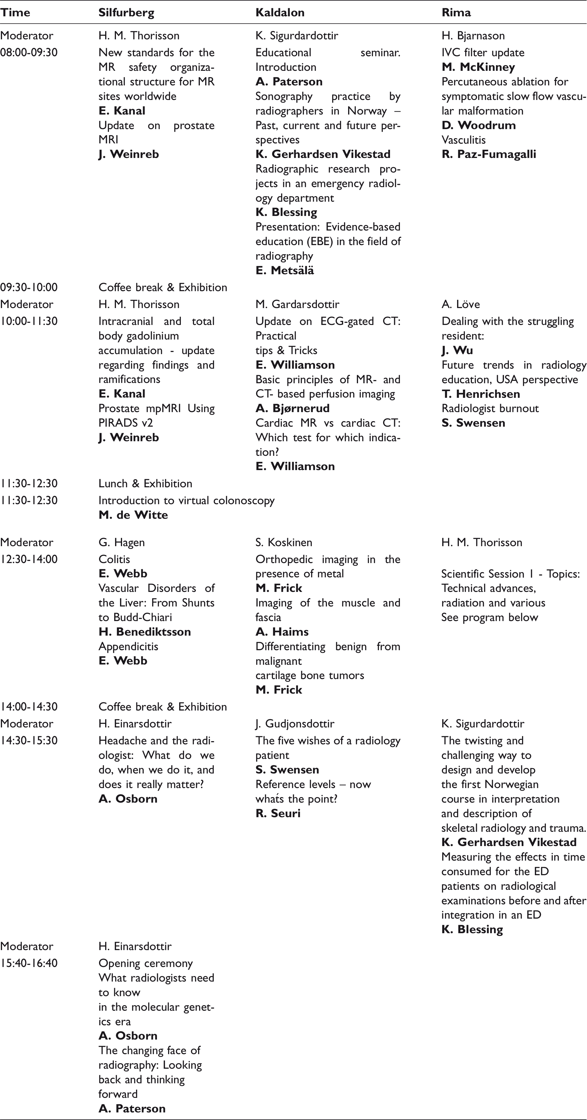

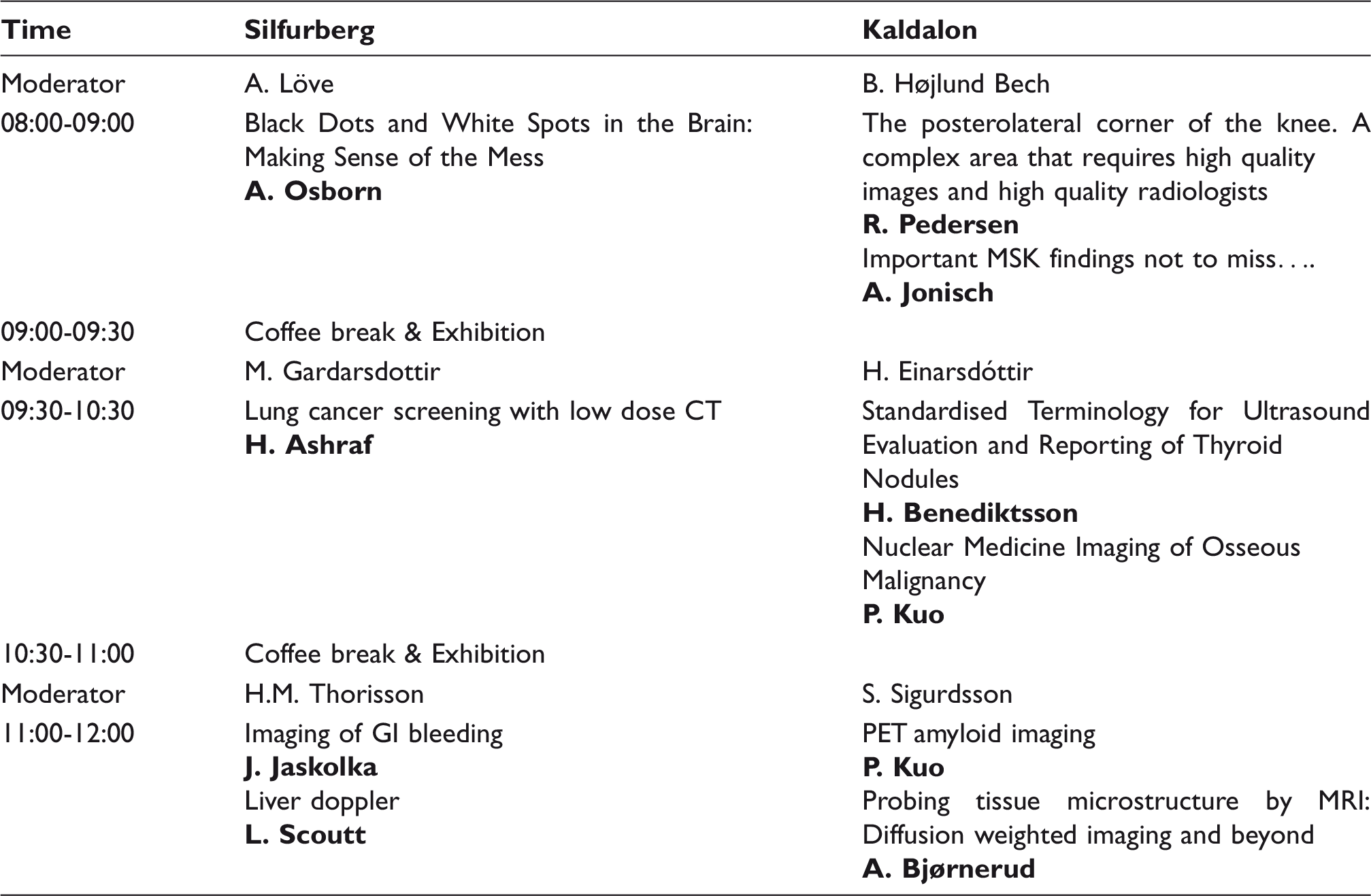


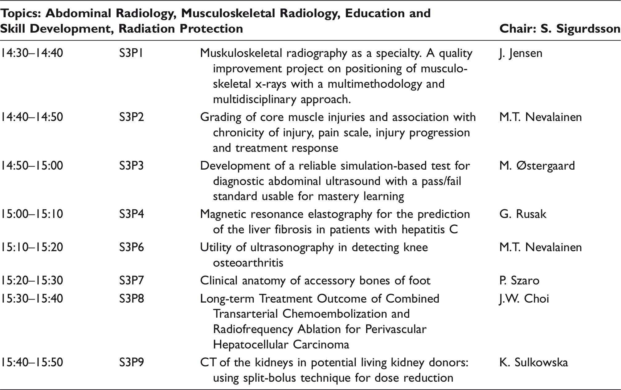
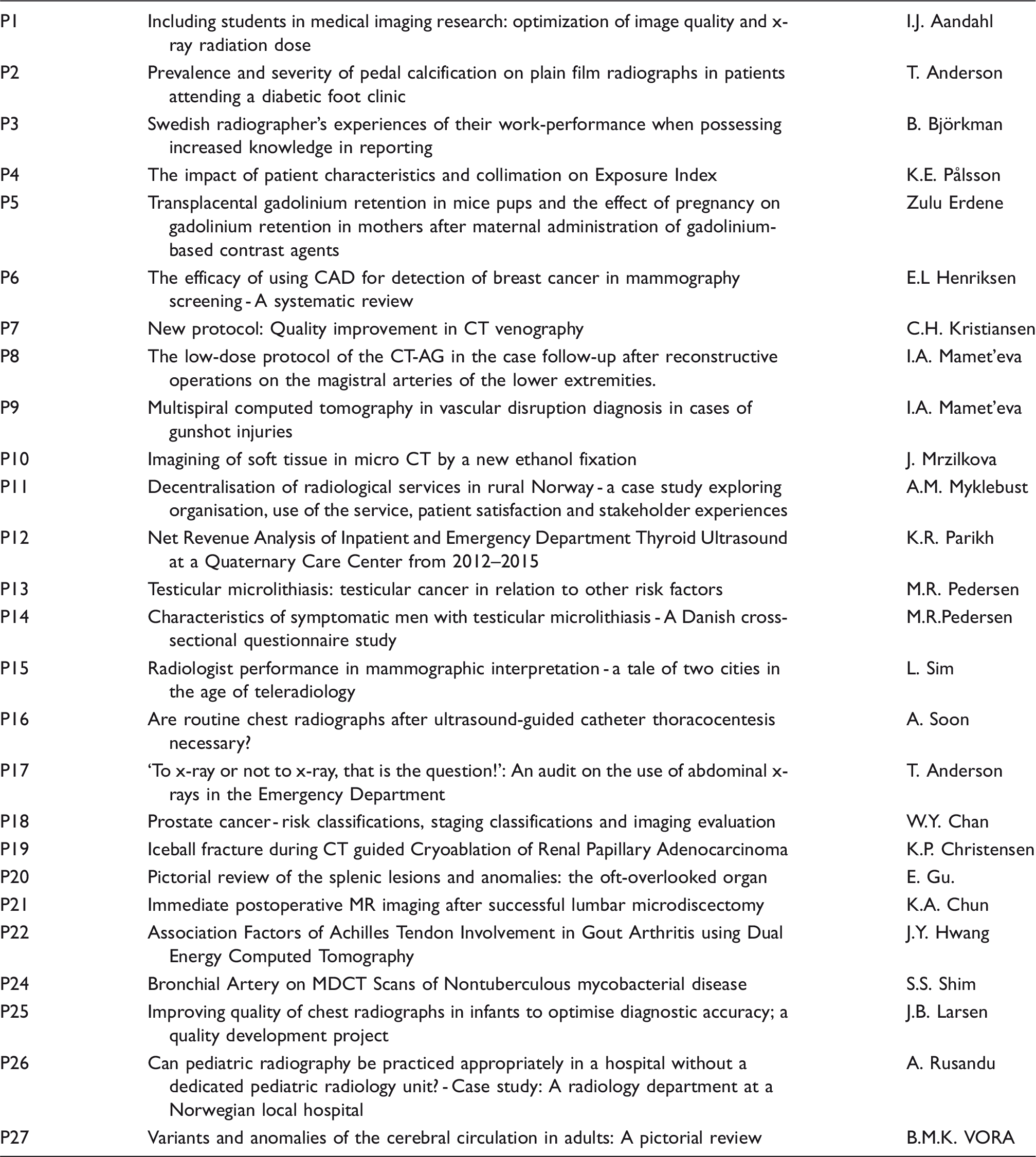