



Abstract
Background
Dedicated blood-pool contrast agents combined with optimal angiographic protocols could improve the diagnostic accuracy of thoracic magnetic resonance angiography (MRA).
Purpose
To assess the clinical utility of Gadofesveset-enhanced imaging and compare an optimized steady-state (SS) sequence against conventional first-pass dynamic multi-phase (DMP) imaging.
Material and Methods
Twenty-nine patients (17 men, 12 women; mean age = 42.7, age range = 18–72 years) referred for MR thoracic venography were recruited. Imaging was performed on a 1.5T MRI system. A blood-pool contrast agent (Gadofesveset) was administered intravenously. Thirty temporal phases were acquired using DMP. This was immediately followed by a high-resolution SS sequence. Three radiologists in consensus reviewed seven thoracic vascular segments after randomizing the acquisition order. Image quality, stenoses, thromboses, and artifacts were graded using a categorical scoring system. The image quality for both approaches was compared using Wilcoxon’s signed-rank test. McNemar’s test was used to compare the proportions of stenosis grades, thrombus and artifacts.
Results
SS had significantly better image quality than DMP (3.14 ± 0.73 and 2.92 ± 0.60, respectively; P < 0.001). SS identified fewer stenoses (>50%) than DMP; the differences in stenosis categorizations was statistically significant (P = 0.013). There was no significant difference in the proportions of vessels with thromboses (P = 0.617). DMP produced more artifacts than SS (101 versus 85); however, the difference was not statistically significant (P = 0.073).
Conclusion
Gadofesveset-enhanced thoracic angiography is clinically feasible. SS imaging produces better image quality and fewer artifacts than conventional DMP imaging.
Introduction
Establishing the patency of the central veins is an integral part of assessment of patients undergoing dialysis, parenteral nutrition, and where complex venous access is required, such as in multi-visceral transplantation. Repeated previous central venous catheter insertion and other venous interventions frequently leads to varying degrees of thrombo-occlusive disease and related stenoses (1). The role of imaging is to provide an accurate assessment of the site and extent of any venous disease and to identify possible etiologies in order to plan subsequent management and avoid related complications. Intravenous digital subtraction angiography (IV-DSA), color Doppler ultrasonography (CDUS), and computed tomography (CT) venography are commonly used, often in combination, for the evaluation of central venous patency.
Magnetic resonance imaging (MRI) of the vascular system is increasingly utilized due to improved MR availability and the advantage of avoiding risks secondary to iodine-based contrast media and ionizing radiation. Contrast-enhanced MR venography was first described in 1997 for lower extremity deep venous system evaluation (2), and MR venography of the upper extremity was later demonstrated with results consistent with conventional X-ray venography (3).
Dynamic high-spatial-resolution contrast-enhanced MR venography has been shown to be equally sensitive and specific for detecting stenoses and occlusions as conventional venography of the central veins of the chest (3–9). However, an intravenous injection of gadolinium-based contrast agent is required, and the majority of these conventional contrast agents are extracellular agents that rapidly leave the vascular compartment. The concentration difference between the veins and background tissues is often substantially reduced as these studies typically rely on peripheral arm vein injection of the contrast agent and subsequent circulation through the arterial and venous systems. Although adequate to characterize the general morphology and degree of stenosis, the signal available, and hence both spatial resolution and image quality of time-resolved images, is often sub-optimal.
Over the past decade “long dwell” gadolinium-based contrast agents have been developed that persist in the intravascular space due to reversible binding to albumin (10). Following injection and redistribution of contrast agents in the circulatory system, vascular imaging can be performed during the equilibrium or “steady-state” (SS) phase which typically lasts several hours. These techniques permit longer acquisition times as they are not limited by the first-pass temporal limitations of conventional methods, and are particularly attractive for venous imaging as the contrast agent does not pass rapidly into the surrounding soft tissues.
The aim of this study was to evaluate and compare an optimized SS sequence against a conventional first-pass dynamic multi-phase (DMP) sequence using a blood pool contrast agent (gadofosveset trisodium) for evaluating the thoracic region in patients with suspected venous disease.
Material and Methods
This was a prospective open-label feasibility study which received clinical trial authorization (11). Ethics approval was obtained from the South-East Research Ethics Committee, UK (ethic application number: 07/H1102/109). Twenty-nine patients (17 men, 12 women; mean age = 42.7, age range = 18–72 years) routinely referred for MR angiography (MRA) of the central veins were recruited. Exclusion criteria included: contraindications to MRI (e.g. pacemaker, aneurysm clips, orbital metal); contraindications to intravenous gadolinium agents, including patients with liver transplants and patients with impaired renal function with estimated glomerular filtration rate < 30 mL/min); and pregnant or lactating women. Informed written consent was obtained from each patient. Clinical trial registration details are as follows: European Medicines Agency: EU Clinical Trials Registry; Trial Registration Number: 2007-002730-11, URL: https://www.clinicaltrialsregister.eu/ctr-search/trial/2007-002730-11/GB.
MRI protocol
Pulse sequence parameters.

The DMP acquisition employs TRICKS temporal acceleration.
Slice thickness is reconstructed to 1.3 mm and 0.8 mm for DMP and SS, respectively.
Image analysis
The DMP and SS datasets were anonymized and reviewed on a PACS workstation (Centricity, GE Healthcare, Waukesha, WI, USA). The images were assessed in consensus by three consultant radiologists with 20 (DJL), seven (TCS), and two (ES) years of experience in MRA reporting. The readers were allowed to individually adjust window centering and level settings of the MR datasets. The DMP and SS images were assessed independently and the order in which they were viewed was randomized to reduce recall bias. The readers were blinded to all clinical and demographic information. The DMP images were used as the reference standard for correct diagnosis. Seven venous segments were assessed in each patient: superior vena cava (SVC), left and right branches of brachiocephalic (LB, RB), subclavian (LS, RS), and the internal jugular (LI, RI).
On each examination and for each venous segment, assessments were made of the following four areas:
image quality in terms of vessel conspicuity using a four-point scale: excellent (optimal visualization of the vessel with no signal loss); good (slight signal loss but good overall visualization of the vessel); moderate (decreased signal intensity but images remain diagnostic); or poor (insufficient signal intensity such that the vessel is not completely identifiable and the image is not diagnostic); presence of stenosis using a six-point scale: no stenosis (0% occlusion); mild (1–30%); mild-to-moderate (31–50%); moderate (51–75%); severe (76–99%); or total vessel occlusion. Each venous segment was evaluated for the highest degree of stenosis within that segment. Assessment of the degree of stenosis was not performed if a vessel could not be fully evaluated due to poor image quality; presence of thrombosis: no thrombosis or partial/complete thrombosis. Assessment of the presence of thrombosis was not performed if a vessel could not be fully evaluated due to poor image quality; presence of artifacts: none or mild/major artifacts impairing diagnosis.
Statistical analysis
The DMP and SS images were compared separately in terms of image quality, presence of stenosis, degree of thrombosis, and presence of artifacts. Normality assumptions were tested using the Shapiro–Wilk test. The image quality of both respective methods was compared using Wilcoxon’s signed-rank test. The specificity and sensitivity of the SS approach in detecting a stenosis > 50% was performed relative to DMP. The specificity and sensitivity of the SS approach in detecting either a partial or complete thrombus was performed relative to DMP. McNemar’s test was performed to determine if the proportions of stenosis, thrombus and artifacts reported using SS were equivalent to DMP. A P value < 0.05 was defined as statistically significant. All the statistical analysis was performed using the R programming language (version 3.1.1, The R Foundation for Statistical Computing, Vienna, Austria).
Results
All 29 patients completed the DMP and SS examinations. No adverse events or reactions were reported following the administration of gadofosveset. A total of 203 venous segments were assessed for both DMP and SS imaging (i.e. seven vascular territories were analyzed in 29 separate patients).
Image quality
For SS and DMP the image quality from all venous segments was 3.14 ± 0.73 (range = 1–4) and 2.92 ± 0.60 (range = 2–4), respectively. The improvement in image quality using SS was statistically significant (P < 0.001). A comparison of image quality in all vessels (Fig. 1a) illustrates that the SS technique had over twice as many vessel segments categorized as excellent when compared to DMP (67 versus 29). However, three vessel segments (two in the left subclavian and one in the right subclavian) were categorized as having poor image quality using the SS technique and were subsequently excluded from stenosis classifications. A comparison of image quality for each vessel segment is shown in Table 2. Table 2 illustrates that the SS technique produced better image quality in each vessel segment and in two of the vessel segments the improvement was statistically significant (right branch of the brachiocephalic P = 0.042; and right subclavian P = 0.019).
(a) Image quality categorizations incorporating all 203 vessel segments, and (b) stenosis categorizations for mild stenosis and above (DMP [red] and SS [green]). Comparison of image quality using DMP and SS. Mean ± standard deviation. Statistically significant (P < 0.05).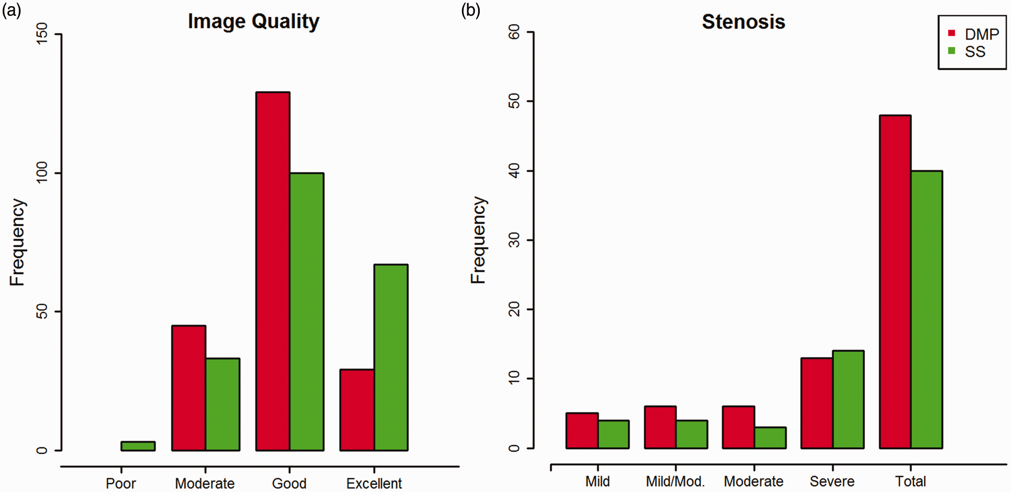

Stenosis categorizations
Comparison of stenosis categorizations using DMP and SS.

n, number of interpretable images for stenosis grading.

A discrepancy in stenosis characterization was noted between techniques in several patients. This example, of a 31-year-old female, illustrates a probable false positive characterization whereby DMP (a) identified a severe stenosis within the left subclavian vein whereas the corresponding SS images (b) identified a patent vessel proximal to a valve.
Thrombus detection
In total, using DMP imaging, 189 vessels were characterized as none, partial, or complete thrombus, as 14 vessels were uninterpretable. Using SS, 191 vessels were characterized for thrombus as 12 vessels were uninterpretable. There was no significant difference in the proportions of thrombus detected (Χ2 = 0.25, P = 0.617). SS has good specificity (81.3%) and sensitivity (99.5%) in detecting thrombus relative to DMP imaging. An example of thrombus detection is shown in Fig. 3. In this instance, the SS acquisition shows wall enhancement which in retrospect was also apparent on the last phase of the DMP acquisition.
A discrepancy in thrombus characterization, on a 56-year-old female, in the right internal jugular vein. The blind review of the initial first pass of DMP (a) defined the internal jugular as not assessable. The review of the SS images (c) identified a large thrombus with wall enhancement in the internal jugular. Subsequent retrospective analysis of the DMP dataset noted less obvious wall enhancement in the last phase (LP) of the DMP acquisition (b).
Artifacts
The SS technique reported fewer artifacts than the conventional DMP approach; however, the difference was not statistically significant (Χ2 = 3.214, P = 0.073). A representative example of mild artifacts that limit the ability for diagnosis using DMP is shown in Fig. 4. In this instance, the additional SS acquisition facilitated diagnosis in all vessels except the SVC.
A 32-year-old male patient in which the DMP images (a) present moderate to good image quality but demonstrate mild artifacts across most vessels (SVC, RB, RS, LB, LS, LI). The corresponding SS images (b) were rated good to excellent with mild artifacts present involving the SVC.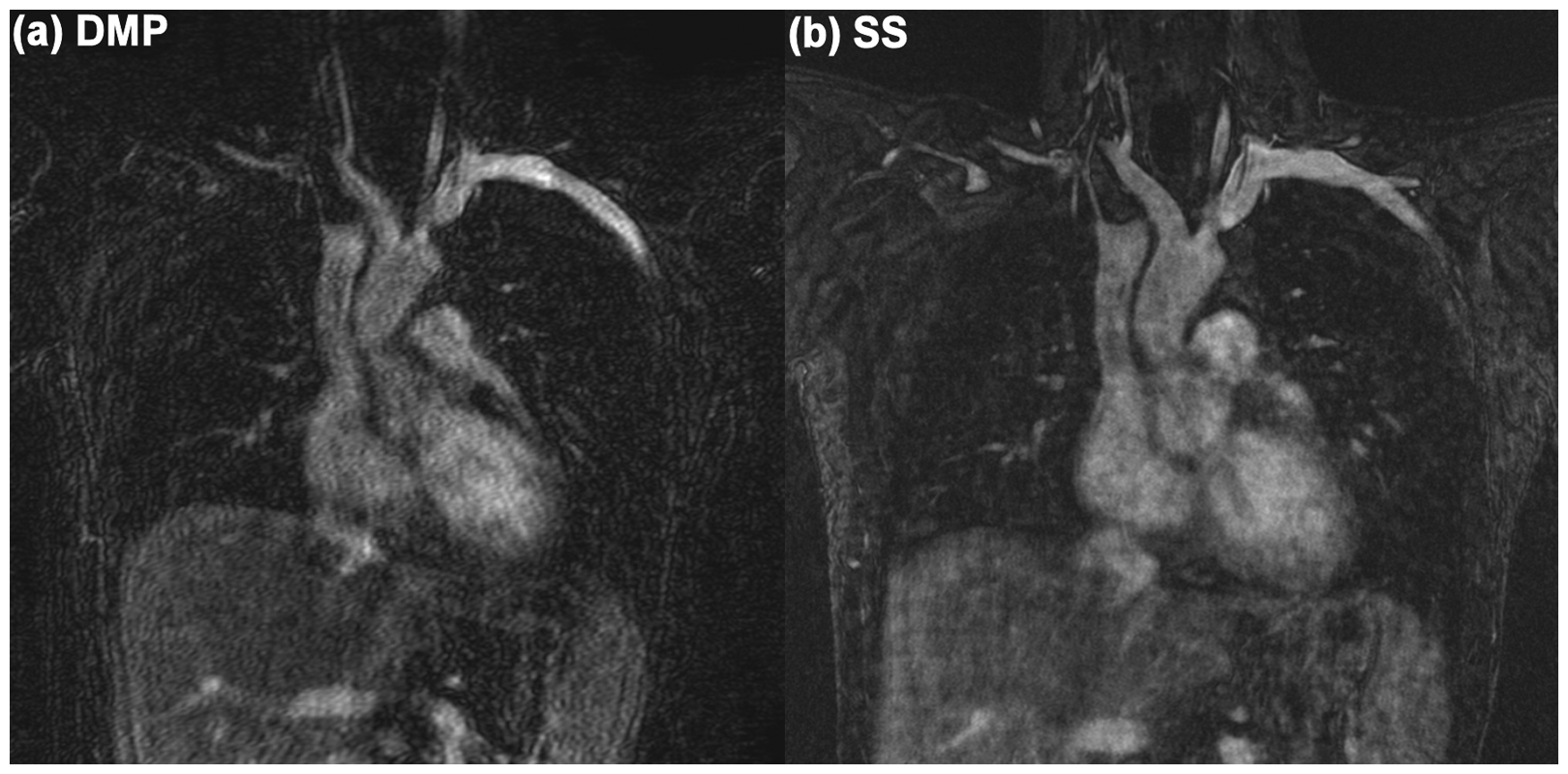
A representative example showing an instance when the DMP and SS images were equivalent is shown in Fig. 5. In this example, the vessels were rated with good-to-excellent image quality and the artifacts were rated as none-to-mild.
A 56-year-old male patient in which the DMP (a, b) and SS (c) images were rated with equivalent image quality (characterized as good to excellent). The presence of artifacts was also equivalent (rated as none to mild). Note: the patient has a catheter in situ (transiting the right subclavian and the right brachiocephalic vein).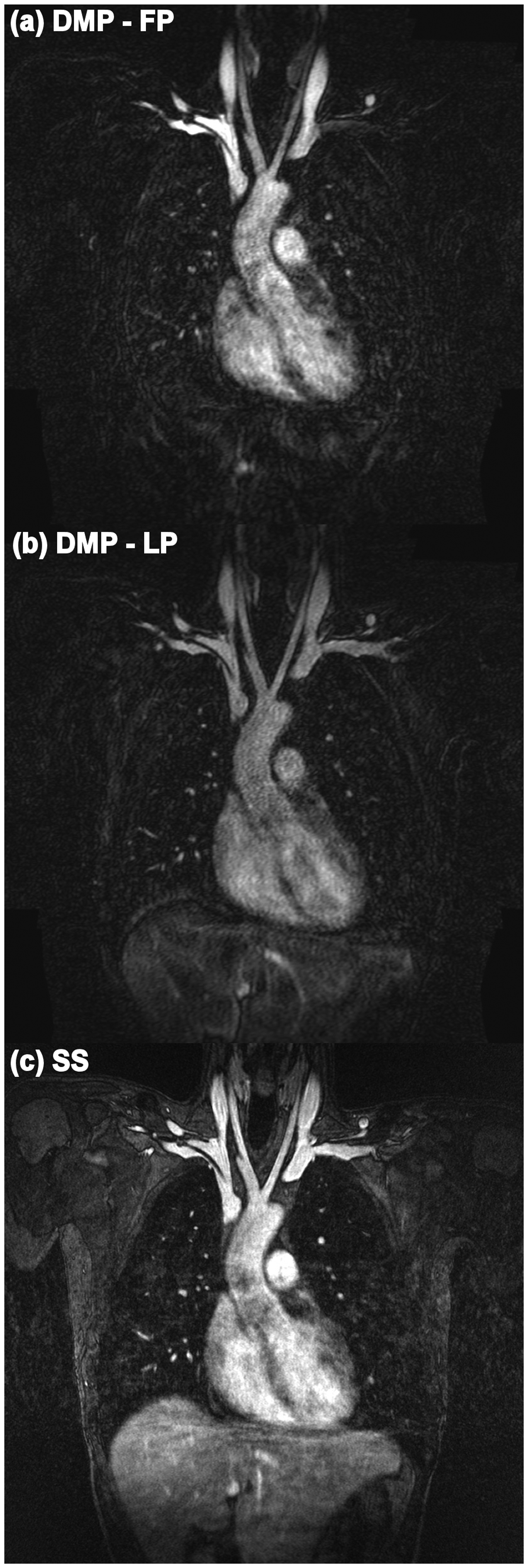
Discussion
This study suggests that the SS approach produces overall better imaging quality and fewer artifacts than DMP imaging. This is encouraging as the longer scan time required for SS makes it more susceptible to motion. Our results also suggest that DMP and SS are in general consistent in detecting thrombus.
Gadofosveset owes its intravascular retention to a strong but reversible affinity to albumin, which extends the vascular lifetime of the contrast agent. As a result, lower doses are required, but this agent can still be used for the multiphase first pass imaging of blood vessels as it has similar relaxivity properties to conventional gadolinium agents. This occurs as gadofoveset is rapidly bound to albumin after injection, producing similar T1 shortening as conventional gadolinium contrast agents residing in extracellular space.
Hadizadeh et al. previously reported on the utility of gadofosveset in peripheral angiography and compared SS and DMP against DSA as the gold standard (13). They found that the SS stenosis grades were more consistent with the gold standard DSA and that conventional DMP tended to overestimate stenosis grading. Our study also notes that DMP tends to overestimate the stenosis grade relative to SS. However, without DSA as a gold standard to reference it is impossible to infer which technique was most accurate.
The feasibility of a combined protocol for the MRI diagnosis of deep vein thrombosis and pulmonary embolism using gadofosveset trisodium has previously been reported (14). Its use for MR venography of the leg veins and inferior vena cava using fat-suppressed 3D gradient echo volume interpolated breath-hold examination showed high diagnostic image quality with no cases of moderate, poor, or non-diagnostic image quality. Additionally, an excellent inter-rater reliability was observed (15).
Although the SS-MRA offers all the advantages of a near isotropic 3D sequence, no information on flow dynamics can be obtained from the SS-MRA. Its use as a standalone technique is clinically adequate to assess central veins, but a combination of DMP and SS techniques would complement each other and in our experience may avoid the need for conventional venography. The two different imaging sets allow for more detailed assessment of any ambiguous venous segments. Future studies of the central veins are warranted to formally assess if the combined protocol can improve diagnostic confidence and accuracy. Anzidei et al. reported on the assessment of carotid artery stenosis using gadofosveset-enhanced MRA and found that the SS image reading was superior to first-pass image reading, but noted that a combined reading proved most accurate (16).
The use of gadofosveset with SS imaging has also been found favorable in the assessment of other thoracic vasculature. In an analysis of 25 patients, ECG-gated, motion-compensated high-resolution SS-MRA of the thoracic vasculature (left superior pulmonary vein, left pulmonary artery, and aortic arch) with gadofosveset offers significantly higher image quality and vessel sharpness when compared to standard first-pass DMP MRA (17).
Separately, the development of non-contrast alternatives remains critical to address issues with gadolinium agent administration, such as imaging patients with renal impairment. A notable study includes Tomasion et al.’s navigated SS approach to compensate for respiratory motion which reported good image quality metrics in the central veins in patients with vascular and cardiac disease (18). Further work in this area is warranted.
There are some limitations to our study. Consensus reading did not address individual variation in subjective assessment, but this approach may enhance overall accuracy compared to independent single observers, thus leading to a maximum advantage of the techniques studied. Furthermore, use of consensus reading was deemed acceptable since the aim of this study was not to determine the general sensitivity and specificity of MR datasets compared with conventional venography datasets, but rather to elucidate the relative degree of diagnostic information obtained with DMP and SS images.
Conventional venography, which has its own limitations, has not been used as the reference standard as the clinical information obtained from MR venography was considered sufficient and IV-DSA was not required in any of the cases following the MR venography examinations. The average interpretation time was not evaluated in our study. However, it has been shown in other work that there was no significant increase in interpretation time when reading both datasets together compared with reading the high-spatial-resolution dataset alone, despite the fact that there were more images overall to interpret (19).
In conclusion, we have found that MR venography with gadofosveset SS imaging in the equilibrium phase produced significantly higher quality images with less artifacts than a conventional DMP time-resolved technique, and was equivalent for the demonstration of venous stenosis and thrombosis.
Footnotes
Acknowledgments
The authors thank Addenbrooke’s Charitable Trust and the MRIS unit staff for their support. The study was supported by the imaging theme of the NIHR Cambridge Biomedical Research Centre in partnership with the University of Cambridge.
Declaration of Conflicting Interests
The author(s) declared no potential conflicts of interest with respect to the research, authorship, and/or publication of this article.
Funding
The author(s) received no financial support for the research, authorship, and/or publication of this article.