



Abstract
Background
Recent studies have suggested that the quantity and quality of adipose tissue and muscle, assessed on non-contrast computed tomography (CT), may serve as imaging biomarkers of survival in patients with and without neoplasms.
Purpose
To assess body composition measures that could serve as predictors of therapy response in patients with extremity soft tissue sarcomas treated with radiation therapy and surgery.
Material and Methods
The study was IRB-approved. Sixty patients had a history of extremity soft tissue sarcoma and underwent FDG-PET/CT prior to radiation therapy and surgical resection. Cross-sectional areas and CT attenuation (HU) of abdominal subcutaneous adipose tissue (SAT), visceral adipose tissue (VAT), and psoas muscle were assessed on non-contrast CT. Clinical information on predictors of tumor recurrence and post-surgical wound infections were recorded. Cox proportional hazard models were used to determine longitudinal associations between body composition and tumor recurrence/wound infections.
Results
Twenty-three tumor recurrences occurred over a follow-up period of 43 ± 35 months. Higher SAT and lower psoas attenuation were associated with tumor recurrence which remained significant after adjustment for covariates (P ≤ 0.01). There were 13 post-surgical wound infections. Higher VAT and SAT attenuation were associated with post-surgical wound infections (P < 0.04); however, VAT attenuation lost significance after adjustment for covariates.
Conclusion
Abdominal adipose tissue and psoas muscle attenuation assessed on non-contrast CT may predict tumor recurrence and post-surgical infections in patients with extremity soft tissue sarcomas.
Keywords
Introduction
Soft tissue sarcomas represent 1% of all adult malignancies and approximately 10,000 patients are diagnosed in the United States with a soft tissue sarcoma each year (1). About 50–60% of soft tissue sarcomas occur in the extremities and these tumors often recur and metastasize, despite complete resection with negative margins (2). Predicting outcome and designing management approaches in patients with soft tissue sarcomas is challenging given the multiple subtypes with different biological behaviors, some showing early progression, recurrence, and death, while others presenting with multiple or late recurrences over decades (3).
While in the past soft tissue sarcomas were routinely treated with amputation, advances in radiation therapy and surgical techniques have made limb-sparing surgery the standard treatment for the majority of localized extremity soft tissue sarcomas (4). External beam radiation therapy is usually administered preoperatively, intraoperatively, postoperatively, or in combination (5). Advantages of preoperative over postoperative radiation therapy include a decrease in radiation dose and radiation volume, improved tissue oxygenation, and decrease of metastatic potential of cells that may be spread at the time of surgery (6–8). However, radiation therapy may increase the risk of postoperative wound infections (9–11). Identification of biomarkers that could predict tumor recurrence and post-surgical wound infections would be valuable in the management of patients with extremity soft tissue sarcomas.
Recent studies have suggested that the quantity and quality of adipose tissue and muscle assessed on non-contrast CT may serve as imaging biomarkers of survival in patients with and without neoplasms. In these studies, abdominal adiposity and high CT attenuation of abdominal fat and lower attenuation of muscle were associated with increased mortality (12–15).
Patients with soft tissue sarcomas often undergo FDG-PET/CT for staging and surveillance, and adipose tissue and muscle measures, assessed on the non-contrast attenuation-correction CT, may be used as imaging biomarkers to predict tumor recurrence and post-surgical infections. In addition, adipose tissue and muscle characteristics could provide insights into cancer-related body composition abnormalities, such as cancer cachexia. The purpose of our study was therefore to assess abdominal adipose tissue cross-sectional areas and CT attenuation of abdominal adipose tissue and psoas muscle on non-contrast CT in patients with soft tissue sarcomas of the extremities undergoing radiation therapy and surgical resection. We hypothesized that increased CT attenuation of abdominal adipose tissue and decreased attenuation of muscle may serve as imaging biomarkers of tumor recurrence and post-surgical wound infections.
Material and Methods
Our study was IRB-approved and complied with HIPAA guidelines with exemption status for individual informed consent.
Patients
A retrospective search was performed to identify patients with a history of extremity soft tissue sarcoma who had undergone FDG-PET/CT prior to onset of any therapy at our institution between January 2005 and June 2016. Inclusion criteria were: age ≥18 years; pathologically proven soft tissue sarcoma of the extremities; diagnostic attenuation-correction non-contrast PET/CT; and radiation therapy followed by surgical resection of the soft tissue sarcoma. Exclusion criteria were malignancy other than soft tissue sarcoma, abdominal surgery or other pathology which could affect abdominal adipose tissue and psoas muscle measurements, such as ascites or metastases. We identified 127 patients with extremity soft tissue sarcoma who had a diagnostic PET/CT available. Forty patients were excluded because of therapy regimen (palliative chemotherapy/radiation therapy without surgical resection or surgical resection and/or chemotherapy but no radiation therapy). Twenty-seven patients were excluded due to tumor recurrence and/or metastatic disease at time of initial PET/CT. Sixty patients met inclusion criteria and comprised the study group.
Clinical data
Clinical data were obtained from electronic medical records: age, sex, BMI, types of pre-existing co-morbidities (cardiovascular disease, renal disease, type II diabetes mellitus, or a combination of those), sarcoma type (adipocyte, fibroblastic, smooth muscle, skeletal muscle, extraskeletal chondrogenic, vascular, or uncertain differentiation), sarcoma grade (grades 1 to 3, French Federation of Cancer Centers Sarcoma Group), sarcoma size (cm) determined as largest diameter on cross-sectional imaging, sarcoma stage (stages I to IV, TNM system of the American Joint Committee on Cancer), treatment regimen including radiation dose and use of adjuvant chemotherapy, presence of positive resection margins, local tumor recurrence, development of wound infection, and time between PET/CT and tumor recurrence/wound infection.
Body composition assessment
Whole-body 18F-FDG-PET/CT (Siemens Biograph 16 or 64; Siemens, Erlangen, Germany, or GE Discovery; GE Healthcare, Milwaukee, WI, USA) was performed per standard clinical protocol. Attenuation-correction CT was performed in the mid-expiration phase without intravenous contrast (slice thickness = 5 mm; table feed per rotation = 18 mm; time per table rotation = 0.5 s; tube voltage = 120 kVp; tube current = 11 mAs; field of view = 50 cm). Images were reconstructed to a slice thickness of 2.4 mm. The CT scanners used in this study were tested on an annual basis according to American Association of Physicists in Medicine (AAPM) and American College of Radiology (ACR) guidelines (AAPM report #74 and #96 and ACR CT QC manual) and standard clinical quality assurance measures were performed to assess for reproducibility of scans over time.
Non-contrast attenuation-correction CTs were used for assessment of abdominal adipose tissue cross-sectional areas (cm2) and abdominal adipose tissue and right psoas muscle attenuation (Hounsfield Units [HU]). Measurements were performed in the abdomen at the mid-portion of the fourth lumbar vertebra remote from the site of sarcoma, to avoid the influence of the primary tumor and changes from prior biopsy on body composition measures. In addition, measurements performed at the L4 level have been shown to correlate with whole body adiposity (16). Automated thresholding methods were applied using a threshold set for –50 to –250 HU to identify abdominal adipose tissue (17), and –29 to 150 HU to identify muscle tissue (18) (Osirix software version 3.2.1; www.osirix-viewer.com/index.html) (Suppl. Fig.). Intra-reader variability coefficients of variation (CV) for these measurements at our institution are 0.6–3.8% and interclass correlation coefficients (ICC) are 0.98–1.0 (95% confidence interval [CI] = 0.83–1.0). For inter-reader variability, CVs are 3.1–3.3% and ICCs are 0.98–1.0 (95% CI = 0.85–1.0). This was determined by analyzing five cases twice by the same reader and by analyzing five cases by two different readers (SAB and JV, with one year of experience under the supervision of MAB with 11 years of experience).
Statistical analysis
Statistical analysis was performed using JMP statistical software version 11.0 (SAS Institute Inc., Cary, NC, USA). For assessment of tumor recurrence and post-surgical wound infection, Cox proportional hazard models were used to assess the longitudinal association between abdominal adipose tissue and psoas muscle measures and tumor recurrence and post-surgical wound infections and the results are presented as hazard ratios (HR) with 95% CIs. Multivariate analysis was performed by subsequently adding three different covariate models, which included known risk factors for sarcoma recurrence (3,19,20). The first model adjusted for age and pre-existing co-morbidities, model 2 consisted of model 1 plus sarcoma type, size, and stage, model 3 included model 2 plus preoperative radiation dose and the presence of positive resection margins. For post-surgical wound infection, three different covariate models which included known risk factors for post-surgical wound infections were added (11). The first model adjusted for age and BMI. Model 2 included Model 1 plus co-morbidities and model 3 included model 2 plus tumor stage, size, and preoperative radiation dose. For all analyses, a two-sided P value of <0.05 was considered statistically significant. Data are shown as mean ± standard deviation (SD).
Results
Patient characteristics. Data presented as mean ± SD for continuous variables and n (%) for categorical variables.

Association of adipose tissue and muscle measures and tumor recurrence. Data are presented as hazard ratio (95% CI) per 1 SD increase in cm2 and HU, respectively.

Model 1: adjusted for age and co-morbidities.
†Model 2: adjusted for Model 1 plus sarcoma type, size, and stage.
‡Model 3: adjusted for Model 2 plus preoperative radiation dose and positive resection margins.
VAT, visceral adipose tissue; CSA, cross-sectional area; SAT, subcutaneous adipose tissue.
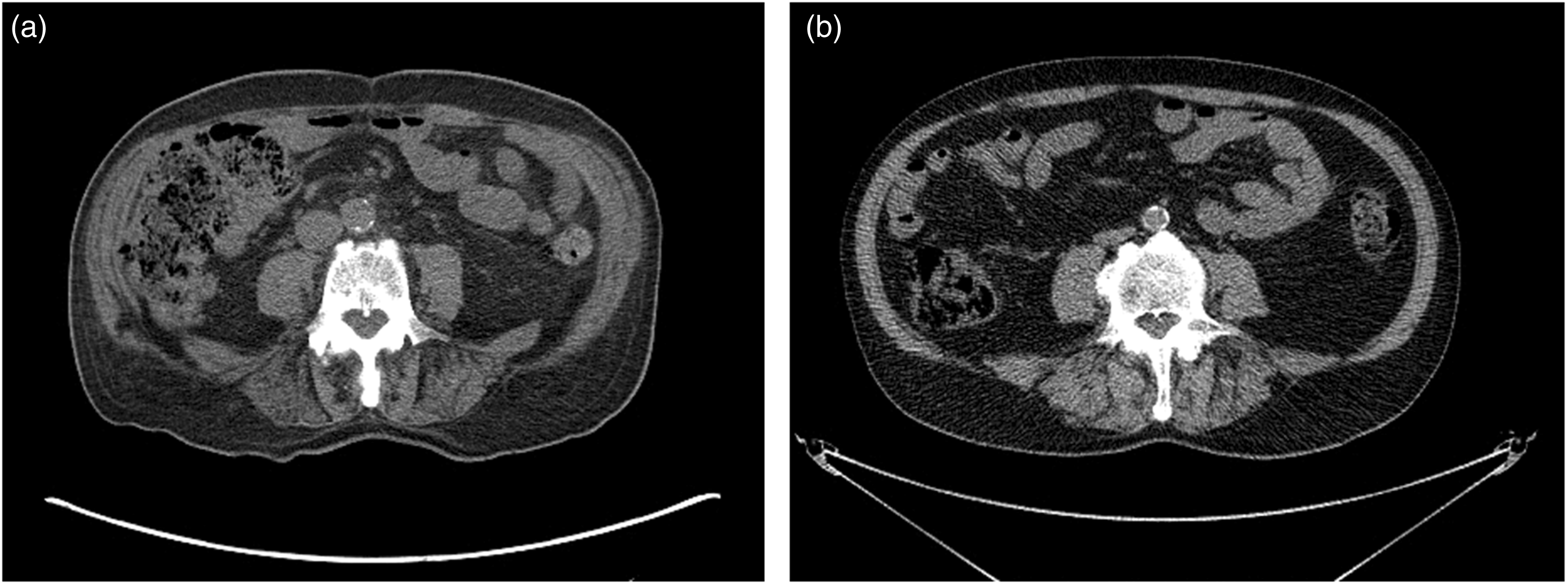
Axial non-contrast CTs at the level of L4 in two men with thigh liposarcoma treated with preoperative radiation therapy followed by surgical resection. (a) A 76-year-old man who developed tumor recurrence demonstrates increased adipose tissue attenuation (–76 HU) and low psoas muscle attenuation (33 HU) compared with (b) 72-year-old year old man who did not developed tumor recurrence and who had lower CT attenuation of adipose tissue (–103 HU) and higher psoas muscle attenuation (52 HU). Images are presented using the same window and level.
There was a significant association between CT attenuation of psoas muscle and tumor recurrence (Table 2, Fig. 1). Decreased attenuation of psoas muscle was associated with tumor recurrence (P = 0.0002). The association remained significant after adjusting for covariates (P ≤ 0.01).
Association of adipose tissue and muscle measures and postoperative wound infection. Data are presented as hazard ratio (95% CI) per 1 SD increase in cm2 and HU, respectively.

Model 1: adjusted for age and BMI.
Model 2: adjusted for Model 1 plus co-morbidities.
Model 3: adjusted for Model 2 plus tumor stage, size, and preoperative radiation dose.
VAT, visceral adipose tissue; CSA, cross-sectional area; SAT, subcutaneous adipose tissue.
Discussion
Our study showed that abdominal adipose tissue and psoas muscle attenuation, assessed on non-contrast attenuation-correction CT, may serve as imaging biomarkers for tumor recurrence and post-surgical infections in patients with extremity sarcomas treated with radiation therapy and surgical resection. Higher abdominal SAT and lower psoas muscle attenuation were associated with increased risk of tumor recurrence and higher abdominal adipose tissue attenuation was associated with post-surgical infections.
Soft tissue sarcomas are a diverse group of malignancies with the majority occurring in the extremities. The different histologic subtypes, varying natural history and response to therapy makes the prediction of outcome and the design of therapeutic regimens challenging (3). Goals of treating patients with soft tissue sarcomas are long-term survival, avoidance of local recurrence, maximizing function, and minimizing morbidity (4). Limb-sparing surgery combined with radiation therapy is considered to be effective for local control in patients with extremity soft tissue sarcomas (4–6). However, tumor recurrence remains a known risk, especially in the setting of positive resection margins. In addition, post-surgical wound infections are frequently encountered following preoperative radiation therapy (9–11). Therefore, biomarkers that could predict tumor recurrence and post-surgical wound infections would be useful in the management of patients with extremity soft tissue sarcomas.
Recent studies have focused on the assessment of body composition by CT to predict outcome in patients with cancers. Studies in patients with gastrointestinal cancers showed that increased abdominal adiposity and muscle loss were independent predictors of tumor recurrence and mortality (12,21,22). A study in patients with pancreatic cancer undergoing pancreatoduodenectomy showed that increased VAT cross-sectional areas was an independent predictor of post-surgical complications (23).
In our study, there were no associations between abdominal fat cross-sectional areas and tumor recurrence or post-surgical infection. However, not only the quantity of adipose or muscle tissue but also the quality of these tissues has been shown to independently predict survival in patients with and without cancer. Two large multicenter studies demonstrated increased all-cause mortality in patients with high CT attenuation of abdominal adipose tissue, independent of known risk factors, such as age, smoking, or co-morbidities (13,14). A study in patients with bone and soft tissue sarcomas undergoing a variety of therapies has demonstrated a positive association between mortality and high CT attenuation of abdominal SAT and low attenuation of psoas muscle, independent of known risk factors, such as age, BMI, co-morbidities, tumor stage, and grade (15). In our study, focusing on patients with soft tissue sarcomas of the extremities treated with a standard regimen, radiation therapy, and surgery, high abdominal SAT and low psoas muscle attenuation were positive predictors of tumor recurrence, independent of tumor size, stage, radiation dose, and other risk factors. In addition, higher abdominal SAT and VAT attenuation were positive predictors of post-surgical infections, independent of known risk factors for SAT attenuation and independent of age and BMI for VAT. These observations might provide insights into the pathophysiology of cancer-related body composition abnormalities, such as cancer cachexia.
The role of adipose tissue in cancer has received recent attention given the positive association between obesity and cancer incidence and mortality (24). Adipocytes present an important part of the tumor microenvironment and studies have shown that cancer cells can cause dedifferentiation of adipocytes into pre-adipocytes. This dedifferentiation is associated with increased lipolysis and loss of lipids, as well as changes in the expression of genes regulating energy turnover, cytoskeleton, and extracellular matrix (25). These cancer-associated adipocytes can have systemic metabolic effects and promote tumor growth (26–28). Studies in monkeys have shown that increased CT attenuation of abdominal adipose tissue corresponds to smaller adipocytes and increased extracellular matrix fibrosis (13). The observed association between increased CT attenuation of adipose tissue, remote from the site of sarcoma, and tumor recurrence may reflect smaller cancer-associated adipocytes and/or an adipocyte microenvironment promoting tumor growth.
Previous studies have elucidated cross-talk between adipose tissue and muscle in cancer. Adipose and muscle tissue secrete inflammatory cytokines, myokines, adipokines, and free fatty acids, which regulate adipose and muscle metabolism (25,29). The observed association between tumor recurrence and low CT attenuation of psoas muscle in addition to the high attenuation adipose tissue may reflect this cross-talk. Interestingly, a study in mice has shown that ablation of adipocyte lipase results in preservation of skeletal muscle, suggesting that the breakdown of fat in cancer precedes that of skeletal muscle and that the breakdown of adipocytes affects the breakdown of muscle (25,30). However, further studies are necessary to investigate molecular mechanisms mediating tissue attenuation and tumor recurrence in patient with soft tissue sarcomas.
The study had several limitations. First, the retrospective study design limits the ability to infer causality. In addition, we were dependent on medical records for clinical information. Second, we did not obtain adipose tissue or muscle biopsies to investigate underlying molecular pathways of adipose and muscle tissue and attenuation mediating tumor recurrence and post-surgical infections. Third, the study was performed using different CT scanners over a long time period. However, quality assurance measures according to the AAPM and ACR were performed to assess for reproducibility of scans over time. Strengths of the study include the large number of patients with extremity soft tissue sarcomas with pre-therapy imaging available and who underwent a standard treatment regimen, namely surgical resection followed by radiation therapy, and detailed measures of tissue attenuation on a standardized non-contrast CT protocol. In addition, the analyses were controlled for multiple covariates, known to predict tumor recurrence and post-surgical wound infections. We did not aim to assess threshold values to assess the sensitivity and specificity of body composition measures to detect tumor recurrence but rather used this pilot study to shed light into the regulation of tumor cells and body composition.
In conclusion, increased abdominal adipose tissue and lower psoas muscle attenuation are associated with increased risk of tumor recurrence and higher abdominal adipose tissue attenuation is associated with the development of post-surgical infections in patients with soft tissue sarcomas of the extremities. Adipose tissue and muscle CT attenuation can be easily assessed on any clinical workstation and these measurements could provide additional information on prognosis in patients with sarcomas. Shedding light into the biology of adipocytes and muscle cells in the tumor microenvironment may help identify metabolic targets that allow for development of new therapies for the treatment of sarcomas.
Footnotes
Declaration of Conflicting Interests
The author(s) declared no potential conflicts of interest with respect to the research, authorship, and/or publication of this article.
Funding
The author(s) disclosed receipt of the following financial support for the research, authorship, and/or publication of this article: This study was supported by NIH grant 5P30DK040561-18 and NIH grant K24DK109940.
References
Supplementary Material
Please find the following supplemental material available below.
For Open Access articles published under a Creative Commons License, all supplemental material carries the same license as the article it is associated with.
For non-Open Access articles published, all supplemental material carries a non-exclusive license, and permission requests for re-use of supplemental material or any part of supplemental material shall be sent directly to the copyright owner as specified in the copyright notice associated with the article.
