



Abstract
Background
Identification of the source of postpartum hemorrhage (PPH) is important for embolization because PPH frequently originates from non-uterine arteries.
Purpose
To evaluate the clinical importance of identifying the non-uterine arteries causing the PPH and the results of their selective embolization.
Material and Methods
This retrospective study enrolled 59 patients who underwent embolization for PPH from June 2009 to July 2016. Angiographic findings and medical records were reviewed to determine whether non-uterine arteries contributed to PPH. Arteries showing extravasation or hypertrophy accompanying uterine hypervascular staining were regarded as sources of the PPH. The results of their embolization were analyzed.
Results
Of 59 patients, 19 (32.2%) underwent embolization of non-uterine arteries. These arteries were ovarian (n = 7), vaginal (n = 5), round ligament (n = 5), inferior epigastric (n = 3), cervical (n = 2), internal pudendal (n = 2), vesical (n = 1), and rectal (n = 1) arteries. The embolic materials used included n-butyl cyanoacrylate (n = 9), gelatin sponge particles (n = 8), gelatin sponge particles with microcoils (n = 1), and polyvinyl alcohol particles (n = 1). In 13 patients, bilateral uterine arterial embolization was performed. Re-embolization was performed in two patients with persistent bleeding. Hemostasis was achieved in 17 (89.5%) patients. Two patients underwent immediate hysterectomy due to persistent bleeding. One patient experienced a major complication due to pelvic organ ischemia. One patient underwent delayed hysterectomy for uterine infarction four months later.
Conclusion
Non-uterine arteries are major sources of PPH. Detection and selective embolization are important for successful hemostasis.
Keywords
Introduction
Postpartum hemorrhage (PPH) is the most significant cause of maternal mortality, accounting for 25–30% of all maternal deaths worldwide (1). Frequent causes of PPH include uterine atony, genital tract laceration, retained placental tissue, abnormal placentation, and coagulopathy (2). In most cases, PPH can be managed with conservative treatment, such as administration of uterotonic agents, uterine massage, uterine packing, and blood transfusion. However, when conservative treatments fail, surgical or endovascular treatment should be considered as a treatment option. Recently, transcatheter arterial embolization (TAE) has been developed as a first-line treatment for intractable PPH and exhibits a high success rate (3,4).
When performing TAE, rapid identification of the cause and source of PPH is quite important because the culprit of PPH is often not confined to the uterine artery in various situations. Sometimes, embolization of the uterine artery is unnecessary when targeted embolization is successfully performed for PPH originating from a non-uterine artery. In addition, the arterial anatomy of the pelvis is the most complex one in the body, and precise catheterization of the bleeding artery is critical for successful treatment of potentially life-threatening hemorrhage (5,6). Therefore, the purpose of this study was to evaluate the clinical importance of detecting the non-uterine arteries causing PPH and the results of their selective embolization.
Material and Methods
Patients
Our institutional review board approved this retrospective study. Informed consent was waived due to its retrospective nature. The medical records of 59 patients (age range = 24–43 years, mean age = 32.9 years) who underwent emergency TAE for PPH in a municipal hospital that served as a secondary referral center were retrospectively reviewed from June 2009 to July 2016. Among these, 19 (32.2%) patients who underwent embolization of non-uterine arteries were included in this study. The initial laboratory findings and basic characteristics of these patients are summarized in Suppl. Table 1.
Embolization procedure
Before transfer to the Angiography Department, all patients were treated with uterotonic agents, vaginal gauze packing, and blood transfusion. Intrauterine balloon tamponade was attempted at the obstetrician’s discretion. Dual-phase abdominal computed tomographic (CT) angiography was performed before TAE with a slice thickness of 1.3 mm to evaluate the bleeding focus in possible cases.
TAE was performed by one of two interventional radiologists with 10 and 13 years of clinical experience, respectively. A 5-F vascular sheath was introduced via the right or left common femoral artery using the Seldinger technique. When the bleeding focus was identified on the CT scan, initial angiography was performed at the relevant arteries with 5-F angiographic catheters (Davis, Cobra and Simmons catheter; Cook, Bloomington, IN, USA). In cases where CT scan was not performed, pelvic aortography was performed with a 5-F pigtail catheter (Cook) with the tip of the catheter 3–4 cm above the aortic bifurcation to evaluate the pelvic arterial anatomy and to identify the bleeding focus. When the bleeding focus was identified on pelvic aortography, selective angiography of the bleeding arteries was performed with 5-F angiographic catheters. After that, super-selective arteriography was performed with a 2.3 or 2.0-F microcatheter (Microferret, Cook; Progreat, Terumo, Tokyo, Japan). When there were arteries showing extravasation or hypertrophy accompanying uterine hypervascular staining, embolization was performed (7). After embolization of the target arteries, completion angiography at the proximal portion of the target artery and a vaginal examination were performed to ensure adequate control of the bleeding. If vaginal gauze packing or an intrauterine balloon was used, they were removed before vaginal examination. If contrast material extravasation on angiography or persistent vaginal bleeding was observed, angiography including aortography with the tip located above the renal arteries was performed to identify another bleeding focus or collateral supply. If another bleeding focus or collateral supply was observed, additional embolization was performed. Embolic materials, such as gelatin sponge particles (Upjohn, Kalamazoo, MI, USA), n-butyl cyanoacrylate (NBCA) (Histoacryl; B. Braun, Melsungen, Germany), polyvinyl alcohol particles (PVA) (Contour; Boston Scientific, Natick, MA, USA), and platinum coils (Tornado; Cook), were used at the discretion of the attending interventional radiologist.
Data analysis and definition
Clinical and angiographic findings, procedure-related complications, and technical and clinical success rates were evaluated. Technical success was defined as complete embolization of the target arteries on angiography after TAE. Clinical success was defined as the instant cessation of bleeding on vaginal examination after TAE without the need for additional surgical management during the patient’s admission period. Patients who showed the following criteria were considered to have coagulopathy: a prothrombin ratio >1.5, a partial thromboplastin time greater than 45 s, or a platelet count of <80,000/µL (8,9). Complications were categorized as major or minor. Major complications included those requiring therapy or hospitalization, or those causing permanent adverse sequelae, or death. Minor complications were those requiring no or nominal therapy without consequences (10).
Results
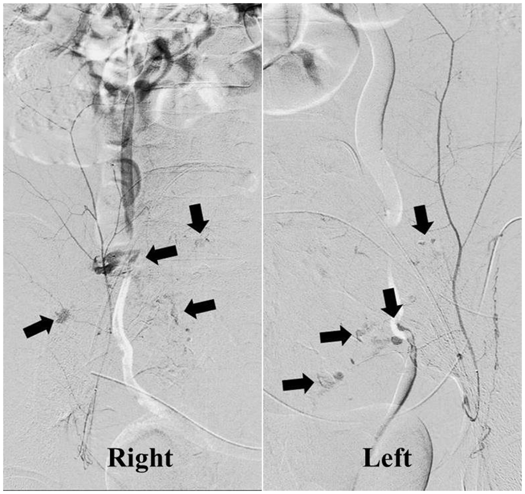
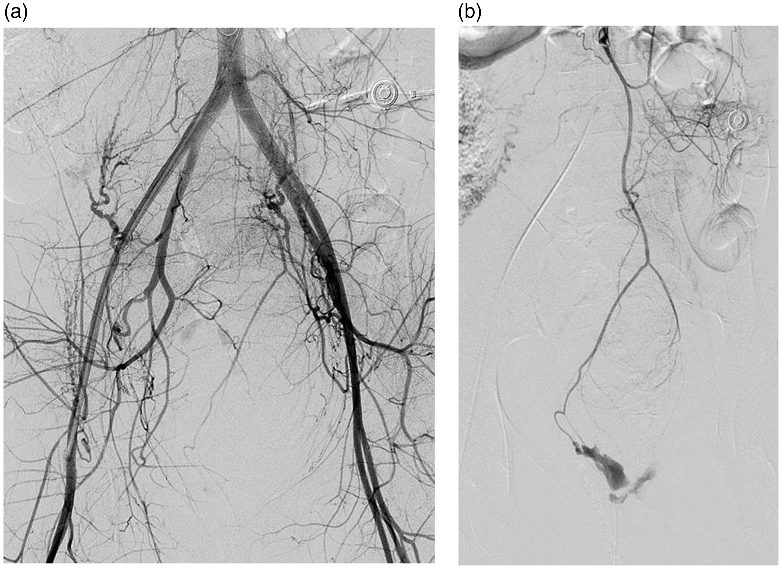
A total of 19 (32.2%) out of 59 patients underwent embolization of non-uterine arteries. Before transfer to the Angiography Department, intrauterine balloon tamponade was attempted in two patients. The initial hemoglobin levels of 19 patients before TAE were 2.9–13.4 g/dL (mean = 8.7 g/dL). Coagulopathy was present at the time of TAE in six (31.6%) patients. Patient characteristics and laboratory and angiographic findings are summarized in Suppl. Table 1. PPH was identified by CT scan (n = 5) and angiography (n = 14). Extravasation from non-uterine arteries, hypertrophied non-uterine arteries accompanying uterine hypervascular staining, and a combination of both were found in 10, six, and three patients, respectively. Non-uterine arteries that contributed to PPH included ovarian (n = 7), vaginal (n = 5), round ligament (n = 5), inferior epigastric (n = 3), cervical (n = 2), internal pudendal (n = 2), vesical (n = 1), and rectal (n = 1) arteries (Figs. 1–4). The embolic materials that were used included NBCA (n = 9), gelatin sponge particles (n = 8), gelatin sponge particles with microcoils (n = 1), and PVA particles (n = 1). NBCA was mixed with iodized oil (Lipiodol, Guerbet, Aulnay-Sous-Bois, France) at a ratio of 1:3. Gelatin sponge particles were prepared in a slurry of approximately 1–2 mm3 gelatin sponge cubes soaked in contrast media. The size of the PVA particles used was 355–500 µm. In 13 patients, additional embolization of the bilateral uterine artery was performed with gelatin sponge particles. Due to persistent bleeding, re-embolization was performed in two patients. Technical and clinical success was achieved in 17 (89.5%) patients. One patient with technical and clinical failure underwent hysterectomy due to persistent uterine bleeding from an atonic uterus. Another patient with multifocal bleeding from non-uterine arteries ultimately underwent hysterectomy following repeated embolization with major complications due to pelvic organ ischemia. One patient with technical and clinical success underwent delayed hysterectomy due to uterine infarction four months later.
Pelvic aortography of a 34-year-old woman who presented with postpartum hemorrhage after vaginal delivery showing hypertrophied bilateral uterine arteries (thin arrows), ovarian arteries (open arrows), and round ligament artery (thick arrows). After embolization of uterine and hypertrophied non-uterine arteries, vaginal bleeding stopped and the patient’s vital signs were stabilized. A 35-year-old woman presented with postpartum hemorrhage after Cesarean delivery. This patient underwent bilateral uterine arterial embolization due to extravasation from the uterine artery. However, the patient’s vital signs were not stabilized although vaginal bleeding stopped. After angiography to evaluate other bleeding foci, multifocal active bleeding was detected at the bilateral inferior epigastric arteries (arrows). A 27-year-old woman presented with postpartum hemorrhage after vaginal delivery. (a) On pelvic aortography, uterine artery hypertrophy and extravasation from the uterine artery were not detected. (b) After repeated arteriography to detect bleeding foci, the rectal artery was confirmed as a bleeding focus. A 39-year-old woman presented with postpartum hemorrhage after vaginal delivery. (a) Left internal iliac arteriography shows suspicious bleeding from the left vaginal artery (arrow). (b) Selective left vaginal arteriography showed extravasation (arrows). After selective vaginal arterial embolization with PVA particles, the bleeding was successfully controlled.

Discussion
Obstetric hemorrhage is a major cause of maternal death. Severe PPH is responsible for approximately 25–30% of all maternal death. Conservative treatments for PPH include uterotonic agents, uterine massage, uterine packing, and blood transfusion. Recently, intrauterine balloon tamponade has been used in attempts to control PPH, and the reported success rate was approximately 83% (11). However, traditional conservative treatment or intrauterine balloon tamponade has limited efficacy in patients with coagulopathy or if the cause of PPH is not uterine atony.
During the past two decades, TAE has been widely accepted as the primary treatment for patients with medically intractable life-threatening PPH (3,4). The cumulative success rate has been reported to be up to 90.7% (3,12–16). The main source of PPH is the uterine artery, arising from the anterior division of the internal iliac artery. However, collateral circulation should also be considered as a bleeding source during TAE as numerous collateral arteries develop during pregnancy. Among the collateral vessels, the ovarian artery is one of the main vessels that contribute to PPH (17). Considering the complex pelvic arterial anatomy and the potential vascular anastomotic network around the uterus, non-uterine pelvic arteries are also possible sources of PPH (6). Moreover, non-uterine arteries are major sources of PPH in certain clinical situations such as cases of birth canal laceration (18,19).
In the present study, 19 (32.2%) out of 59 patients with PPH underwent TAE for non-uterine arteries. The incidence of PPH from non-uterine arteries was higher in cases of birth canal injury or abnormal placentation than in cases of uterine atony. PPH among patients with birth canal injury or episiotomy site bleeding was ceased by the embolization of bleeding non-uterine arteries without uterine arterial embolization, except in one patient. Based on the retrospective review of the angiographic finding, the consensus was that additional uterine arterial embolization was unnecessary even in this patient. According to previous reports of PPH from birth canal injury, bleeding could be successfully controlled by super-selective target arterial embolization without uterine artery embolization (18,19). Therefore, it would be desirable to make more efforts to find causative non-uterine arteries when the PPH is not due to uterine atony.
With respect to embolic materials, NBCA was used in nine out of 13 patients with extravasation from a non-uterine artery. In eight patients, bleeding from a non-uterine artery stopped immediately after embolization. In one patient with coagulopathy, hemostasis failed because of persistent uterine atony. NBCA was first introduced for the treatment of PPH by Pelage et al. (20). Since then, several reports have suggested that NBCA is effective in addressing life-threatening PPH (21–24). According to previous reports, NBCA is effective in treating PPH from extravasation or pseudoaneurysm (8,13,21). In our experience, embolization with NBCA was successful, especially for PPH caused by extravasation from a single non-uterine artery. The results of our study are in agreement with the results of previous studies showing that NBCA can be applied as a primary embolic material for PPH (8,13,21–24).
In our study, a CT scan was performed in five patients. CT findings could be used to predict the bleeding focus, resulting in rapid embolization. In real practice, TAE for PPH is often performed without CT scans due to the patients’ unstable vital signs. However, the procedure time can be decreased by predicting the bleeding focus with a CT scan. According to previous reports (25–27), CT angiography and multidetector cone beam CT (MDCT) have a significant value in detecting and localizing PPH. In addition, these examinations provide supplementary information of extrauterine abnormalities. Among our cases, active extravasation was observed by CT in four patients. Based on these findings, selective arteriography of the suspected bleeding artery was done directly without diagnostic aortography. By doing this, the procedure time was decreased and successful embolization was achieved.
In our study, one patient underwent hysterectomy due to uterine infarction four months after embolization. This patient underwent embolization of the ovarian artery and the round ligament artery due to persistent vaginal bleeding after uterine arterial embolization. In this patient, vascularity of the uterus completely disappeared after post-embolic angiography. A CT scan performed four months later showed intrauterine air with fluid accumulation and diffuse thinning of the myometrium suggestive of uterine necrosis. Uterine necrosis is one of the rarest complications following TAE for PPH. Several hypotheses have been suggested to explain the cause of uterine infarction, including the use of particles being too small, use of PVA, absence of an anastomotic vascular system between the uterus and ovarian or pelvic arteries, embolization performed with high pressure, absence of antibiotic treatment, or existence of sepsis (28). In the present case, embolization of the bilateral uterine, right ovarian, and right round ligament arteries were performed with a slurry of approximately 1–2 mm3 gelatin sponge cubes. This might have resulted in the deprivation of a potentially important supplementary blood supply to the uterus, even though the gelatin sponge particle size used was not too small and the left ovarian and round ligament arteries, which were not major collateral arteries of the uterus, were not embolized. Therefore, the risk of uterine infarction should be considered when performing additional non-uterine arterial embolization.
We also experienced major complications due to pelvic organ ischemia after repeated embolization in one patient. This patient presented with intractable massive bleeding caused by placenta previa combined with placenta percreta. Even after bilateral uterine arterial embolization with NBCA and gelatin sponge particles, there remained numerous collateral vessels to the uterus from the branches of the bilateral internal iliac arteries. Due to deteriorated vital signs of the patient, embolization of multiple bleeding arteries were performed, including bilateral internal iliac arterial embolization with gelatin sponge particles. Three hours after the initial embolization, massive vaginal bleeding recurred and additional embolization of the bilateral internal iliac and round ligament arteries was performed. Despite repeated embolization, the patient underwent a hysterectomy due to persistent vaginal bleeding. After embolization and hysterectomy, the patient experienced impairment of the anal and bladder sphincter and skin necrosis at the bilateral buttock area following colostomy and skin flap surgery. During embolization, multiple bleeding branch and bilateral internal iliac arterial embolization was inevitable due to massive bleeding. However, massive pelvic arterial embolization can lead to disastrous results. Therefore, if uncontrollable massive bleeding is expected, predelivery treatment could be considered, such as temporary internal iliac artery balloon occlusion (29–32).
Two patients who had cervical arterial embolization were included in our study. During those procedures, selective embolization of the cervical artery with NBCA without additional uterine arterial embolization was performed. Originally, the cervical artery is a branch of the uterine artery (33). However, the cervical artery is a small proximal branch of the uterine artery that possesses quite different morphological characteristics than the tortuous ascending uterine branch. For this reason, we included these cases in our study. However, this enrollment was based on the clinical aspect rather than the traditional anatomical classification.
Our study has some limitations that warrant consideration. First, it was limited by its retrospective nature. The interventional protocol was not uniformly designed for each interventionist. Therefore, the decision of whether to perform additional embolization of the uterine artery after non-uterine arterial embolization was subjective and operator dependent. Second, our study was limited by the small number of patients. Therefore, additional prospective studies with more cases are needed to confirm the clinical importance of detecting non-uterine arterial bleeding during TAE for PPH.
In conclusion, non-uterine arteries are major sources of PPH. Therefore, their detection and selective embolization are important for successful hemostasis in patients with PPH.
Footnotes
Declaration of Conflicting Interests
The author(s) declared no potential conflicts of interest with respect to the research, authorship, and/or publication of this article.
Funding
The author(s) received no financial support for the research, authorship, and/or publication of this article.