



Abstract
Background
Whole spine localizers (WS-loc) of magnetic resonance imaging (MRI) are performed for enumeration of the vertebrae but they can be also used for the evaluation of the spine.
Purpose
To assess the accuracy of fracture detection using WS-locs of MRI and compare the findings with standard high-resolution short tau inversion recovery (STIR) sequences, and to determine whether the review of WS-locs is useful and if additional information can be gained by assessing the thoracic spine section of the WS-locs.
Material and Methods
A total of 298 magnetic resonance (MR) examinations of the lumbar spine with WS-locs were evaluated. Two independent readers reviewed the images. In case of fracture detection, further characterization of the fracture was performed. To assess inter-reader agreement, unweighted Cohen’s kappa with 95% confidence intervals (CI) and Phi coefficients were calculated.
Results
The study sample included 187 female and 111 male patients (age range = 65–94 years; median age = 75.0 years). The WS-locs detected 42 fractures of the lumbar spine and 36 of the thoracic spine. Inter-reader agreement for fracture detection in the lumbar and thoracic spine was strong (K = 0.87, 95% CI = 0.78–0.95, Phi = 0.87, and K = 0.88, 95% CI = 0.79–0.96, Phi = 0.88, respectively).
Conclusion
WS-locs from MR examinations of the lumbar spine provide a good diagnostic tool for the detection and evaluation of unsuspected vertebral fractures. WS-locs show strong inter-reader agreement for fracture detection in the thoracic and lumbar spine.
Introduction
Despite the rapid development of modern radiological techniques and evolving imaging possibilities, the diagnosis and therapy of vertebral fractures remain challenging (1). Up to 40% of symptomatic vertebral fractures are not diagnosed (2). Vertebral fractures are the most frequent of all osteoporotic fractures; their incidence in postmenopausal women is approximately 20%. Especially in elderly patients, the clinical manifestations of vertebral fractures may be obscured by additional co-morbidities. When postmenopausal women sustain one vertebral fracture, the risk for further vertebral fractures increases fivefold (3). However, it is unclear whether the risk for further fractures is also increased in patient groups other than postmenopausal women.
In patients aged > 70 years with low back pain (LBP), radiographs are initially performed, even if the radiographic detection of the vertebral fractures is rather poor (4). To exclude some rather rare reasons for LBP such as infection, tumor, or metastases, magnetic resonance imaging (MRI) can be additionally obtained (5). MRI may also help in fracture characterization (sub/acute versus old) and in the detection of unexpected fractures. Due to an increased risk of additional fractures (hip or other spine segments), associated medical and economic impact, and subsequently impaired function and disability, the relevance of unexpected vertebral fractures has been emphasized (5–9).
Recent studies have assessed the reliability of localizers from computed tomography (CT) and MRI examinations for the recognition and evaluation of vertebral fractures (10–13). MRI localizers of the lumbar spine (LS) were used to detect vertebral fractures and showed excellent inter- and intra-observer agreement (14). Due to the recent development of coils used in MRI, nowadays the whole spine localizers (WS-loc) are performed at the beginning of the MRI of the lumbar spine for enumeration of the vertebrae (15).
The aims of this study were as follows: to assess the accuracy of fracture detection of the lumbar spine using WS-locs of MRI and to compare the findings with standard high-resolution short tau inversion recovery (STIR) sequences; and to determine whether the review of WS-locs is useful for detection of additional fractures in thoracic and cervical spine
Material and Methods
Study design
This study was approved by the Ethical Review Committee (EKNZ Project-ID: 2016-02141).
A total of 298 MRI consecutive examinations of the lumbar spine with WS-locs performed between January and June 2017 were selected from the local Picture Archiving and Communication System (PACS) in the radiology digital archive. Patients aged >65 years who had an MRI of the lumbar spine with WS-locs were included in the study. The exclusion criteria were: (i) missing WS-locs; (ii) missing information on the referral reason; and (iii) metal hardware resulting in non-evaluable image quality. To investigate a representative patient population, the study was performed on all consecutive patients aged >65 years and not only >70 years as previously described (4).
Image analysis
Two independent blinded readers with experience in spine imaging (reader 1: radiologist (MK) with two years of experience; reader 2: orthopedic surgeon (JMB) with four years of experience) evaluated the WS-locs. Lumbar, thoracic, and cervical spine segments of the sagittal and coronal WS-loc were reviewed for fractures. In case of disagreement, a senior radiologist was consulted (FA).
After assessment of the WS-locs, comparison with the lumbar spine STIR sequences was performed. STIR sequences were used as the internal reference standard sequences for fracture detection in the lumbar spine. The fractures in the thoracic and cervical spine have been evaluated on the WS-locs; there was no additional sequence of the thoracic or cervical spine performed.
The reading sessions were done using GE Centricity RA 1000 PACS software, Version 4.0.11 (GE Healthcare, Barrington, IL, USA). Image quality was separately evaluated by both readers as 1 (poor), 2 (medium), or 3 (good).
Fracture characterization
When one or more fractures were detected, both readers performed further characterization of the most severe fracture, separately for each segment of the spine. The following criteria were analyzed: (i) height loss (>50% or <50%); and (ii) spinal canal stenosis.
MR examination
MR examinations were performed with 1.5-T and 3-T systems (Avanto Fit/Aera and Skyra, respectively; Siemens Medical Solutions, Erlangen, Germany) using integrated body coils with patients in the supine position. MR examinations of the lumbar spine that included WS-locs in the sagittal and coronal planes were used for fracture detection. Sagittal STIR sequences of the lumbar spine were used as the internal reference standard method to verify whether there was a fracture in the lumbar spine that correlated to the previously detected finding on the WS-locs. Detailed MR parameters of the WS-locs and STIR sequences are shown in Table 1.
MRI parameters.

TR/TE, repetition time/echo time; FOV, field of view; STIR, short tau inversion recovery; 3×, three blocks.
Statistical methods
Nominal variables are presented as numbers with appropriate percentages, while continuous variables are presented as medians (Me) with interquartile ranges (IQR). To evaluate inter-reader agreement, unweighted Cohen’s kappa with 95% confidence intervals (CI) and Phi coefficients were calculated and interpreted as previously described (16–18). STATISTICA 13.1 (Statsoft, Tulsa, OK, USA) was used for statistical analyses (19).
Results
In total, 298 patients (187 women, 111 men) were included in the study (age range = 65–94 years; median age = 75.0 years). The flow chart presenting patient selection is shown in Fig. 1.
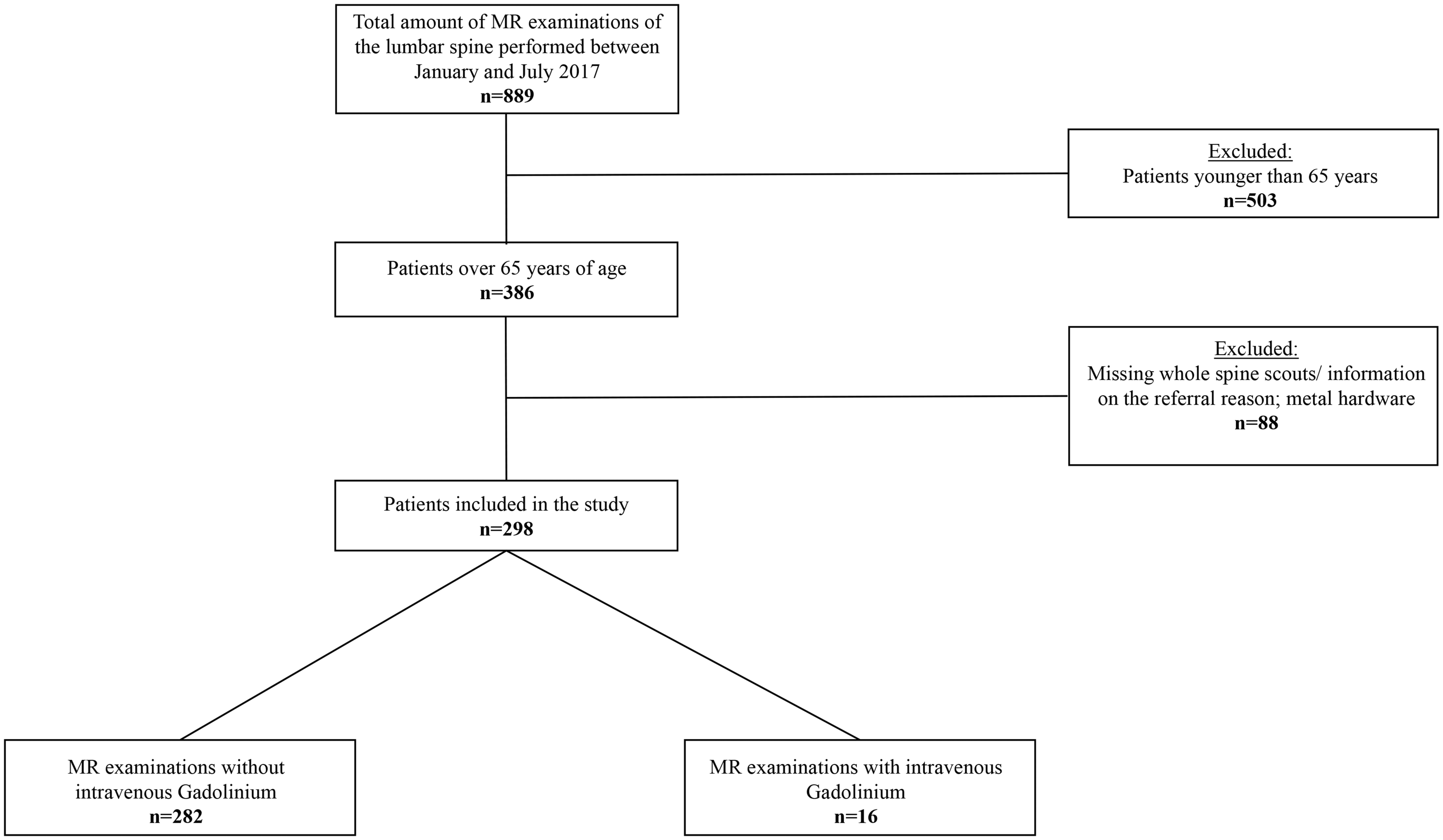
Flow chart demonstrating the selection process of patients included in the study.
The most common referral reason for performing MRI of the LS was lumbar pain (152/187 and 87/111 in female and male patients, respectively). The second most common referral reason for MRI of the lumbar spine was clinically suspected or known vertebral fracture (11/187 and 5/111 in female and male patients, respectively). Detailed patient characteristics of all patients are found in Table 2.
Detailed patient characteristics of all patients (n = 298).

Lumbar fractures
Reader 1 detected 42 fractures in the WS-locs; however, four of them turned out to be false positives in sagittal STIR sequences (sensitivity = 100%, specificity = 98%). Reader 2 detected 45 fractures of the LS in WS-locs, but two of them were false positives. Moreover, there were two false-negative findings in WS-locs identified by reader 2. Inter-reader agreement for the detection of fractures of the LS was strong (K = 0.87, 95% CI = 0.78–0.95, Phi = 0.87). Detailed fracture characteristics such as affected vertebral body, height loss, and spinal canal stenosis and inter-reader agreement are presented in Table 3. L1 was the most commonly affected vertebral body (25/42 of all LS fractures).
Detailed fracture characteristics for lumbar spine (LS) such as affected vertebral body, height loss, and spinal canal stenosis with inter-reader agreement.

Thoracic fractures
Reader 1 detected 36 fractures in the sagittal WS-locs; 33 of these were also visible in the coronal WS-locs. Reader 2 identified 33 fractures of the TS in sagittal WS-locs, with 32 of them evident in the coronal WS-locs. Inter-reader agreement for fracture detection was strong (0.88, 95% CI = 0.79–0.96, Phi = 0.88). Detailed fracture characteristics such as affected vertebral body, height loss, and spinal canal stenosis with inter-reader agreement are presented in Table 4. Th12 was the most commonly affected vertebral body (15/36 of all identified TS fractures).
Detailed fracture characteristics for thoracic spine (TS) such as affected vertebral body, height loss, and spinal canal stenosis with inter-reader agreement.

In 17 of patients who were diagnosed with a fracture in the LS, an additional fracture in the TS was identified. Moreover, in 22 out of 216 patients who did not have a fracture of the LS, at least one fracture of the TS was found. Examples of the MRI WS-loc with fractures of the LS and TS are presented in Figs. 2–4.
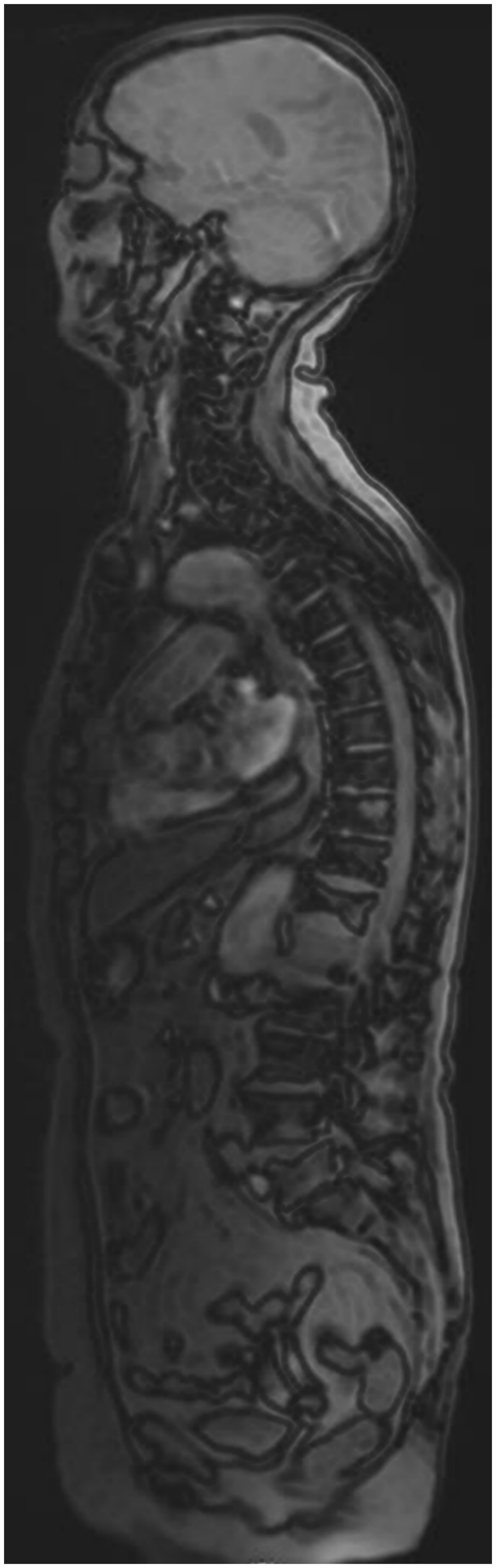
A 67-year-old male patient; WS-loc of MRI with a sub/acute fracture of L1 and an old fracture of Th12.
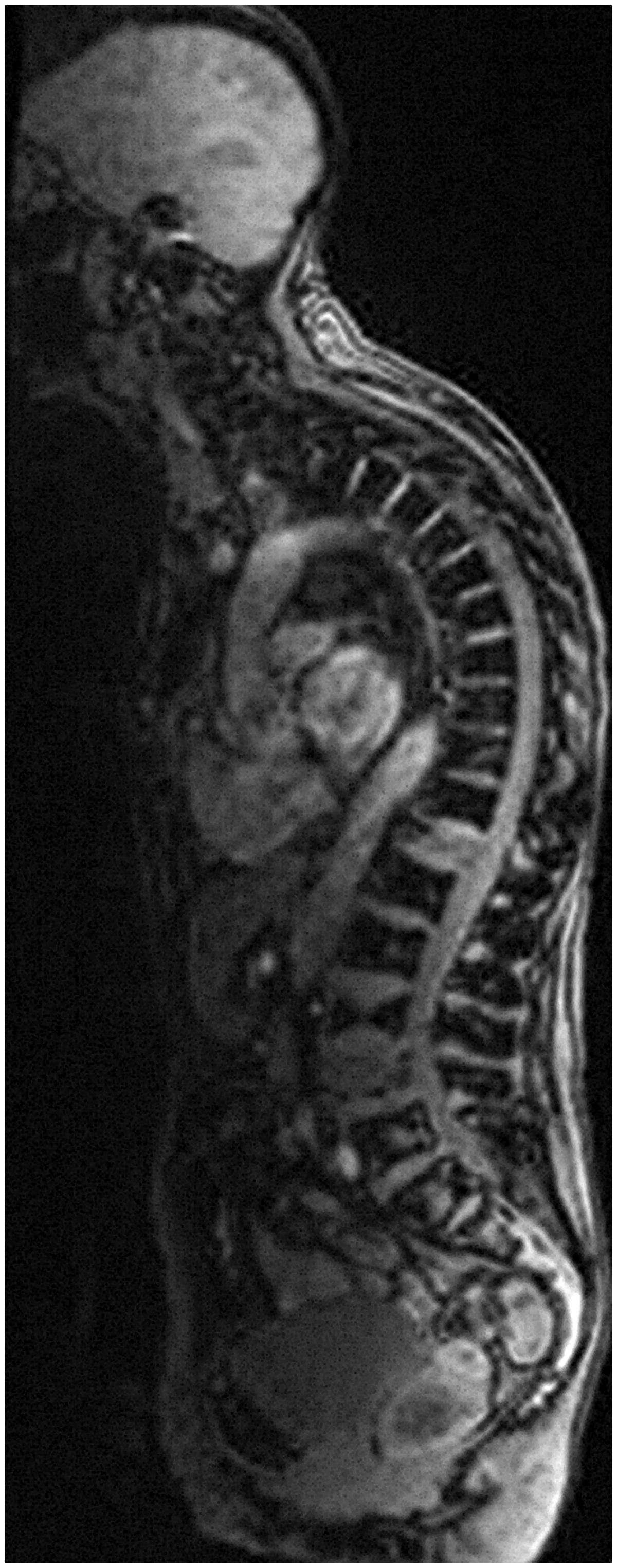
An 83-year-old female patient; WS-loc of MRI with a sub/acute fracture of L4, an old fracture of L3, and a sub/acute fracture of Th12.
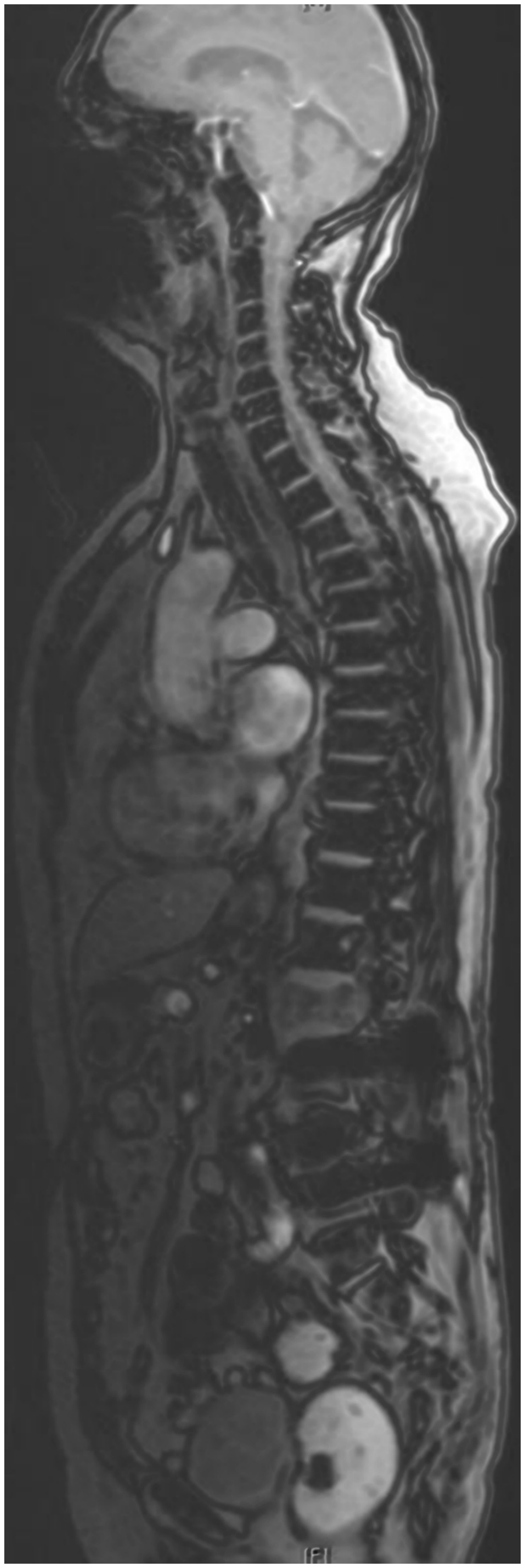
A 75-year-old female patient; WS-loc of MRI with a sub/acute fracture of L2.
Cervical fractures
There were no fractures or any pathologic signal abnormalities identified in the cervical spine.
Image quality
Image quality was good in 69.0%, medium in 25.3%, and poor in 5.7% of all cases.
Additional information
In all analyzed cases (n = 281), all findings of the LS and TS were mentioned in the final radiology report and there was no need to call the referring physician. A total of 245 patients underwent the MR examination via 1.5-T and 36 patients via 3-T scanners.
Discussion
This study provides a comprehensive evaluation using the WS-locs of MRI. The results suggest that WS-locs of MRI can be used for fracture detection and these outcomes are consistent with previous studies that focused on only one section of the spine, rather than the whole spine (10, 12, 14). Our study shows that WS-locs of MRI provide a reliable tool for the detection of vertebral fractures in the thoracic and lumbar spines. Despite a complete depiction of the cervical spine offered by the WS-locs, there were no fractures or pathologic findings observed in this region.
WS-locs of MRI depict the entire spine in both sagittal and coronal planes. Once a fracture is detected in any of the planes, it is possible to scroll for a correlative finding in both planes. As this study demonstrates, the agreement between sagittal and coronal WS-locs for fracture detection in the thoracic spine is very high (K= 0.95, 95% CI = 0.9–1). The possibility of scrolling between images, both in the sagittal and coronal WS-locs allows for even higher confidence during the evaluation of vertebral fractures.
WS-locs of MRI are obtained as three different blocks of the cervical, thoracic, and lumbar spine and then automatically reconstructed into the WS-locs. They have been described as a reliable and time-efficient tool for enumeration of the vertebrae; their implication in spine MR protocols has been primarily suggested for this reason (15).
In this study, parallel imaging was used to acquire the WS-locs in the sagittal and coronal planes, and the acquisition time was 86 s (approximately 28 s for each part of the spine). Because this method is performed using reduced volume of k-space data, the acceleration of acquisition time by factors of 1.5–3 is provided (20,21). The increased acquisition speed is the main advantage of this technique; however, its application is limited by artefacts and reduced image quality, such as signal-to-noise ratio (20, 21).
Our study shows that parallel imaging can be applied for acquisition of the WS-locs and it provides reliable fracture detection in the thoracic and lumbar spine. However, this approach may not be applicable in patients with diffuse osseous metastases with hyperintense signal in the whole spine, thus impeding detection of acute fractures.
The study of Xu et al. found that vertebral body height loss is an important indicator for the presence of vertebral fractures (22). For this reason, we sought to characterize the height loss of detected fractures in the WS-locs and applied such criteria as height loss of < 50% or > 50%. The inter-reader agreement was moderate (K = 0.68, 95% CI = 0.42–0.94) and strong (K = 0.80, 95% CI = 0.58–1) for characterization of the height loss in the LS and TS, respectively. However, malignant metastatic osseous disease may lead to kyphotic deformity and spinal malalignment, so in this case the loss of vertebral height cannot be used as a fracture indicator in the WS-locs. It must be also stated that the differentiation between benign and malignant vertebral fracture is not possible using the WS-locs.
To investigate a representative patient population, the study was performed on all consecutive patients aged > 65 years, without any additional information on patient medical history and co-morbidities (such as osteoporosis, metabolic bone disease, or medication). Reviewing the WS-locs, we found 42 and 36 fractures of the LS and TS, respectively. Sagittal STIR sequences of the lumbar spine were used as a reference standard method to verify whether the fracture of the LS visible in the WS-locs was truly positive. Only four of these turned out to be false positives in sagittal STIR sequences, resulting in 100% sensitivity and 98% specificity of the WS-locs. Moreover, 36 fractures of the thoracic spine were detected in the sagittal WS-locs, 33 of which were also visible in the coronal WS-loc. They were correctly described in the final report; however, they may have been considered unexpected findings, as the primary imaging focus was the lumbar spine.
As the study shows, the WS-locs may reveal additional fractures of the thoracic spine; these findings may require further imaging and focused clinical examination. The study of Kim et al. found that if there is a fracture involving > 2 vertebral bodies, MRI is required to make the diagnosis, because the possibility of confirming the hot uptake lesion as a new fracture by bone scanning is low (23). In this situation, the WS-locs can help in exact planning of the MRI exam with focus on the affected areas, without the need for the whole spine imaging.
The coronal WS-locs were used only for evaluation of the spine hence incidental findings such as renal and liver cysts were reported separately but not included in the study.
Our findings suggest that time efficient WS-locs are a helpful tool for the detection of unexpected vertebral fractures, including in the spine segments other than those initially suspected.
Our study has several limitations. First, the included patient group was relatively heterogeneous, with patients aged 65–94 years. Decreased bone mineralization and loss of muscle mass and function in elderly patients, known as sarcopenia, are potential risk factors for vertebral fractures, especially in women (24,25). However, we were not able to validate this hypothesis as there was no control group of younger patients. Second, all MR examinations were performed without specific information on bone diseases, for example, osteoporosis. Finally, we used sagittal STIR sequences of the lumbar spine to verify whether the detected fractures of LS were true positives. There was no additional sequence of the thoracic spine performed to verify whether the suspected findings were truly positive. The MR examination was performed primarily due to lumbar pain and the WS-locs were a usual part of the MR study. Detected fractures of the thoracic spine were reported separately and were subsequently followed by the referring clinicians. The addition of verifying sequences of the thoracic and lumbar spine in future studies with WS-locs of MRI may be valuable and could potentially provide supplementary results.
In conclusion, time-efficient WS-locs from MR examinations of the lumbar spine provide information for the evaluation of vertebral fractures and can be used not only for enumeration of the vertebrae, but also for the detection of unsuspected vertebral fractures. These fractures may need further imaging and should be reviewed separately. The WS-locs of MRI show a strong inter-reader agreement for fracture detection of the thoracic and lumbar spine and can be helpful for both radiologists and orthopedic surgeons.
Footnotes
Declaration of Conflicting Interests
The author(s) declared no potential conflicts of interest with respect to the research, authorship, and/or publication of this article.
Funding
The author(s) received no financial support for the research, authorship, and/or publication of this article.