



Abstract
Background
Varicocele is among the most common causes of male infertility because of various mechanism, including hypoxia.
Purpose
To evaluate testicular vascularization (TV) with ultrasensitive Doppler before and during the Valsalva maneuver (VM) and correlate TV changes to varicocele’s spermatic venous reflux graduated by color Doppler.
Material and Methods
From January to February 2016, 69 men (135 testis) referred for scrotal ultrasound were prospectively enrolled. An institutional review board approved the study. As gold standard, the spermatic venous reflux in the supratesticular region was assessed with color Doppler ultrasound and graded from 0 to III. A new ultra-sensitive Doppler (USD) sequence was performed on testicular parenchyma in an axial view. The TV changes before and during the VM were qualitatively graded from 0 to 2 using a visual scale by consensus. The vessels surface was quantified using customized MATLAB® software, and compared to the testicular delineated surface, resulting in a vessels surface ratio (VSR). The absolute and relative VSR difference before and during the VM was calculated. The qualitative scale and the quantitative VSR changes were compared with the spermatic venous reflux grade using an analysis of variance.
Results
A strong correlation has been found between the spermatic venous reflux grade and TV decrease during the VM using USD, for qualitative graduation as well as for quantitative measurement (P < 0.0001).
Conclusion
TV assessed by USD decreased significantly during the VM in patients with varicocele; this decrease was significantly associated with spermatic venous reflux grade.
Introduction
The incidence of varicocele in the general population is about 15% and can reach 30% in infertile men (1–3). Various pathological mechanisms have been suggested to explain why varicocele affects male fertility, including an increase in scrotal temperature, retrograde flow of adrenal or renal metabolites (4), and hypoxia (5). These changes lead to spermatozoa receiving an elevated amount of reactive oxygen species which is in turn associated with lipid peroxidation and sperm DNA fragmentation (6,7). Varicocele is defined as the presence of blood reflux within the spermatic veins resulting (or not) in vessel dilatation. Although varicocele is graded clinically (8), some studies report the presence of subclinical varicoceles, such as spermatic venous reflux detected by conventional color Doppler ultrasound (CDUS) or spectral Doppler that may also affect fecundity (9,10). Several classifications have been developed using B Mode/CDUS/spectral Doppler, based on vein size or topography, flow reverse during Valsalva maneuver (VM) (11–15). Solving the difficulties in defining the men in which varicocele negatively affects spermatogenesis would allow clinicians to better select those who have the most to benefit from treatment. Some authors previously mentioned the possibility that varicocele may induce intra-testicular vascular disorders, hypoxia, and possibly have harmful effects on spermatogenesis, as assessed using ultrasound (US) or magnetic resonance imaging (MRI) (6,16–18).
Ultrasensitive Doppler (USD) is an innovative US modality that provides a significant improvement in color flow imaging performance in terms of temporal resolution and sensitivity. Its first research application was published on brain functional imaging in rats (19) and neonatal brain in humans (20).
Our first published experience in testicular USD using this novel method found that testicular vascularization (TV) was reduced in non-obstructive azoospermia compared to obstructive azoospermia in a normal population. In this study, we assessed the intra-observer reproducibility based on three repeated TV measurements on 43 testes with an interclass correlation (ICC) of 0.801 (95% confidence interval [CI] of 0.695–0.879) (21). The effect of the VM was not evaluated.
The present study evaluated: (i) the changes in testicular vascularization (TV) estimated by USD during VM in men referred for scrotal US; and (ii) the correlation of TV with the spermatic cord venous reflux grade based on Oyen’s classification.
Material and Methods
Patients and study design
This prospective monocentric study was approved by the hospital ethics committee and each patient signed an informed consent form (Ethics Committee, #13-046). From January to February in 2016, 80 men referred for scrotal US examination were consecutively enrolled. Eleven patients with either a testicular focal lesion or acute scrotum injuries, such as torsion or epididymo-orchitis, were excluded, leaving a total of 69 patients enrolled in the study. Clinical indications were the following: infertility (60/69); pain (6/69); hypogonadism (2/69); and erectile dysfunction (1/69). Age and clinical data including infertility status and diagnosis of varicocele following physical examination were recorded. In case of infertility, patients were classified based on semen analysis as either azoospermia (no spermatozoon in semen) or oligo-astheno-teratospermia (according to the World Health Organization [WHO] criteria, oligozoospermia if spermatozoa < 15 million/mL, asthenozoospermia if < 32% progressive motile spermatozoa, teratozoospermia) (22). There may be multiple etiologies of infertility.
US examination
The examination was conducted by a single radiologist (LR), using an ultrafast US system (Aixplorer, SuperSonic Imagine, Aix-en-Provence, France) with a high frequency linear transducer (SL15-4, 7.5 MHz central frequency). First, USD was performed on testicular parenchyma, which was then followed by conventional B mode and CDUS on supratesticular veins before and during the VM.
Ultrasensitive Doppler protocol
A specific US Doppler sequence was developed in order to acquire USD information on the testis parenchyma. The USD sequence was adapted from previous research and was performed with the following parameters: time for insonification = 1 s; frame rate = 9600 frames/s using plane waves imaging; number of angles of insonification: 6 (from –5° to 5° [2° steps]); central frequency = 7.5 MHz; at 50 V; imaging depth = 20 mm. The USD algorithm based on singular value decomposition was applied to the raw US data. Next, a numerical filter was applied to remove the first 50 eigenvectors over the compounded raw radiofrequency data for each pixel, resulting in the cancellation of tissue signal. After these steps, only the blood vessel signal remained. By integrating the filtered data over time on each spatial pixel, we obtained a power Doppler image of the testicular parenchyma (Fig. 1). Raw frequency data were post-processed on a personal computer using customized MATLAB® software (2015a, MathWorks, Natick, MA, USA).
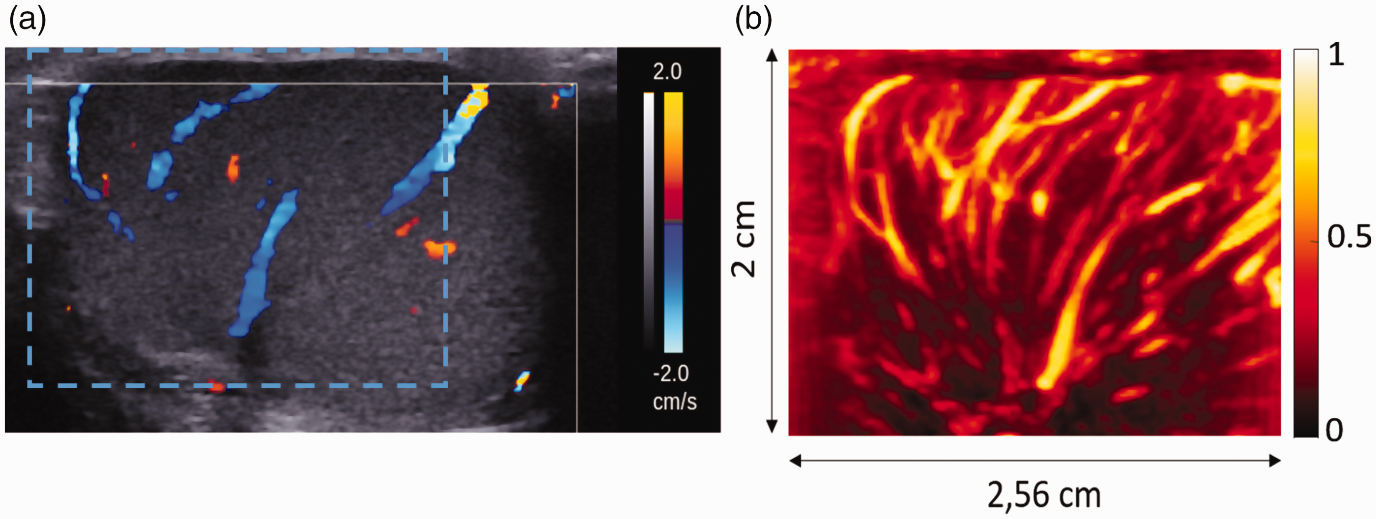
Conventional color Doppler (CDUS) and USD images. Conventional CDUS (a) with velocities set at the lowest available level, 2 cm/s. The dashed square represents the USD image position. The normalized USD of the same testis (b) shows an increased sensibility in detecting slow flowing vessels.

Qualitative scale based on subjective visual perfusion decrease. USD images before (a, c, e) and during (b, d, f) VM on normalized USD acquisitions for three different patients. (a, b) Patient 1 = grade 0 (no change); (c, d) Patient 2 = grade 1 (slight decrease); (e, f) Patient 3 = grade 2 (intense decrease).
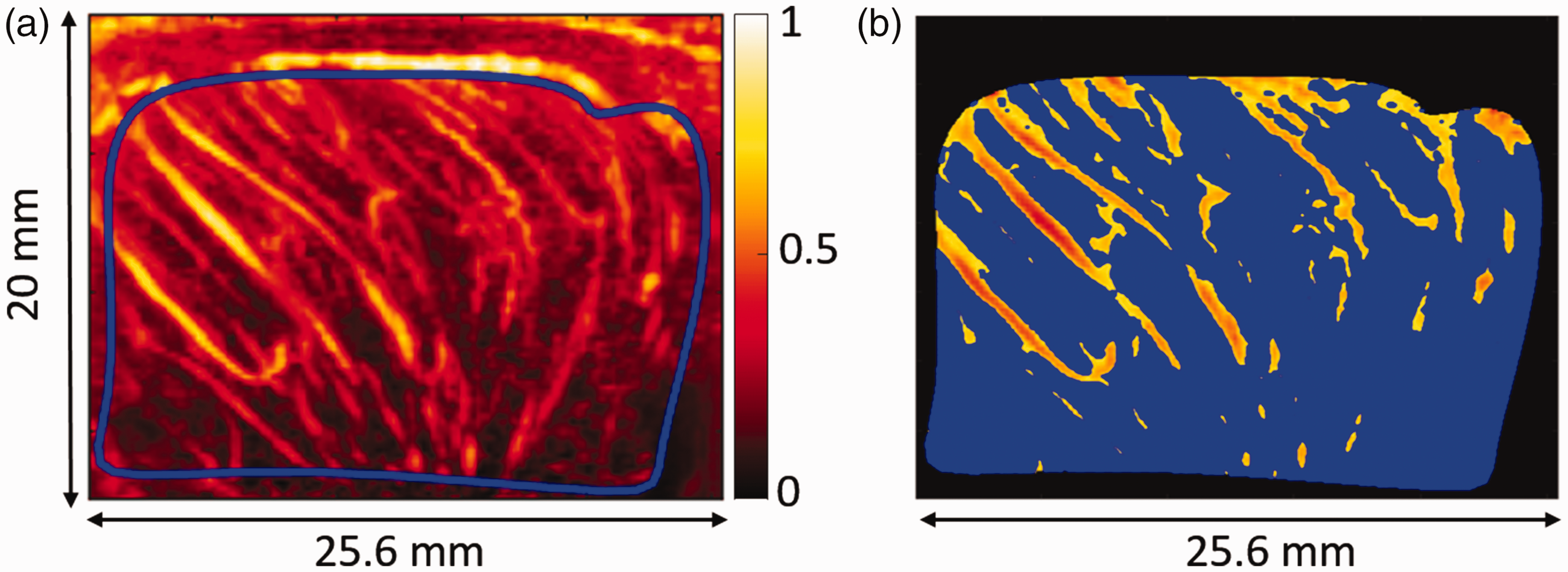
Segmentation. (a) Manual segmentation of testicular parenchyma avoiding capsular vessels using a custom algorithm. (b) Resulting segmented vessels.
The same operator (LR) performed all USD image processing before the conventional US exam. Two consecutive transverse USD acquisitions were performed on the same plane: the first while the patient was free breathing at rest and the second at least 3 s after starting a VM. If the acquisition failed because of motion, it was repeated. The testicular vascularization changes before (at rest) and during VM were assessed both qualitatively and quantitatively:
The qualitative evaluation was performed by visually grading TV changes on USD acquisitions on a scale of 0–2 by consensus (LR and JLG). Grade 0 was assigned when TV appeared unchanged by the VM (Fig. 2a and 2b). Grades 1 (Fig. 2c and 2d), and grade 2 (Fig. 2e and 2f) corresponded to a slight or marked visual decrease in TV during the VM, respectively; The quantitative evaluation of TV based on blood vessel surface was performed using a MatLab algorithm described in a previous feasibility study (21): first, a region of interest (ROI) was defined delineating the boundaries of testicular parenchyma in the USD images, taking care to avoid the capsular vessels. Second, TV was calculated. In the ROI, a common adequate threshold on the maximum intensity Doppler signal was defined to quantify the vessel surface. This threshold was defined as 75% (chosen after several tests on the first 20 patients). This 75% threshold represented the most accurate value across all patients that reflected the vascular vessel surface according to a visual control based on the radiologist’s (LR) experience. Within the ROI, the vessel surface was defined as the number of pixels with a higher intensity than the threshold (Fig. 3). All intensity points below this threshold were not taken into account in the vessel surface calculation. Then, the ratio between the vessel surface and the delineated testicular parenchyma surface, called the vessels surface ratio (VSR) was derived as a percentage. The same threshold was applied in all patients, both before and during VM.
Conventional US imaging protocol (including spermatic vein reflux detection)
The same operator (LR) performed all the conventional US exam. Standard B-mode and CDUS imaging at low PRF (velocities settled at 2–6 cm/s) was performed and patients were examined in the supine position. Testicular volume was determined using computer-assisted calculation in B-mode US based on the following formula: length × width ×height × 0.523. Spermatic vein flow was studied in the supratesticular region, specifically in the spermatic cord using conventional CDUS at rest and during VM. All men were graded bilaterally, in four groups in the supine position, based on Oyen’s classification of venous reflux (15). This was performed according to venous flow reversal during VM and videos were analyzed to assess the duration of the reflux The four groups were as follows: Grade 0 = no reflux before or during VM; Grade I = slight reflux during VM (<2 s); Grade II = significantly prolonged reflux (>2 s) but intermittent; Grade III = reflux at rest during normal respiration; and lastly continuous during the entire VM (Video 1 and 2). If the assessment of reflux analysis was not clear, the VM was repeated.
Statistical analysis
Testicular volume and VSR before and during VM were compared between bilateral testes using a Student’s t-test. As the intra-patient correlation of testicular surface was not significant, each testis was analyzed independently. A paired Student’s t-test was used to compare VRS before and during VM for both classifications derived from spermatic venous reflux grade and qualitative TV changes using USD acquisitions. The correlation between the spermatic venous reflux grade and the absolute and relative difference in VRS before and during VM (evaluated by USD) was tested with an analysis of variance (ANOVA). Lastly, a similar analysis was performed to test the associations between the qualitative classification in TV changes and the absolute and relative differences in VSR before and during the VM.
Results
The study population included 69 men (mean age = 36 ± 8.8 years). Semen analysis showed azoospermia (n = 14; in three cases after chemotherapy, one with Klinefelter Syndrome, the other from unknown origin), cryptozoospermia (n = 3, unknown origin), or oligoasthenoteratospermia (n = 49). One patient previously underwent an orchiectomy (necrosis after testicular torsion). Clinical examination found a palpable varicocele in 11 patients on the left side and never found one on the right side. USD evaluation before and during VM was then performed on 135 testes. For two patients, analysis was only completed on one side due to US attenuation.
Table 1 shows the following results presented by side: testicular volume; presence of palpable varicocele on examination; VSRs before and during VM; classification of patients according to Oyen’s spermatic venous reflux grade [14]; and the grade assigned according to qualitative TV changes observed by USD acquisitions as defined in the previous section.
Testicular volume, testicular surface vessels surface, patient’s classification according to spermatic cord venous reflux grade, patient’s classification according to qualitative TV changes; the results are presented side by side.
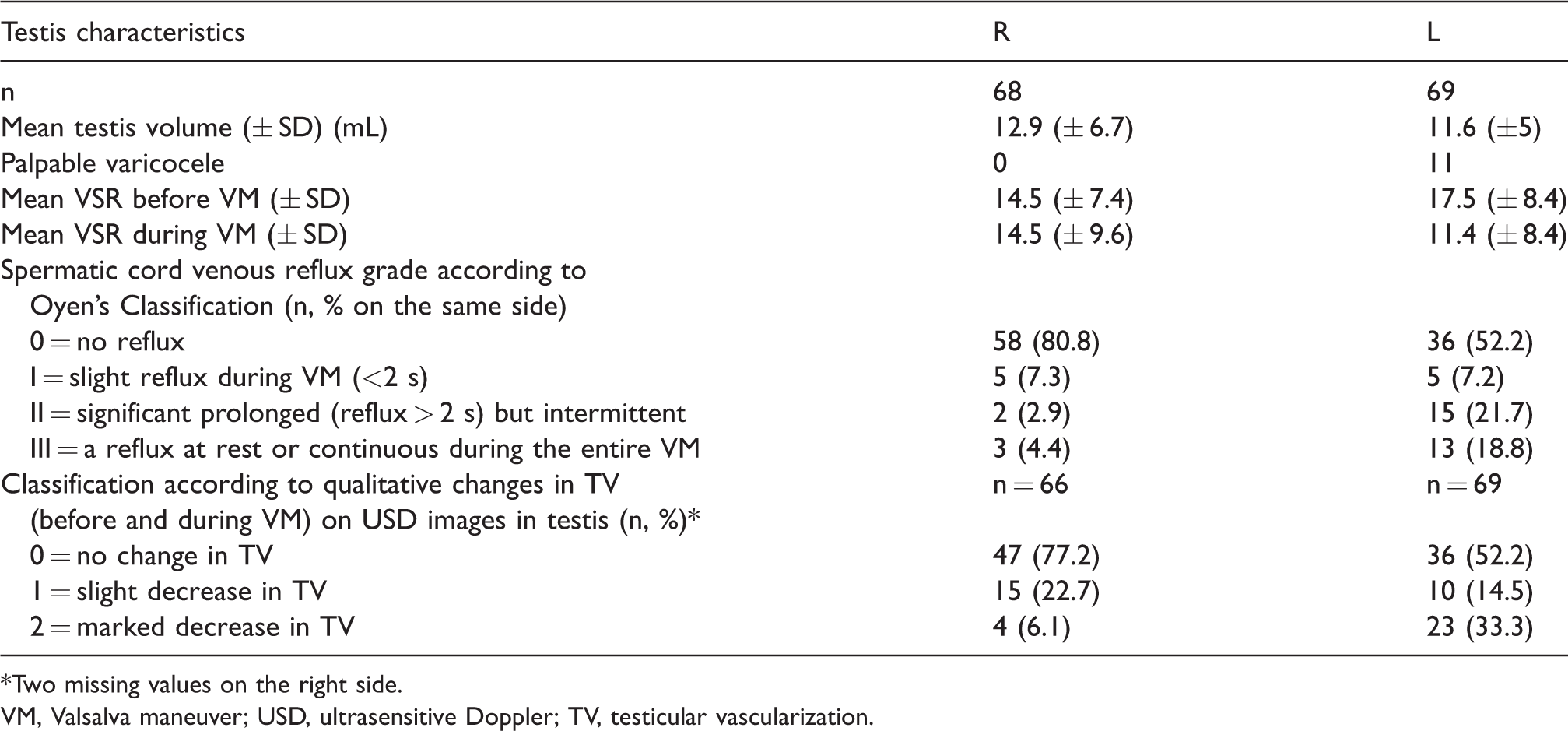
*Two missing values on the right side.
VM, Valsalva maneuver; USD, ultrasensitive Doppler; TV, testicular vascularization.
Varicocele with sonographically detected spermatic venous reflux grade ≥1 was found in 33/69 patients on the left side and bilaterally in 10 patients, although clinical varicocele was only detected 11 patients (all presenting with grade III). The mean testicular volume (n = 135) was 12.2 ± 5.9 mL. A slight difference in testicular volume was observed between right (12.9 mL ± 6.7 mL) and left side (11.6 mL ± 5 mL) (t-test, P = 0.02).
Table 2 presents the results of the comparative analysis between the grade of spermatic venous reflux and both the relative or absolute VSR and qualitative TV changes evaluated by USD.
Comparison of the vessel’s surface ratio (VSR), mean absolute difference, and mean relative difference for both patient classifications: Oyen’s classification according to spermatic veins reflux and subjective changes of vascularization observed in USD images, before and during VM.
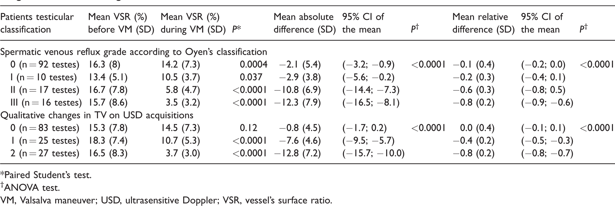
*Paired Student’s test.
†ANOVA test.
VM, Valsalva maneuver; USD, ultrasensitive Doppler; VSR, vessel’s surface ratio.
VSR was significantly reduced during VM compared to baseline measurements for Grade II and III varicoceles (60% ± 30% and 80% ± 20%, respectively; P < 0.0001). A significant reduction was also found in the qualitative changes determined by USD evaluation for grades 1 and 2 (Fig. 4, P < 0.0001).
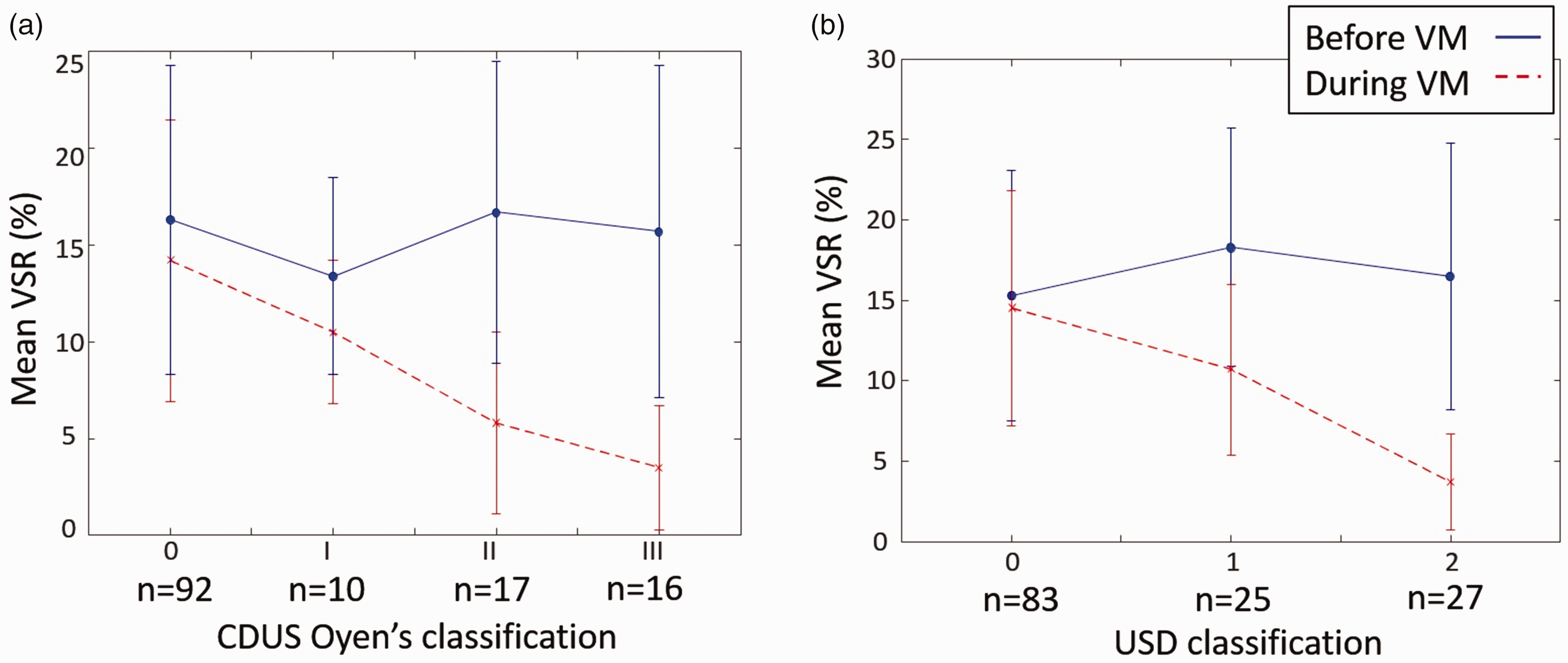
Vessel surface ratio variation before (blue line) and during (red line) VM. It has been correlated to (a) the venous reflux CDUS classification and (b) the qualitative USD scale defined above.
Both classifications showed that the changes in VSR quantification provided a strong difference when there is a significant varicocele and when the qualitative USD graduation increased. VM induced a moderate but significant VSR decrease for patients without varicocele (Grade 0) (P = 0.0004). Finally, a significant correlation was found between spermatic venous reflux grade and VSR decrease during VM (P < 0.0001) using both relative and absolute comparisons. The decrease in quantitative VSR measurements was also significantly associated with changes in qualitative TV classification (P < 0.001). Figure 5 shows an extinction of TV during VM, with surrounding dilated veins.

USD native images of an azoospermic man (with bilateral varicocele) with grade III left varicocele, and testicular hypotrophy before VM (a); (b) during VM, the testicular surface is reduced, due to compression surrounding the ectatic peripheral veins. Notice also the TP extinction (classified as grade II on the qualitative visual scale).
Discussion
Our study suggests that TV decreases during VM in patients presenting with varicocele. Moreover, this decrease is correlated to the grade of spermatic venous reflux. We also demonstrate that qualitative TV changes are also strongly correlated to quantitative evaluation. It is clear that USD detected both veins and arterial vessels, and one speculated that VM could induce an increase in testis vein surface, but on the contrary, we found that the increased pressure transmitted through the veins seemed to prompt a decrease and even extinction in arterial and venous flow, because, as shown in Fig 5, no vessels could be identified with USD in many patients during VM. This result is important to keep in mind in case of further clinical or experimental studies using the testicular vascularization as a parameter.
In fact, VM is supposed to reproduce, when applied in the supine position, the physiological process of an increase in abdominal pressure leading to venous reflux. Subsequent ischemia could gradually impact testicular volume and may damage spermatogenesis. This result seems in accordance to Zampieri’s study, showing that sports practice (supposed to increase the abdominal pressure) seemed to make progress a sub clinical varicocele into a clinical varicocele concerning a pubertal boys’ cohort (23).
Previous studies have assessed the potential role of US (24–27), testicular contrast harmonic imaging (28), elastography (29), and MRI (18) in evaluating the functional status of the testes in men with varicocele or testicular dysfunction (30). There are also studies reporting an improvement in the testicular blood supply after varicocelectomy via spectral Doppler analysis. In addition, Harrison et al. assessed the relationship between intracompartmental pressure and intrastesticular blood flow [27].
Since the first clinical classification system was created (8), several Doppler classification systems have been developed to improve the selection of patients that require treatment (13,31). At present there is no gold standard to confirm the presence of a varicocele, because clinical examinations and even venography may miss significant venous reflux. Moreover, even with techniques such as spectral Doppler and CDUS, many factors can influence the performance of venous reflux diagnosis, including the position and angulation of the probe, and motion during VM. Reflux duration (> 1 s in Iosa’s (13), > 2 s in Oyen’s (15)) is often subjectively calculated. The use of USD images during VM may help patient management as a complement to the classification system used. A significant difference in VSR was found for Groups II and III: the treatment for Grade I varicocele can be argued, particularly if TV is not affected by VM. A slight decrease in VSR was observed for Grade 0 varicoceles. This can be explained by two main factors: patients may have been wrongly classified in Group 0 or their reflux may have been underestimated. We may also suppose that VM may physiologically induce a slight TV decrease that cannot be visually assessed with USD.
In our study, the infertile patients had various known (such as cryptorchidism, chemotherapy, hypogonadism) and unknown causes for their infertility. Each cause may affect both the vascularity and testicular volume differentially. It is the reason why we only focused on TV changes before and during VM and not on the quantification of TV according to infertility status. We also did not define a “control” group, because even men with both a normal spermogram and a normal scrotal exam may have an asymptomatic venous reflux during VM diagnosed using CDUS.
Mean testicular volume was lower than the normal reference regarding the patient’s age. Further, this is a frequent finding in the infertile population [18].
Our study has several limitations. First, inter-observer reproducibility was not assessed. Nevertheless, we previously studied our intra-observer reproducibility based on three repeated measurements on 43 testes and established an interclass correlation (ICC) of 0.801, with a 95% CI of 0.695–0.879 (21). This cohort could be considered small. The clinical findings of varicocele were not compared to our present results because of variability induced by the different physicians who prospectively performed the clinical work. We noticed that there were fewer patients with clinical varicocele (16/69) compared to patients with spermatic venous reflux, and right varicocele was never clinically suspected.
Results were also not compared with spermatic vein venography since it was only performed in five patients. The venography results confirmed reflux in the pampiniform plexus and allowed for an embolization. A control using the same USD technique was performed in only one patient, because this research USD device was only loaned to us for a short period. A restitution of similar perfusion before and during VM was established.
In daily practice, quantitative TV evaluation may be complex using USD, and we are aware of the limitations and variation in quantifying vascularization with CD/PD images, as the chosen algorithm is not considered to be an absolute marker. Rather, it is a more precise tool compared to a subjective visual scale. Our study assessed patient classification based on the visual inspection of vascularization in USD images and was close to vessels quantification. Therefore, we expect that real-time qualitative analysis of USD/CDUS during the VM will be useful, since extinction or decrease of TV was quite obvious. Further studies should be conducted in order to evaluate the delay between start of VM and USD signal extinction as well as the effect of standing during the duration of signal extinction during VM.
In conclusion, TV assessed by USD appeared to decrease during VM in patients with varicocele; this decrease was associated with the degree of venous reflux measured by conventional Doppler. This may represent a physio-pathological explanation for the progressive decrease in testicular volume and potential infertility and may help, as a marker of potential testicular ischemia, in future selection of patients eligible for treatment, especially in infertile men or adolescents.
Footnotes
Acknowledgments
The ultrasound scanner was provided by Supersonic Imagine.
Declaration of Conflicting Interests
The author(s) declared no potential conflicts of interest with respect to the research, authorship, and/or publication of this article.
Funding
The author(s) received no financial support for the research, authorship, and/or publication of this article.