



Abstract
Background
Due to the broad variability of the prostatic artery (PA), its origin, small calibers, and tortuous courses, prostatic arterial embolization (PAE) is challenging, time-consuming, and results in high radiation doses.
Purpose
To evaluate the accuracy of PA detection using cone-beam computed tomography (CBCT) performed from the aortic bifurcation in combination with a semi-automatic detection software in comparison to oblique view digital subtraction angiography (DSA) with internal iliac artery (IIA) injection.
Material and Methods
Twenty-two consecutive patients were included in this retrospective, IRB-approved study between July and December 2017. CBCT from the aorta and 30° oblique-view DSA from both IIAs were obtained for PA detection. Results of suggested PAs from the semi-automatic vessel detection software after CBCT and IIA DSA were compared. Moreover, dose area product (DAP) was recorded. Statistical analysis included Spearman’s correlation, Mann–Whitney U test, and the Wilcoxon test considering P<0.05 as significant.
Results
PA type was classified significantly better with CBCT compared to DSA (P=0.047). In IIA DSA, PAs could not be identified in 18% on the left and in 17% on the right side. CBCT detected all PAs, although truncation occurred in 59% because of the limited field of view. Mean DAP of the whole procedure was 257,161.32±127,909.36 mGy*cm2. Mean DAPs were for a single DSA 14,502.51±9,437.67 mGy*cm2 and for one CBCT 15,589.23±2,722.49 mGy*cm2. A mean of 14.82 DSAs and only one CBCT were performed. CBCT accounted for 6% and DSA for 84% of the entire DAP of the procedure.
Conclusion
CBCT with semi-automatic feeding vessel detection software detects PAs more accurately than IIA DSA and may reduce radiation dose.
Introduction
Prostatic artery embolization (PAE) is a new therapeutic approach for the treatment of non-neurogenic lower urinary tract symptoms (LUTS) due to benign prostatic hyperplasia (BPH) (1). Recent studies showed that the technique is not inferior to the gold standard, the transurethral resection of the prostate (TURP), in terms of International Prostate Symptom Scoring (IPSS) improvement and offers shorter hospitalization times, faster returns to normal activities as well as comparable levels of immediate postoperative pain and lower levels of pain at 12-month follow-up compared to TURP (2, 3) and avoids retrograde ejaculation (4).
However, the procedure remains challenging because of a variable prostatic artery (PA) anatomy with small calibers and tortuous courses (5). So far, DSA serves as the gold standard for the detection of the PA. On the other hand, it has been shown that the intervention is time-consuming and requires long fluoroscopy times, multiple oblique views, and employs cone-beam computed tomography (CBCT) to assess target vessels to avoid non-target embolization resulting in high radiation doses (6, 7).
Preprocedural imaging such as CT (5) and magnet resonance imaging (MRI) (8) were suggested for target vessel detection to reduce intervention times. However, preprocedural multislice CT (MSCT) showed lower detection rates of PAs (9, 10). Moreover, MSCT is associated with less signal-to-noise and contrast-to-noise ratios as well as higher radiation doses compared to intraprocedural CBCT (10). It could be shown that MR angiography is able to correctly detect PA in about 76.5% of cases compared to DSA (8, 11). Although pre-interventional cross-sectional imaging showed a reduction in the required amount of contrast medium, image fusion with a native CBCT would not increase detections rates of PAs.
On the other hand, contrast-enhanced CBCT proved to be a useful adjunctive, peri-interventional tool for the detection of PAs (12). Chiaradia et al. (13) reported the utilization of a detection software using selective dual-phase CBCT (DP-CBCT) performed separately from the left and right internal iliac artery (IIA) to identify the pelvic vascular anatomy in a small case series. This approach is already established in other embolization procedures such as transarterial chemoembolization (TACE) (14). The software uses an algorithm to detect the shortest track from the marked catheter tip to a previously segmented target volume (15).
The aim of the present study was to evaluate the accuracy for PA detection and potential dose savings of single-phase CBCT performed from the aortic bifurcation in combination with a semi-automatic detection software in comparison to oblique view digital subtraction angiography (DSA) with IIA injection serving as gold standard.
Material and Methods
Twenty-two consecutive patients referred by urologists for PAE were included in this retrospective, Institutional Review Board-approved, single-center, single-arm study between July and December 2017. All procedures were performed by the same interventional radiologist with the experience of 20 patients previously treated with PAE (>7 years of interventional experience) on the same angiography system (Allura FD20 with XperCT Roll protocol; Philips, Best, The Netherlands). Utilization of the semi-automatic detection software (EmboGuide, Philips, Best, The Netherlands) was performed by one separate radiologist. Inclusion and exclusion criteria for PAE are listed in Table 1. Patients’ characteristics are summarized in Table 2. Institutional ethics committee approval (EA2/005/19) and written informed consent were obtained.
Inclusion and exclusion criteria.

IPSS, International Prostate Symptom Scoring.
Patients’ characteristics.
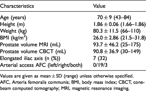
Values are given as mean ± SD (range) unless otherwise specified.
AFC, Arteria femoralis communis; BMI, body mass index; CBCT, cone-beam computed tomography; MRI, magnetic resonance imaging.
Procedure and CBCT imaging
All patients received a multiparametric MRI compliant with the Prostate Imaging Reporting and Data System version 2.0 (PIRADS v2) standards (16) to exclude prostatic cancer and to visualize the gross vascular anatomy on the day before the procedure to gain an impression whether elongated iliac axes were to be expected.
First, a 10-cm, 5-F sheath (RadioFocus Introducer II, Terumo, Shibuya, Tokyo, Japan) was, in most cases, introduced in the right common femoral artery, unless vascular anatomy demanded a left-sided approach.
Second, a 0.035-in. guide wire (145 cm Fixed Core Guide Wire Guide Safe-T-J® Curved, Cook Medical, Bloomington, IN, USA or 180 cm Radiofocus Guide Wire M – angled or Stiff type angled, Terumo, Shibuya, Tokyo, Japan) was advanced into the abdominal aorta, followed by a diagnostic catheter (65 cm, 4 F RIM, Cordis, Miami Lakes, FL, USA).
Third, CBCT was performed from the aortic bifurcation using the XperCT ND Roll protocol on the Allura FD20 (Philips, Best, The Netherlands) for CBCT acquisition, allowing for a lateral positioning of the C-arm in all procedures. A 180° rotation was performed in 10 s with a frame rate of 30 images/s at 120 kVp. The acquired three-dimensional (3D) volumetric CBCT images had an isotropic resolution of 0.6 mm. The flat panel detector covered a field of view (FOV) of 250 × 250 × 194 mm with a matrix size of 384 × 384 × 296 pixels. The CBCT injection protocol included an injection of 50 mL of an iodine-based contrast agent (Imeron® 300, Bracco Imaging, Milan, Italy) without dilution at an injection rate of 5 mL/s with a delay of 2 s followed by a bolus of 10 mL saline at the same injection rate. The injection was performed with the Accutron HP-D-HT® (Medtron, Saarbrücken, Germany) injector.
Immediately after CBCT acquisition, the prostate was segmented within several seconds and the catheter tip was selected using the semi-automatic vessel detection software. The software calculated the target vessels using a minimal path-optimization algorithm (Fig. 1) and a 3D overlay to the fluoroscopic images was displayed (15).

(a, b) Cone-beam computed tomography planning separately due to aortic bifurcation truncation for the left and right side on the same dataset. (c, d) Internal iliac artery DSA. (e, f) Pretreatment DSA. * reference point semi-automatic vessel tracking; + manually segmented prostate volume; → chosen PA; ⇒ microcatheter tip placement for treatment. DSA, digital subtraction angiography.
Additional oblique view DSA with 2 frames/s was manually performed with 3–5 mL of contrast agent from the IIA as recommended by Bilhim et al. (17) over a 5-F UAC or Pisco Prostatic Catheter (Merit Medical, South Jordan, UT, USA) and was later used as the gold standard for detection of PAs.
As soon as the PAs were identified, the feeding vessel was catheterized using microcatheters, i.e. the 130-cm Progreat alpha 2.0 F (Terumo, Shibuya, Tokyo, Japan) in combination with microwires such the 0.016-in. Sagitta - Wire Guide AQ® Hydrophilic Coating 180 cm (Cook Medical, Bloomington, IN, USA) or 180-cm Radiofocus - GuideWire GT 45° or the 200-cm Radiofocus - GuideWire GT 90° (both Terumo, Shibuya, Tokyo, Japan).
After reaching the target vessel, another DSA was performed with manual contrast injection to rule out non-target embolization sites. If potential non-target embolization was suspected, additional CBCT acquisition was allowed.
Embolization was performed using the 100–300 µm or 300–500 µm Embospheres (Merit Medical, South Jordan, UT, USA) after injecting 200 µg nitroglycerine in the target vessel. Post-embolization DSA documented therapy success, if no typical prostate blush was detected.
CBCT analysis
A descriptive analysis was performed for the evaluation of CBCT coverage to assess whether the FOV displayed the pelvis from the aortic bifurcation to the apex of the prostate. Moreover, expected PA type was retrospectively compared between the CBCT performed from the aortic bifurcation in combination with a semi-automatic detection software and the 30° left or right anterior oblique view DSA with injection performed from the left and right IIA. First, all IIA DSA scans were reviewed by two raters and classified as described in detail by de Assis et al. (18): type 0 = not visible; type 1 = inferior vesical artery (IVA) originating from anterior division of IIA, in a common trunk with superior vesical artery (SVA); type 2 = from anterior division of IIA, inferior to SVA; type 3 = from obturator artery; type 4 = from internal pudendal artery; and type 5 = less common origins. All detected vessels in CBCT scans were categorized without consultation of the IIA DSA by the interventional radiologist in a second reading performed after two weeks. The interrater reliability was analyzed using Cohen’s kappa test. Finally, the expected PAs from CBCT and IIA DSA were correlated with the actual embolization side. Moreover, an intramodality comparison between the left and right side was carried out.
Dose analysis
The manufacturer’s dose area product (DAP) meter was used to acquire dose measurements. Cumulative DAP, cumulative kerma (Air), and DAP values of each DSA as well as the total and cine specific fluoroscopy times (FT) were recorded. Moreover, DAP values of all CBCT acquisitions were documented and analyzed.
Statistical tests
All statistical analysis was performed using IBM SPSS Statistics 25 (Armonk, NY, USA) including the Shapiro–Wilk test, Spearman’s correlation, Cohen’s kappa test, the Mann–Whitney U test, and the Wilcoxon test. P values < 0.05 were considered significant. Descriptive values are given as mean ± SD (range).
Results
All procedures could be successfully completed without major complications according to the Society of Interventional Radiology reporting guidelines (19).
CBCT analysis
Type of PA was classified significantly different between DSA performed from the IIA compared to the semi-automated CBCT feeding vessel detection (P = 0.047; Fig. 2). There was no significant difference in artery type classification between the left and right side within the modalities (Table 3). Furthermore, there was a good interrater reliability with κ = 0.722 agreeing in DSA PA type classification. After a careful plausibility check, the semi-automatic detected arteries proved to be the right choice in subsequent, more selective DSA during the procedure. Typical plausibility mistakes of the software were vessel cross-overs in cases, in which two vessels lay right next to each other. Another detection failure occurred if the superior ramus of the pubic bone was right next to the target vessel, so that the pelvic rim was suggested to be a vessel.

Boxplot comparing assumed prostatic artery types in digital subtraction angiography and cone-beam computed tomography.
Comparison of the expected prostatic artery type: DSA vs. CBCT.

Values are given as n (%).
*IVA and its prostatic branches: type 0 = not visible; type 1 = IVA originating from anterior division of IIA, in a common trunk with SVA; type 2 = from anterior division of IIA, inferior to SVA; type 3 = from obturator artery; type 4 = from internal pudendal artery; and type 5 = less common origins.
CBCT, cone-beam computed tomography; DSA, digital subtraction angiography; IIA, internal iliac artery; IVA, inferior vesical artery; SVA, superior vesical artery.
On IIA DSA, PAs could not be rated sufficiently in 18% of cases on the left side and in 17% of cases on the right side, whereas all PAs could be detected on CBCT. Even the external iliac origin of a right-sided prostate artery could be detected using the semi-automatic vessel detection software with an aortic injection protocol (Fig. 3) that was missed in DSA due to the more selective injection protocol.

Type 5 pulmonary artery on the right, type 2 on the left side. (a) 3D cone-beam computed tomography segmentation and semi-automatic vessel tracking, aortic bifurcation in the field of view. (b, e) 3D overlay for guidance. Pre- and post-treatment digital subtraction angiography on the left (c, d) and on the right side (f, g).
Anatomy truncation occurred in 59% of cases using CBCT due to the detector size (Table 4). However, all acquired CBCTs were sufficient enough for semi-automatic feeding vessel detection. Aortic bifurcation truncation (36%) led to a second target vessel assessment on the same dataset, whereas distal truncation (18%) carries the risk of missing potential feeding arteries and more important fails to display potential downstream arteries at risk for non-target embolization. In one large patient, neither the aortic bifurcation nor the distal portion of the prostate fitted in the FOV.
CBCT coverage.

CBCT, cone-beam computed tomography; FOV, field of view.
Dose analysis
The mean DAP of the whole PA embolization procedure was 257,161.32 ± 127,909.36 mGy*cm2 (range =102,587.00–577,067.00 mGy*cm2; Table 5). A mean of 14.82 DSAs and only one single CBCT were performed in the reported procedures. The mean DAP for a single DSA was 14,502.51 ± 9437.67 mGy*cm2 (range =20.00–61,776.00 mGy*cm2) and slightly lower compared with one CBCT acquisition with 15,589.23 ± 2722.49 mGy*cm2 (range = 9585.00–19,944.00 mGy*cm2). However, CBCT accounted for only 6% of the cumulative DAP (Fig. 4), whereas DSA was responsible for 84% and another 10% for fluoroscopy guidance. The mean fluoroscopy time for the whole procedure was 17 min 57 s (range = 5 min 56 s – 34 min 54 s).
Dose metrics.

Values are given as mean ± SD (range).
CBCT, cone-beam computed tomography; DAP, dose area product; DSA, digital subtraction angiography.

Dose area product and kerma (Air) percentages of cone-beam computed tomography, digital subtraction angiography, and guidance.
Discussion
The presented analysis has shown that the use of CBCT with semi-automatic feeding vessel detection software reliably depicts PAs even with contrast injections performed proximal to the aortic bifurcation compared to 30° left and right anterior oblique DSA performed from the IIA. Fluoroscopy time and DAP can potentially be decreased using the proposed protocol.
In PAE, identifying the PA remains a major challenge due to the complex inter- and intra-individual anatomic diversity and is still the most time-consuming part of the procedure (20). There is consensus that ipsilateral oblique views of 25°–55° are most suitable to depict the inferior vesicle and PAs. Moreover, cranial angulations of 10°–20° can be used to differentiate vesicle branches (21).
Most PAs (one-third of cases) proved to be type 1, which in other studies was only the second most common artery type, which could be related to the relatively small sample size (17, 18, 22). In this study, eight PAs were not identified by IIA DSA (18% left, 17% right). One PA originated from the right external iliac artery which could technically not be seen in DSA. The other missed PAs were type 1 (n = 1), type 4 (n = 1), and, in a greater proportion, type 2 (n = 5). However, all PAs were detected in CBCT using the vessel tracking software, although anatomy truncation occurred in 59% of cases.
Oblique views are necessary for PA detection. However, these acquisitions are associated with an increase in radiation dose for staff and patients (23). Skin doses of 2 Gy can easily be reached, potentially resulting in transient erythema (7). Even a case of radiodermatitis after a complex procedure with fluoroscopy times of 72 min and a DAP of 8,023,949 mGy*cm2 was reported (24). Despite high-dose exposure of otherwise healthy patients, high physician lens doses can be reached hitting the annual equivalent dose limit with one treatment per week if the ceiling mounted screen and leaded eyeglasses are not properly used (7), emphasizing the need of dose reduction associated with PAE. Using CBCT and semi-automatic vessel tracking software early in the procedure allows for low-dose exposure with mean DAPs of 257,161 mGy*cm2 (range = 102,587–577,067 mGy*cm2), which is considerably less compared to other studies without CBCT usage and reported mean DAPs of 2,415,000 mGy*cm2 (range =625,000–9,503,000 mGy*cm2) (25). Andrade et al. (6) use CBCT for target verification, if required, and report average DAPs of 450,700 mGy*cm2 (range =248,300–791,730 mGy*cm2), which is about twice the applicated dose compared to the suggested routine use of CBCT in the beginning of the procedure. In addition, fluoroscopy times could be reduced using the semi-automatic vessel detection software to approximately 18 min, which is again half the fluoroscopy time reported by Andrade et al. (6) and Bagla et al. (26) of about 30 min. Pisco et al. (25) reported fluoroscopy times of about 20 min without the use of CBCT, which might be related to long-term experiences of this study group going back to March 2009. Therefore, initial CBCT acquisition and vessel tracking seems to be particularly favorable for colleagues who are not supervised by eminently experienced physicians to reduce fluoroscopy and intervention times. The significant discrepancy between the assumed PA origin in IIA DSA and the suggested artery of the software (Fig. 2, Table 3), which proved to be the right choice during the interventions, underlines the hypothesis.
Moreover, the sensible use of CBCT could potentially avoid patient follow-ups suggested by the SIR guidelines for patient radiation dose management setting thresholds for DAP > 500,000 mGy*cm2 and fluoroscopy times > 60 min (27). Moreover, new generation angiography suites can additionally reduce radiation exposures using CBCT in the beginning of the procedure down to 53,130 mGy*cm2 (28).
One CBCT accounted for 6% and a mean of 14.8 DSAs per treatment for 84% of the cumulative DAP in this study, which is consistent with previously reported data, where DSA is responsible for 71.5% (6) to 79.8% of the total DAP (7). Garzon et al. (7) used CBCT in 40% of their cases, being 3% of the cumulative DAP.
Chiaradia et al. (13) reported, in a small case series of six patients, that automatic vessel detection with a prototype software is feasible. All patients received a DP-CBCT (arterial phase used for artery detection and delayed phase for parenchymal delineation after a single 24-mL contrast injection) with the catheter placed in the IIAs of both sides resulting in four CBCT acquisitions. The proposed protocol showed that a single injection from the distal abdominal aorta and a single-phase CBCT is sufficient enough to detect the PAs with lower DAPs (454,000 mGy*cm2 [range = 171,500–782,500 mGy*cm2]) and fluoroscopy times (47 min [range = 36–71 min]) (13). In 36%, the aortic bifurcation was truncated; however, a second vessel detection assessment could be performed on the same dataset.
Although the software results are reliable, especially in the peripheral branches, careful revision of the axial CBCT images is important to make sure that there is no crross over to a wrong artery in cases in which two small arteries lay right next to each other. More obvious is the misinterpretation of a superior ramus of the os pubis located close to the small arteries.
Pre-interventional MRI is suggested by Kim et al. (8) to identify the PA using multiplanar and curved planar reconstructions starting to draw the centerline close to the prostate and following the arterial branches back to the IIA. This method avoids a separate CT angiography, reducing radiation dose in the pre-interventional setting and time demand on patients during the procedure.
CBCT is useful for identification of the PAs compared to TACE procedures (12,14). However, non-target embolization cannot be excluded by proximal contrast injections, so that selective CBCT acquisitions after contrast injections with 0.5–1 mL/s through the microcatheter might be helpful to evaluate sites of non-target embolization taking into account that the mean CBCT DAP in this study of 15,589.23 mGy*cm2 is only slightly higher compared to the mean DSA DAP of 14,502.51 mGy*cm2 (Table 5) (12).
The present study is limited by its small sample size and a missing reference group as the presented technique was adopted early to the procedure in our institution. However, all interventions were performed by the same interventional radiologist to avoid an operator bias. Due to the postprocedural analysis, the exact number of software misinterpretations were not recorded, but if it occasionally occurred, it was immediately identified by the careful revision of the software’s proposal. Moreover, truncation of the aortic bifurcation, the prostatic apex, or both occurred in CBCT as the maximum FOV was too small in 59% of patients. However, the acquired dataset could always be used for sufficient bilateral semi-automatic vessel detection. Further studies are necessary to evaluate the lowest contrast amount and flow rates allowing for secure vessel detection in CBCT. Furthermore, the results are specific for the vendor and the angiography suite used in the present study and cannot be extrapolated for other devices.
In conclusion, CBCT with semi-automatic feeding vessel detection software detects the PA rather than IIA. DSA at an early stage of the procedure may reduce the radiation dose of PAE.
Footnotes
Declaration of conflicting interests
The authors declared the following potential conflicts of interest with respect to the research, authorship, and/or publication of this article: AM reports grants from Merit Medical, outside the submitted work. BH reports grants from Bracco Group, Philips Healthcare, and Terumo Medical Corporation outside the submitted work. BG reports personal fees from Philips Healthcare outside the submitted work. MJ reports grants from BIH Clinical Scientist, during the conduct of the study; grants from Philips Healthcare, outside the submitted work. TD, DS, and GW have nothing to disclose in relation to this article.
Funding
The authors disclosed receipt of the following financial support for the research, authorship, and/or publication of this article: MJ participates in the BIH‐Charité Clinical Scientist Program (DM.BIH-04.15) funded by Charité – Universitätsmedizin Berlin and the Berlin Institute of Health.
