



Abstract
Background
Magnetic resonance imaging (MRI) is widely used in patients with multiple sclerosis (MS) for different indications. However, frequent administration of gadolinium in these patients can have some potential complications. So, a more limited approach reducing the use of gadolinium should be considered.
Purpose
To evaluate the additional benefits of contrast-enhanced MRI over non-contrast-enhanced MRI in routine follow-up of patients with MS.
Material and Methods
This is a retrospective cohort study including patients with MS who underwent both contrast-enhanced and non-contrast-enhanced MRI for two time-points with an interval of at least six months. Non-contrast-enhanced images were compared for each patient and interpreted as non-progressive or progressive disease. Then, rate and type of enhancing lesions were analyzed and compared between the groups. All images were reviewed and compared visually by two radiologists.
Results
A total of 462 patients (392 women; mean age = 36 years) were included. Of these patients, 352 were in the non-progressive group and 112 were in progressive group. Comparison of baseline and follow-up contrast-enhanced MRIs revealed that 13 (3.7%) patients in the non-progressive group and 58 (51.8%) patients in progressive group developed enhancing lesions (P < 0.001). All 58 patients in the progressive group developed new enhancing lesions, whereas all those in the non-progressive group revealed persistent or reactivated enhancing lesions without evidence of new lesions.
Conclusion
According to the very low incidence rate of new enhancing lesions in patients with non-progressive disease on follow-up non-contrast-enhanced MRI, routine administration of contrast in follow-up studies is not suggested.
Introduction
Multiple sclerosis (MS) is an inflammatory disorder of central nervous system with increasing incidence in last decade. It more commonly involves women and young adults (1). Although early diagnostic criteria for MS were predominantly clinical-based, para-clinical studies, especially magnetic resonance imaging (MRI) are increasingly applied in conjunction with clinical judgment for optimal diagnosis (2). Brain MRI is considered an essential test in the diagnostic process of MS and should be obtained in all patients (2). Diagnosis of MS is based on the specific criteria that confirm the presence of demyelinating plaque in the white matter of brain or spinal cord (3). MRI is also used in known cases of MS as routine follow-up of silent disease, for therapy monitoring, or in case of unexpected clinical deterioration (4). In these situations, the recommended protocol by standard guidelines includes brain MRI with and without gadolinium-based contrast agents every 6–12 months, depending on the duration of disease and clinical course (4). Gadolinium enhancement occurs when the blood–brain barrier is damaged during active inflammation of new lesions or reactivation of previous lesions. These enhancements usually remain for mean period of three weeks and rarely longer than months (4). However, frequent administration of gadolinium in these patients can be associated with some potential complications. Patients with impaired renal function are at risk of nephrogenic fibrosis. It has been also suggested that there is a potential association between neuronal deposition in dentate nucleus and globous pallidus with gadolinium injection in patients with normal renal function (5–7). Therefore, administration of gadolinium needs a more cautious and reasonable approach, especially in patients with MS who will probably require 1–2 injection per year during their life after diagnosis of MS.
Although most studies and guidelines support the use of both enhanced and unenhanced MRI in the follow-up of patients with MS (4), a few recent studies have suggested that high-quality unenhanced MRI with 3 T or quantitative subtraction software is sufficient to rule out disease activity and, hence, administration of gadolinium could be limited to those patients with evidence of disease progression on non-contrast-enhanced MRI (8,9). However, lower Tesla MRI scanners are more widely used throughout the world and subtraction and quantitative software packages are not commonly applied in most centers. The aim of the present study was to evaluate the role of non-contrast-ehanced 1.5-T MRI with visual assessment in the multiple sclerosis compared to contrast-enhanced MRI.
Material and Methods
Patients
The present study is a retrospective cohort study of patients with MS and was approved by the local ethics committee. We extracted a list of patients with MS from our picture archiving and communication system (PACS) according to the national identifying number between October 2017 and May 2018. Patients who underwent both contrast-enhanced and non-contrast-enhanced MRI during this time period, with a complete follow-up MRI study (including both contrast-enhanced and non-contrast-enhanced MRI) with an interval of at least six months were included. Images and patient data were available in our PACS and were analyzed for T2/fluid-attenuated inversion recovery (FLAIR) imaging and contrast-enhancing lesions.
MRI acquisition
All MRI images were obtained with a 1.5-T MRI system (Siemens Avanto). The full MRI protocol for patients with MS consisted of T2/FLAIR sequences. The scanning protocol included axial and sagittal slices through the brain, T2-weighted images fast spin echo (TR/TE = 4110/100 ms), FLAIR (TR/TE = 7500/92 ms), and T1-weighted (T1W) images spin echo (TR/TE = 450/9 ms) before and after an intravenous injection of gadolinium-based agent.
MRI reporting
All images were reviewed and compared visually by two radiologists. In case of disagreement, a third radiologist also assessed the images. Patients were categorized on the basis of stability or progression of lesions in follow-up non-contrast-enhanced FLAIR sequences compared to baseline non-contrast-enhanced MRI. Progression defined as the presence of at least one new or enlarging lesion on follow up non-contrast-enhanced MR images (Fig. 1e and f). Otherwise, when no change (stable) (Fig. 1c and d) or reduction (regression) in number or size of lesions (Fig. 1a and b) was detected, it was considered non-progressive disease. Then, the T1W follow-up images with contrast were assessed for evidence of enhancing plaques. If any enhancing plaque was found, the images were compared to baseline contrast-enhanced MRI. According to this comparison, three types of enhancement were reported by radiologists as persistent (seen in a previous contrast study) (Fig. 2a), reactivated (seen in a previous non-contrast study, but not enhanced in previous contrast image) (Fig. 2b), and new (not seen in either previous non-contrast or contrast study) (Fig. 2c).

Transverse slices of FLAIR sequences of baseline and follow-up MRI of three different patients with regressive (a, b), stable (c, d), and progressive (e, f) demyelinating plaques. FLAIR, fluid-attenuated inversion recovery; MRI, magnetic resonance imaging.
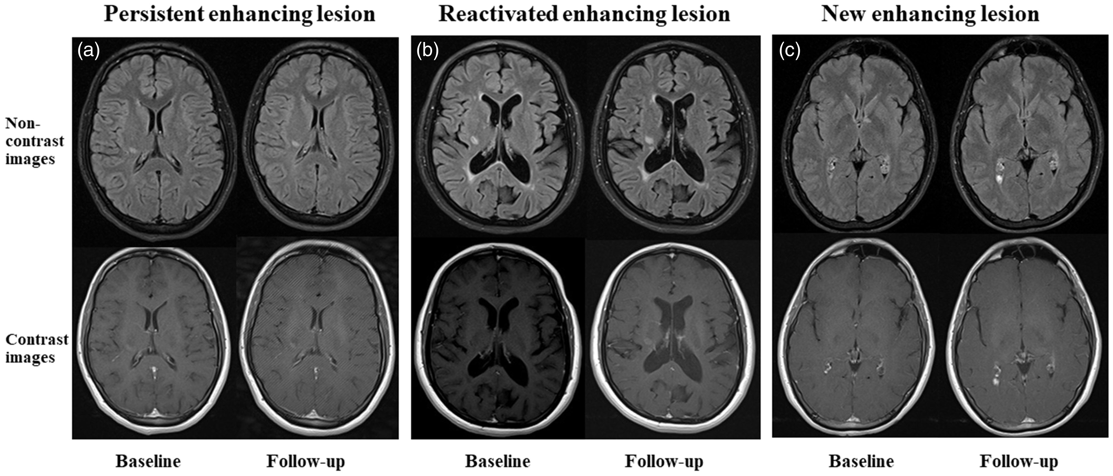
Transvers slices of non-contrast and contrast MRI of three different patients with enhancing lesions on their follow-up studies with persistent (a), reactivated (b), and new (c) enhancing lesions compared to baseline studies.
Statistical analysis
Statistical analysis was performed by SPSS software (SPSS Statistics for Windows, version 18.0). The qualitative and quantitative parameters were represented as number (%) and mean ± SD, respectively. T-test was used for comparison of age between progressive and non-progressive disease. For comparison of qualitative variables between the groups, chi square test was applied. P < 0.05 was considered statistically significant.
Results
We included 464 patients (72 men, 392 women; mean age = 36.2 ± 8.3 years) who met the inclusion criteria with at least two sets of MRI scans with an interval of at least six months. We categorized patients into non-progressive and progressive groups, depending on the presence of disease progression on the follow-up compared to baseline non-contrast-enhanced MRI. Of these patients, 352 cases were in the non-progressive group, including 339 cases with stable disease and 13 patients with evidence of regression, and 112 patients were in the progressive group. The mean age and female/male ratio for each group are shown in Table 1. A comparison of these parameters between the groups revealed no significant difference.
Comparison of demographic features between the non-progressive and progressive groups.

Values are given as n or mean ± SD.
*P < 0.05 is significant.
Of the study participants, 71 showed enhancing lesions in the follow-up examination. Evaluation of baseline and follow-up MRIs revealed that only 13 patients in the non-progressive group (two patients with regressive disease and 11 patients with stable disease) had evidence of enhancing lesions in the follow-up study, whereas in the progressive group 58 (51.8%) patients developed enhancing lesions on follow-up contrast-enhanced MRI (Table 2). The ratios were significantly different between the progressive and non-progressive groups.
Comparison of rate of enhancement between non-progressive and progressive groups.

Values are given as n (%).
*P < 0.05 is significant.
Further analysis of lesions in those with evidence of enhancement in follow-up study (71 cases) showed that all those patients in the progressive group (58 cases) developed new enhancing lesions, while none of those in the non-progressive group (13 cases) had new enhancing lesions at the second MRI (Table 3). In this group, eight patients revealed persistent enhancing lesions and five patients revealed reactivation of previous lesions.
Comparison of the type of enhancement between the non-progressive and progressive groups.
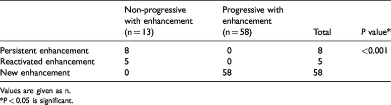
Values are given as n.
*P < 0.05 is significant.
If we consider the enhanced MRI as a gold standard with the presence of enhancing lesions as a marker of a positive test, the sensitivity, specificity, negative predictive value (NPV), and positive predictive value (PPV) of unenhanced MRI with progressive disease as a positive result for prediction of new enhancing lesions are 81.6%, 86.2%, 96.3%, and 51.8%, respectively.
Discussion
The present study reveals that the majority of patients with MS without new T2 lesions on follow-up MRI did not show any enhancing lesions either. Only 3.7% of these patients developed enhancing lesions on follow-up contrast-enhanced MRI, which were all persistent or reactivating lesions. On the other hand, nearly all patients with new enhancing lesions had already shown disease progression on non-contrast-enhanced MRI as new T2 lesions.
Gadolinium is a heavy metal that is widely used as a contrast agent to increase the diagnostic accuracy of MRI studies, especially in the imaging of the central nervous system (10). However, a recent drug safety communication from the U.S. Food and Drug Administration (FDA) warned that these agents can be retained in the body and made a recommendation “to minimize repeated GBCA imaging studies when possible, particularly closely spaced MRI studies.” Although there is no reported adverse effect associated with the retention of gadolinium, lack of long-term assessment warrants further and continued evaluation of the administration of gadolinium, especially in high-risk groups including patients requiring multiple lifetime administration of gadolinium. One of these high-risk groups is patients with MS. MS is an inflammatory and autoimmune disease of the central nervous system with most patients diagnosed at a young age. Since many new lesions are not clinically evident, even the majority of asymptomatic patients with established MS need at least an annual follow-up MRI examination with and without contrast (4). Thus, these patients are expected to undergo frequent contrast-enhanced MRI studies during their lifetime. In recent years, several studies, mostly with 3.0-T MRI, have evaluated the efficacy and necessity of contrast-enhanced MRI in follow-up of patients with MS. Eichinger et al. (8) retrospectively evaluated 507 follow-up images of 359 patients (34% with clinically suspected disease activity). Using 3.0-T MRI and double inversion recovery (DIR)-based subtraction, only four new enhancing lesions were missed in 1992 lesions and none of the patients were missed according to the detection of overall disease progression with either FLAIR or DIR-based non-contrast-enhanced MRI compared to contrast-enhanced MRI. They concluded that contrast-enhanced MRI has no additional benefit in the routine follow-up of patients with MS (8). Another similar study by Mattay et al. (11) also showed that all patients with new enhancing lesions had progressive disease on T2/FLAIR images. Our study with 1.5-T MRI also showed that all patients with new enhancing lesions already had evidence of disease progression on non-contrast-enhanced MRI. Since most of the new enhancing lesions will persist as T2 hyper-intensity after resolution of enhancement, the presence of new enhancement or new/enlarging T2 hyper-intense lesions compared to previous studies of a patient would indicate new inflammatory activity. Thus, among patients with clinically silent MS, only those with new T2/FLAIR or new enhancing lesions need therapeutic measures. In the present study, all patients with new enhancing lesions had new T2/FLAIR lesions as well; contrast-enhanced MRI does not add any additional information except for the timing of new lesions to non-contrast-enhanced MRI. Therefore, in terms of clinical management, administration of contrast can be limited to those patients with progressive disease on non-contrast-enhanced MRI in whom the presence of new enhancing lesions would change the clinical decision.
Karimian-Jazi et al. (12) also evaluated 100 patients with a total of 559 MRI follow-up studies. They found 152 enhancing lesions, of which only seven lesions (three new lesions, one persistent lesion, and three reactivated enhancing lesions) had no evidence of new T2/FLAIR lesions (12). Thus, the probability of missing an enhancing lesion in patients with stable T2/FLAIR images was only 1.7%. This ratio was 3.7% in our non-progressive patients. This small difference in the rate of missed enhancing lesions between the present study and other similar studies might be related to the sampling method. While we included 464 patients with 464 different MRI sets, other studies had no limitation in this regard and might include different sets of MR images of the same patients. This can potentially lead to duplication and re-analysis of the same clinical scenario in these patients and thus affect the distribution of enhancing and non-enhancing lesions.
Rudie et al. (9) implemented a protocol to limit the administration of contrast to just those patients with new T2/FLAIR lesions and reduce additional administration of contrast and imaging in 87% of patients. According to the high NPV (96.4%) of non-contrast-enhanced MRI for the detection of enhancing lesion, avoiding administration of contrast in patients without new T2/FLAIR lesions can potentially lead to a reduction in contrast-enhanced MRI in 75.8% of our cases with a rate of missed lesions of only 2.8% in all patients. It should be considered that all these missed lesions are persistent or reactivating previous lesions. On the other hand, if in a patient with new T2/FLAIR lesions, the presence of enhancing lesions will not change the clinical management decision, the rate of contrast administration can be further reduced and limited to those whose specific clinical course needs further information about the timing of new lesions. In the other words, if the management of patients with new progressive disease on T2/FLAIR images and those with enhancing lesions were clinically the same, considering all patients with progressive and non-progressive disease, contrast-enhanced MRI leads to a change in management decision in only 2.8% of all 464 patients in the present study.
The comparison of clinical and non-contrast imaging features of enhancing and non-enhancing lesions may also help in more precise predictions of enhancement and further limitations in the administration of contrast and a reduction in the rate of missed lesions (13). However, regarding the retrospective nature of our study, our data were not complete regarding the clinical course of patients at the time of the MRI examinations. Therefore, we could not evaluate and compare the clinical course and management of progressive and non-progressive patients with and without enhancing lesions. Hence, we suggest further prospective studies with a focus on the role of different features of non-contrast imaging according to the clinical setting and management of patients with MS.
In conclusion, routine administration of contrast in follow-up studies is not recommended in patients with MS, according to the very low incidence rate of new enhancing lesion in patients with non-progressive disease in follow-up non-contrast-enhanced MRI. Furthermore, considering that all patients with new enhancing lesions already have evidence of progressive disease on non-contrast-enhanced MRI, administration of contrast can be further reduced in these patients if clinical management will not be changed regarding the presence or absence of enhancement.
Footnotes
Acknowledgements
The present article was extracted from the thesis written by Mehrnaz Ghaedian. The authors wish to thank the Center for Development of Clinical Research of Namazi Hospital for their contribution to the statistical analysis.
Declaration of conflicting interests
The author(s) declared no potential conflicts of interest with respect to the research, authorship, and/or publication of this article.
Funding
The author(s) received the following financial support for the research, authorship, and/or publication of this article: The present article was financially supported by Shiraz University of Medical Sciences.
