



Abstract
Background
It is vital to know the anatomical variations of the wrist to avoid iatrogenic injuries during carpal tunnel (CT) surgery.
Purpose
To determine the anatomical variations of the median nerve (MN) and the prevalence of persistent median artery (PMA) on wrist magnetic resonance imaging (MRI).
Material and Methods
A total of 300 wrists evaluated by MRI during 2013–2015 were retrospectively identified. While branching of the MN distal to the CT is accepted as the normal anatomy, proximal to the tunnel and within the tunnel were considered as variations. The prevalence of PMA was also evaluated. The patients were assigned to groups according to age, gender, and wrist side and compared to determine whether there was any significant difference in terms of these variations. All evaluations were assessed with the shared decision of a musculoskeletal radiologist and a radiology resident.
Results
Of the 300 wrists, 38 (12.7%) and 34 (11.3%) had a bifid MN proximal to the CT and within the CT, respectively. Only one nerve trifurcation was seen within the CT. The MN exhibited branching distal to the CT in 227 (76%) patients. PMA was observed in 44 (14.7%) patients. Of the 44 PMA cases, 28 (63.6%) also had a coexisting MN variation. There was no significant difference in the prevalence of MN variations and PMA in the subgroups (P > 0.05).
Conclusion
Nearly one in four patients (24.4%) have MN variations and 14.8% had PMA. Preoperative evaluation of these common anatomical variations with MRI will be protective against postoperative complications of CT surgery.
Introduction
Carpal tunnel syndrome (CTS) is the most common type of peripheral entrapment neuropathy. CTS can develop as a result of median nerve entrapment at the carpal tunnel and it may also be idiopathic (1,2). Today, surgical treatment for carpal tunnel syndrome (CTS) is among the most frequently performed hand surgeries. Although the surgery is shown to be safe and effective most of the time, it is also possible to encounter occasional complications (3). It is vital to know the median nerve variations associated with the carpal tunnel in order to avoid potential iatrogenic nerve injuries during surgery. Iatrogenic injury to the median nerve or either the palmar or motor branch may cause thenar weakness, numbness, paraesthesia, and burning-like pain in the distribution of the median nerve (radial 3.5 fingers).
The carpal tunnel is a fibro-osseous tunnel located at the palmar surface of the wrist. While branches of the median nerve, located within the carpal tunnel, at the distal aspect of the tunnel represent the normal anatomy, branching at the proximal tunnel and branching within the tunnel are considered variations. Vascular feed of the hand can also exhibit anatomical variations. Embryonic development of the median artery goes into regression around the eighth week of gestation in most of the population. However, this does not happen in fewer individuals and the artery remains as the persistent median artery (PMA) during the entire lifetime (4). A patent PMA provides considerable vascular feed to the distal median nerve (5). In addition, a thrombosed or calcified PMA and PMA aneurysm in the carpal tunnel may lead to CTS (6,7). PMA should be evaluated carefully, since PMA itself may lead to CTS and damage to the PMA during CTS surgery may lead to complications. After PMA injury, bleeding or symptoms due to decreased median nerve blood supply may occur.
Most of the previous studies show anatomical variations of the median nerve and PMA on cadaveric specimens.
Ultrasonography was used to identify variations in a limited number of studies employing radiological techniques (8–10). However, studies evaluating the identified anatomical variations using magnetic resonance imaging (MRI) are very scarce (11). Today, wrist MRI is a frequently used imaging modality in patients with wrist pain. In addition, it is an effective imaging modality for evaluating ligament and tendon pathologies as well as the carpal tunnel and median nerve. It may also help identify median nerve variations and presence of PMA with high accuracy besides carpal tunnel and median nerve pathologies due to high soft-tissue resolution. MRI is also superior to ultrasonography in that it lacks user-dependent variability. In one of the very few studies performed with MRI, Pierre-Jerome et al. (11) detected median nerve variation in 24.1% of all cases. In the study conducted by Bayrak et al. (8) and Granata et al. (12) using ultrasonography, this rate was found to be 19% and 18.5%, respectively.
The aim of the present study was to determine the anatomical variations of the median nerve and the prevalence of PMA on wrist MR images and to determine the differences between the groups that were created according to age, gender, and side of the examined wrist in terms of the described anatomical variations. Showing that the median nerve variations and PMA are seen at different frequencies in different age, gender, and wrist side groups may guide the surgeon in which patient group should be more careful during carpal tunnel surgery. Thus, riskier groups in terms of these complications can be identified.
Material and Methods
The present study was approved by the Institutional Review Board of Izmir Katip Celebi University. Due to the retrospective nature of the study, informed consent from the patients and providers was not required.
Participants
A total of 334 patients evaluated by a wrist MRI for any preliminary diagnosis were retrospectively screened between February 2013 and July 2015. Thirty-four patients who had MR images with poor diagnostic quality and resolution, history of hand-wrist surgery, MR images in which slices did not encompass the distal aspect (distal part of the carpometacarpal joint), and missing demographic data were excluded (Fig. 1). Clinical and demographic data including age, gender, and side of the examined wrist were recorded for all patients. The study included 300 patients, i.e. 158 (52.7%) women and 142 (47.3%) men (age range = 18–85 years; mean age = 38 years). There were 157 (52.3%) right wrists and 143 (47.7%) left wrists.
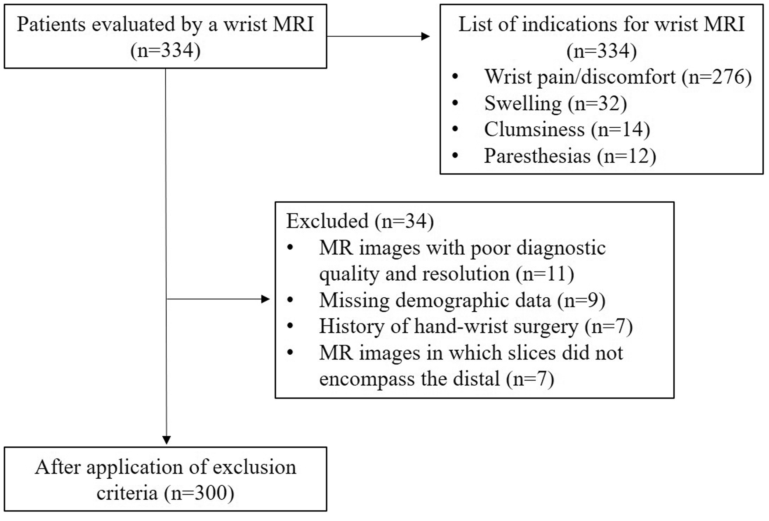
The diagram illustrates the flow of participants through each stage of the study.
MRI scan
All MRI scans were performed with a 1.5-T GE Signa Excite MRI device using a wrist coil. Images were obtained while patients were placed in prone position with the arm in full extension and wrist in neutral position. The evaluations were performed on axial turbo spin echo (TSE) T1, axial fat-suppressed TSE proton density (PD), coronal STIR, and sagittal fat-suppressed T2-weighted images obtained without using a contrast. Slice thickness was 3.2 mm and interslice gap was 0.5 mm. The slices extended from the proximal carpal tunnel to the distal carpometacarpal joint, wherein images encompassing the proximal aspect of this joint were excluded from the study.
MRI interpretation
The median nerve was mainly evaluated on axial TSE T1-weighted and TSE PD fat-suppressed MR images encompassing the section from the distal forearm to the distal carpometacarpal joint. In addition, nerve calibrations and signals were checked from the coronal and sagittal slices obtained from the patients. The flexor retinaculum was considered the proximal end and the carpometacarpal joint was considered the distal end of the carpal tunnel. Median nerve variations were assessed with respect to the carpal tunnel. While branching distal to the carpal tunnel was classified as the normal anatomy (Fig. 2a), branching proximal to the tunnel and within the tunnel were classified as variations (Fig. 2b and c). The median nerve was defined as bifid and trifid median nerve if it divided into two and three separate nerve branches, respectively, within and proximal to the carpal tunnel.
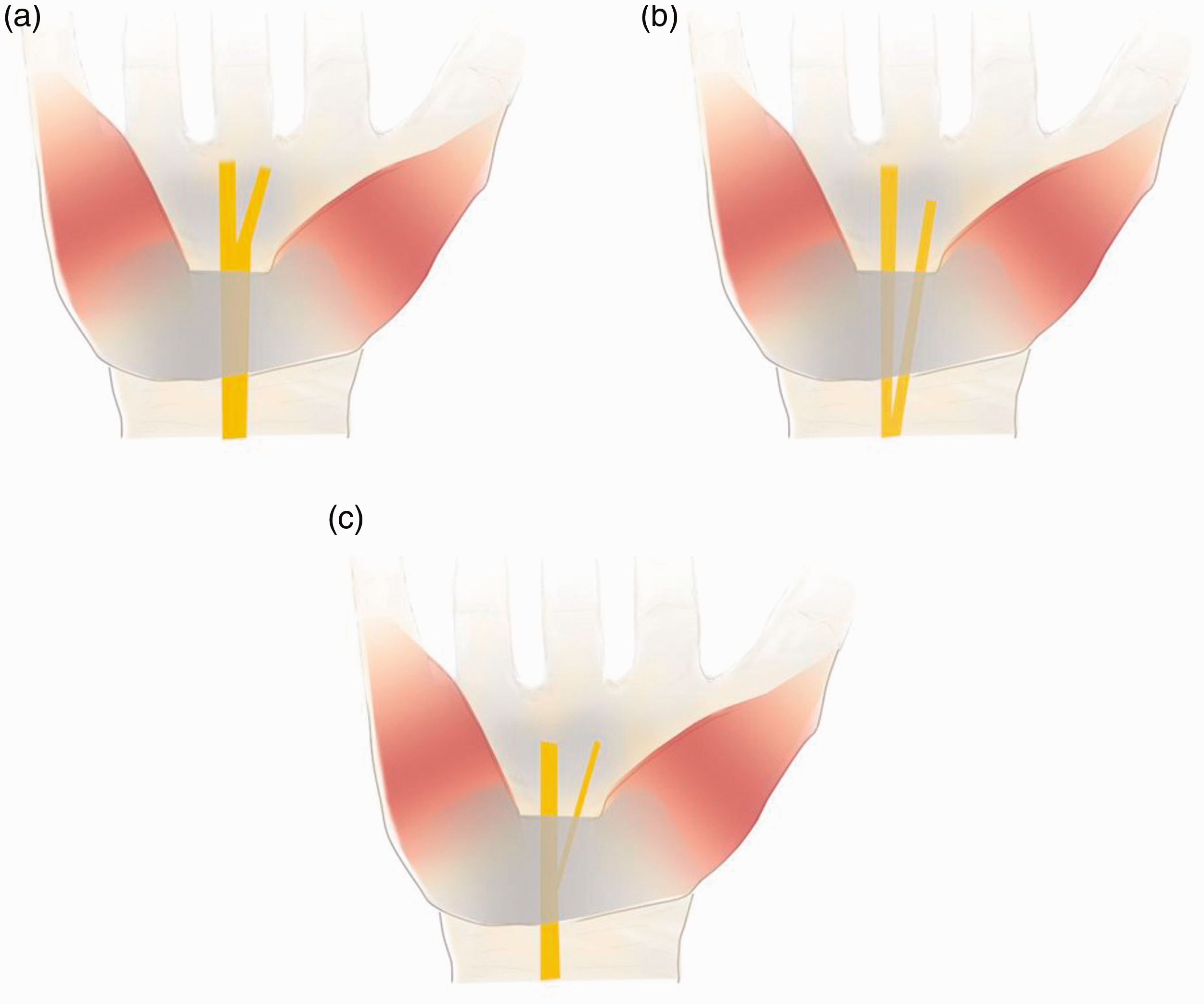
Schematic drawing shows (a) normal bifurcation of the median nerve distal to the carpal tunnel, (b) early bifurcation of the median nerve proximal to the carpal tunnel, and (c) inside the carpal tunnel. Normal anatomy is represented in (a), anatomical variations of the median nerve are represented in (b, c).
Presence of PMA was evaluated from axial TSE T1 and PD fat-suppressed images. Within the carpal tunnel, the PMA can be recognized by its well-defined circular shape that is continuous on consecutive slices, its location close to the median nerve and higher signal intensity compared to the nerve.
Median nerve variations and presence of PMA were assessed with the shared decision of a musculoskeletal radiologist with 15 years of experience and a radiology resident who has been trained on musculoskeletal radiology. They had no knowledge of the patients’ age, gender, and side of the examined wrist while analyzing the images. The patients were assigned to groups according to age (18–39 years, 40–59 years, or 60–80 years), gender (female or male), and side of the examined wrist (right or left). The groups were compared to determine whether there was any statistically significant difference in terms of median nerve variations and prevalence of PMA.
Statistical analysis
All analyses were performed using SPSS (Statistical Package for Social Sciences, Inc., Chicago, IL, USA) version 16.0. The chi-square test was used to determine if there was a statistically significant difference in terms of the frequency of median nerve branching patterns and Fisher’s exact test was used to determine if there was a statistically significant difference in terms of PMA prevalence between the groups that were created according to age, gender, and side of the examined wrist. P < 0.05 was considered statistically significant.
Results
The median nerve exhibited branching distal to the carpal tunnel, which represents the normal anatomy, in 227 of 300 patients (76%) (Figs. 3 and 4). On the other hand, of the 300 wrists, 38 (12.7%) and 34 (11.3%) had a bifid median nerve proximal to the carpal tunnel (Fig. 5) and within the carpal tunnel (Fig. 6), respectively. Median nerve trifurcation within the carpal tunnel was only observed in 1 (0.3%) patient. Accordingly, 73 (24.4%) of the 300 patients had a variation. Distal branching, i.e. normal anatomy, was observed in 227 (75.6%) patients.
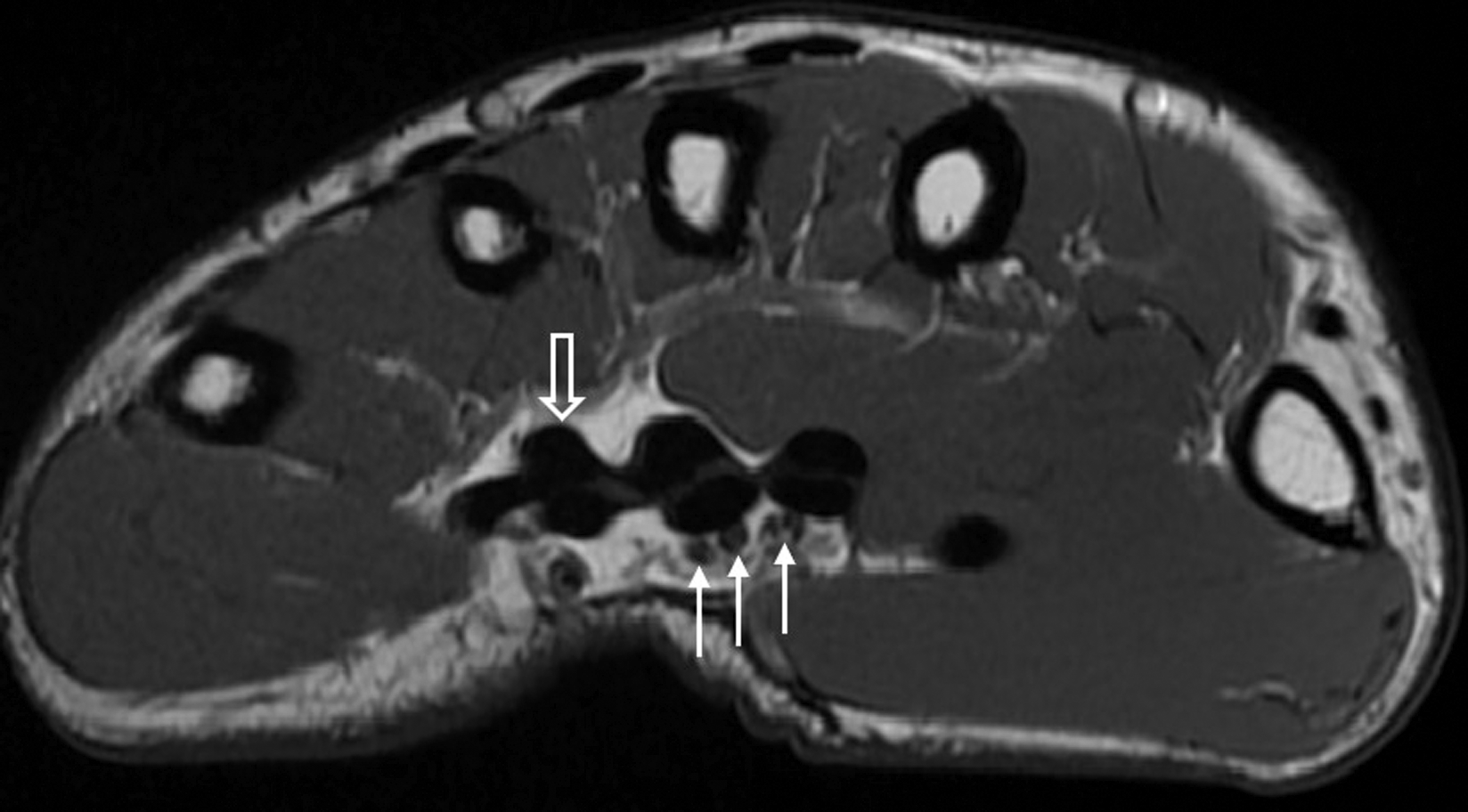
Turbo spin echo T1-weighted axial image shows multiple branches of the median nerve (arrows) distal to the carpal tunnel which represent the normal anatomy. The normal median nerve is observed with higher signal compared to neighboring tendons (open arrow).
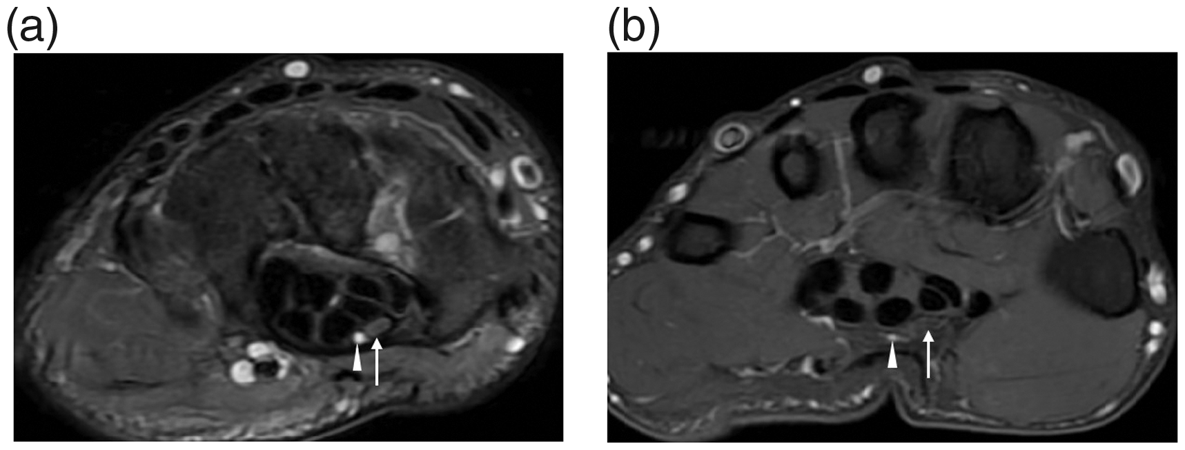
(a) TSE proton density fat-suppressed axial image shows the median nerve (arrow) and PMA (arrowhead) in the carpal tunnel. (b) TSE proton density fat-suppressed axial image of the same patient shows multiple branches of the median nerve (arrow) and PMA (arrowhead) distal to the carpal tunnel. The patient represents the normal anatomy of the median nerve. PMA, persistent median artery; TSE, turbo spin echo.
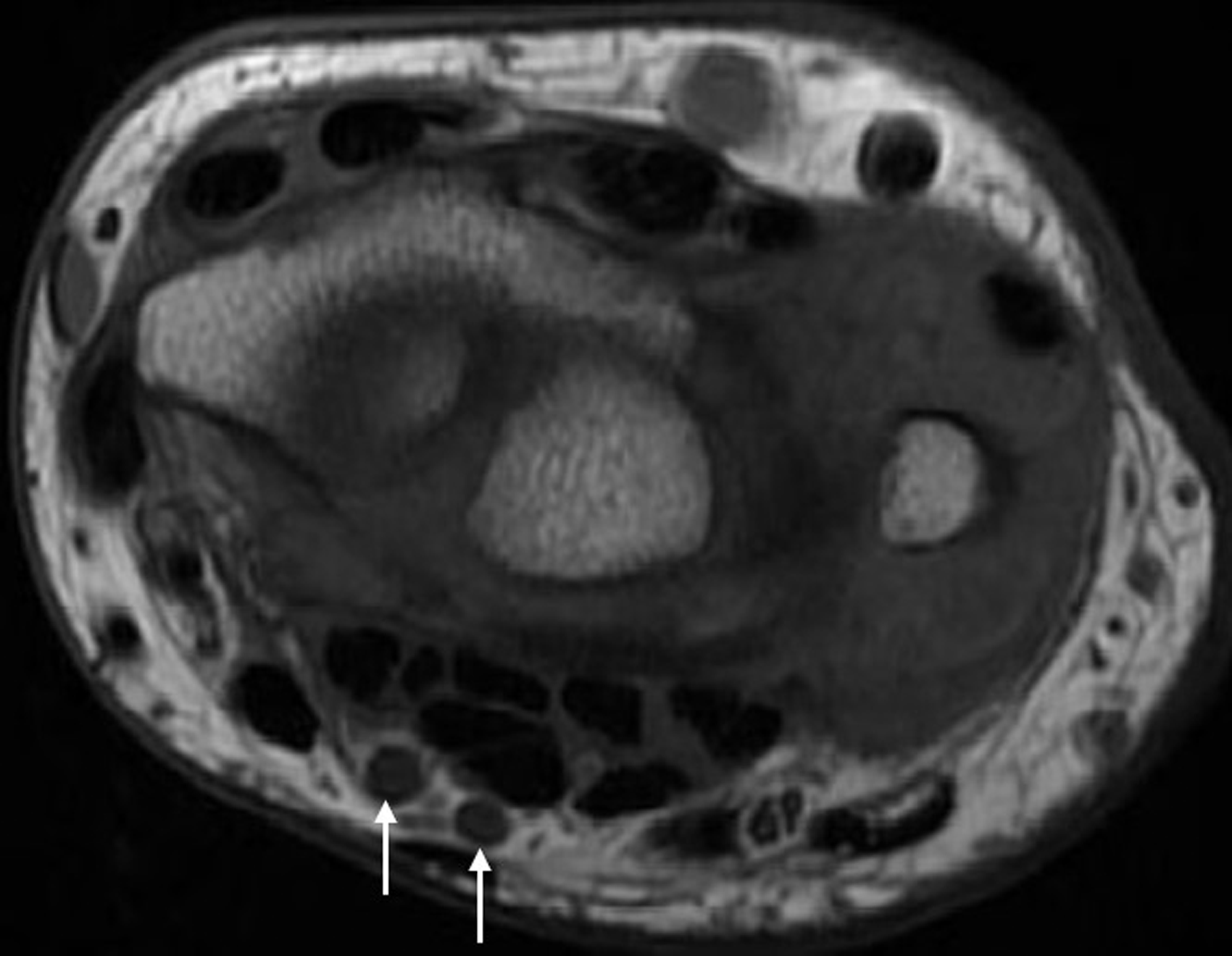
Turbo spin echo T1-weighted axial image shows bifid median nerve (arrows) proximal to the carpal tunnel which represent the anatomical variation of the median nerve.
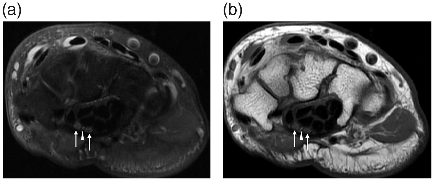
(a) TSE proton density fat-suppressed and (b) TSE T1-weighted axial images show two branches of the median nerve (arrows) and PMA (arrowhead) in the carpal tunnel. PMA travels between two nerve branches. PMA, persistent median artery; TSE, turbo spin echo.
The patients were classified according to age as Group I (aged 18–39 years), Group II (aged 40–59 years), and Group III (aged 60–80 years). The number of patients in each group and patient demographics are provided in Table 1. There was no statistically significant difference between the different age groups in terms of the frequency of median nerve branching patterns (P = 0.337 for proximal to the tunnel, P = 0.677 for within the tunnel, and P = 0.424 for distal to the tunnel).
Demographic findings of the patient population.

Values are given as n or mean (range).
There was also no statistically significant difference between genders in terms of the frequency of median nerve branching patterns (P = 0.284 for proximal to the tunnel, P = 0.902 for within the tunnel, and P = 0.121 for distal to the tunnel) (Table 2).
Prevalence of median nerve branching of subgroups based on age, gender, and sides.
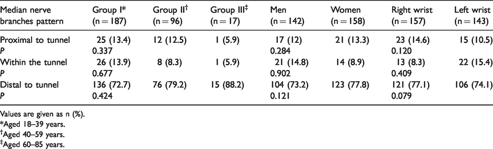
Values are given as n (%).
*Aged 18–39 years.
†Aged 40–59 years.
‡Aged 60–85 years.
There was no statistically significant difference between right and left wrists in terms of the frequency of median nerve branching patterns (P = 0.120 for proximal to the tunnel, P = 0.409 for within the tunnel, and P = 0.079 for distal to the tunnel). Table 2 shows the frequency of median nerve branching patterns according to age, gender, and side of the examined wrist.
PMA was observed in the carpal tunnel in 44 (14.7%) of the 300 wrists. Of the 44 PMA cases, 28 (63.6%) also had a coexisting median nerve variation. Coexistence of median nerve variations and PMA was found to be statistically significant (P = 0.001). None of the patients with PMA had any findings suggestive of thrombosis.
Differences between the different age groups were evaluated in terms of PMA prevalence. Thirty-two (17.1%) of the 187 patients in Group I, 11 (11.4%) of the 96 patients in group II, and 1 (5.8%) of the 17 patients in group III had PMA. There was no statistically significant difference between the age groups in terms of prevalence of PMA (P = 0.103).
Of the 158 female patients, 18 (11.4%) had PMA. Of the 142 male patients, 26 (18.3%) had PMA. There was no statistically significant difference between gender groups in terms of prevalence of PMA (P = 0.103).
Nineteen (13.3%) of the 143 left wrists and 25 (15.9%) of the 157 right wrists had PMA. There was no statistically significant difference between the right and left wrists in terms of prevalence of PMA (P = 0.624). Table 3 shows the PMA prevalence with respect to age, gender, and side of the examined wrist.
Prevalence of PMA based on age, gender, and sides.

Values are given as n (%).
*Aged 18–39 years.
†Aged 40–59 years.
‡Aged 60–85 years.
PMA, persistent median artery.
Discussion
Today, there are studies suggesting that anatomical variations associated with the carpal tunnel (bifid-trifid median nerve, persistent median artery, etc.) also play a role in the etiology of CTS in addition to the pathologies affecting the median nerve (8). On the other hand, some of the recent studies have failed to confirm that anatomical variations lead to increased risk of CTS (13). However, it is indisputable that such “anatomical traps” should be previously identified in order to safely and reliably release the flexor retinaculum in CTS surgery, especially while using the endoscopic approach. Besides the fact that median nerve variations may lead to CTS, it is important that radiologists know the median nerve variations and report such variations using common terminology with orthopedists in order to protect the nerve branches during endoscopic surgical procedures.
The normal median nerve located in the carpal tunnel divides into branches distal to the carpal tunnel. Median nerve branching proximal to the carpal tunnel and within the tunnel are considered variations. Similarly, branching distal to the carpal tunnel was considered normal anatomy and branching proximal to the tunnel and within the tunnel was considered a variation in the present study. In this study, bifid-trifid median nerve was observed proximal to the tunnel in 38 (12.7%) patients and within the tunnel in 35 (11.7%) patients, wherein these rates were considerably high compared to the surgical literature (14,15). Lindley and Kleinert (15) investigated the prevalence of aberrant or unexpected anatomic structures during elective carpal tunnel surgery and detected five median nerve variations in 526 wrists (1%). Beris et al. (16) found intraoperatively variations of median nerve at the wrist in 11 out of 110 patients (10%). In our study overall, 24.4% of all cases had median nerve variations. This shows us that nearly one in four patients have median nerve variations. Such common variations known and reported by radiologists will contribute to explaining the carpal tunnel syndromes of unknown cause. In addition, identification of these variations can help prevent inadvertent injuries associated with median nerve variations during CTS surgery. Therefore, we are of the opinion that the presence of variations in median nerve branching should be specified and described in detail on routine wrist MRI reports.
Studies demonstrating median nerve variations using MRI are very scarce in the literature. The present study includes the highest number of patients among the MRI studies conducted so far. In a study by Pierre-Jerome et al. (11) employing classifications similar to those employed in our study, the rate of bifid median nerve within the carpal tunnel was 18%, which was higher compared to the results of our study (11.7%), whereas the rate of variations proximal to the tunnel was 6.1%, which was lower compared to the results of our study (12.7%). The rate of branching distal to the tunnel, i.e. normal anatomy, was similar in both studies. The mentioned difference between the results of these two studies may stem from different MRI sequences and slice thickness used as well as different populations involved. The fact that the rate of cases representing the normal anatomy was similar suggests that MRI is highly successful in showing normal anatomy. Similar to the study by Pierre-Jerome et al., our study did not show a significant difference in terms of the frequency of median nerve variations between the groups created according to age, gender, and side of the examined wrist.
In a study by Bayrak et al. (8) comparing the prevalence of median nerve variations between patients with and without CTS, it was found that the rate of variation was 19% in the CTS group (8). In another study by Granata et al. (12), conducted with a similar purpose, the same rate was found to be 18.5%. Bayrak et al. (8) and Granata et al. (12) investigated the prevalence of median nerve variation in patients with and without CTS, which is different from our study. Thus, they were able to conclude whether the bifid median nerve is a risk factor for CTS. In the present study, the rate of median nerve variations was approximately 25%, which is higher than the values reported in both the abovementioned studies. Ultrasonography was the preferred imaging modality in both studies mentioned above. In the present study, we used MRI. In addition, our classification was also different compared to these studies. The relationship between the presence of bifid median nerve and clinical findings was reported to be poor in previous surgical studies (15–17). Moreover, considering the high rate of median nerve branching variations (25%) in our study group, it is apparent that every variation in median nerve branching will not cause clinical findings.
The prevalence of palmar type of PMA was reported to be in the range of 0.9%–50% in adults (18,19). On the other hand, the antebrachial type of PMA is much more common with a prevalence of 70%–76%. In the present study, the prevalence of the palmar type of PMA was found to be 14.7%. The difference between these studies in terms of PMA prevalence may be associated with factors such as different patient populations, measurement technique, and definition of PMA.
Carry et al. (20) found a prevalence of PMA of 26.6% in a pediatric population. Our study was conducted with adults, which may be the reason for the lower prevalence of PMA. The fact that the pediatric population had a higher PMA prevalence than the adult population can be explained with the hypothesis that the process of PMA regression continues from embryonic life until adulthood. The same study also showed that age had a negative correlation with the presence of PMA (although not statistically significant), which is supportive of the mentioned hypothesis.
Chen et al. (21) found that the frequency of bifid median nerve was 9.4% and prevalence of PMA was 7.5% in their study using ultrasonography. In our study, the frequency of bifid median nerve (24.4%) and prevalence of PMA (14.7%) were both considerably higher than the same reported by Chen et al. In addition, 9.3% of our patients had coexisting median nerve variation and PMA, wherein the coexistence was statistically significant. The same rate was 6.3% in the study by Chen et al., which was also lower compared to our results. However, Chen et al. concluded that the probability of the coexistence of these two variations was higher than the probability of the variations existing independently. Both of the studies discussed above were conducted using the Doppler US technique. We think that technique differences may have a role in the difference between the results. High soft-tissue resolution and less user dependence of MRI may have also contributed to the higher rates of median nerve variations and PMA in our study. In their case report, Gutowski et al. (22) presented a patient with an arteriovenous malformation of a PMA and a bifurcated median nerve. They stated that surgeons should be aware of median nerve bifurcations, particularly when a PMA is identified. It should be noted that there may be additional structures in the carpal tunnel that can cause nerve compression. Specifically, in such patients undergoing carpal tunnel surgery, it is necessary to identify more than one median nerve accompanying PMA on preoperative imaging. Similar to the case with median nerve variations, there was no significant difference in terms of PMA prevalence between the groups created according to age, gender, and side of the examined wrist in our study.
The present study has some limitations. Primarily, the study was retrospective in design. Another limitation was that the correlation between results and clinical data was not studied. MRI results also could not be compared with the surgical results, since not all patients included in the study underwent wrist surgery. On the other hand, our study is important as it demonstrated prevalence in a large series and used routine wrist MRI. The strength of the present study is that it is the most extensive study conducted with the largest sample size among other studies investigating the anatomical variations of the median nerve and PMA using MRI.
In conclusion, the rates of median nerve variations and PMA were 24.4% and 14.8%, respectively, on the wrist MRI scans. The coexistence of median nerve variation and PMA was 9.3%. We are of the opinion that preoperative evaluation of these anatomical variations, which are considerably common, with MRI will be protective against postoperative complications, particularly in patients planned to be operated for CTS. Further studies to be conducted with large series in order to perform anatomical comparisons on MR images and cadavers may help to create common classifications for surgery, anatomy, and radiology. Therefore, a common terminology can be developed as a guide for orthopedic surgeons in terms of the median nerve and the vascular structures in the relevant region as identified on MRI before carpal tunnel surgery.
Footnotes
Declaration of conflicting interests
The author(s) declared no potential conflicts of interest with respect to the research, authorship, and/or publication of this article.
Funding
The author(s) received no financial support for the research, authorship, and/or publication of this article.
