



Abstract
The calf muscle group is a common area for injury within the professional athlete population. Anatomical and biomechanical differences between the different component muscles vary their individual predispositions to and patterns of injury. However, there is a common unifying factor: injuries involving tendinous components have greater clinical implications with regards to rehabilitation, potential intervention, length of time to return to play, and re-injury rates. As such, accurate understanding of the underlying anatomy and subsequent interpretation of the injury patterns carry significant clinical ramifications. Ultrasound is a useful tool but has limitations, particularly when assessing soleus. As such, magnetic resonance imaging remains the workhorse in calf injury investigation.
Background
Calf strains are common injuries in professional athletes; the largest proportion arise in gastrocnemius, commonly the medial head (1). The muscle is classified as “high risk” for injury due to a combination of two main anatomical factors: it spans two joints (the knee and ankle) allowing rapid lengthening and high tensioning contractions; and is composed of type 2 muscle fibers, which contract rapidly and with force.
Conversely, the soleus muscle has historically been characterized as “low risk” as it only crosses one joint and predominantly comprises type 1 slow twitch muscle fibers (1). As such, soleus injuries have tended to be underestimated, confounded by their more subacute presentation (often presenting with worsening stiffness and tightness over days to weeks and often in older athletes (1,2)) and the limitations of ultrasound for accurate assessment.
The third muscle in the calf completing the triceps surae is plantaris. Historically in the literature, this was understood to be absent in 7%–20% of the population (3); however, more recent cadaveric studies have identified a plantaris tendon in 98%–100% of specimens. Therefore, the proposition that the plantaris is a vestigial or irrelevant rudimentary structure may need to be reconsidered (4,5). Although it spans two joints, it is neither a significant knee flexor nor ankle plantar flexor but is thought to have a proprioceptive function as it contains a high density of muscle spindles. It can be injured in isolation or in combination with injuries to the gastrocnemius and/or soleus (3).
Anatomy
Gastrocnemius
Gastrocnemius has two muscle bellies originating from the posterior distal femur: the medial head arises from the medial supracondylar ridge and lateral head from the posterolateral margin of the lateral femoral condyle. The proximal tendons of gastrocnemius expand and spread as thin fascia on the posterior muscle surface, from which the muscle fibers then arise. Distally, the fibers attach to a broad fascia anteriorly that condenses as it extends caudally. It then unites with the posterior fascia of soleus to form the Achilles tendon.
Soleus
The soleus can be viewed as a structure where muscle fibers arise from an internal scaffold of dense connective tissue (the intramuscular tendons) with external support by a loose connective tissue envelope (the anterior and posterior fascia).
It has a broad origin extending from the posterior surface of the head and proximal third of the fibular diaphysis, across the tibiofibular syndesmosis to the middle third of the medial tibial border. The tibial neurovascular bundle penetrates the fibrous arch formed by the proximal origins of the soleus to enter the deep tissues of the leg.
Proximally, the surrounding epimysium condenses to form two intramuscular aponeuroses, one medial and one lateral, from which the proximal muscle fibers arise (Fig. 1) (6).
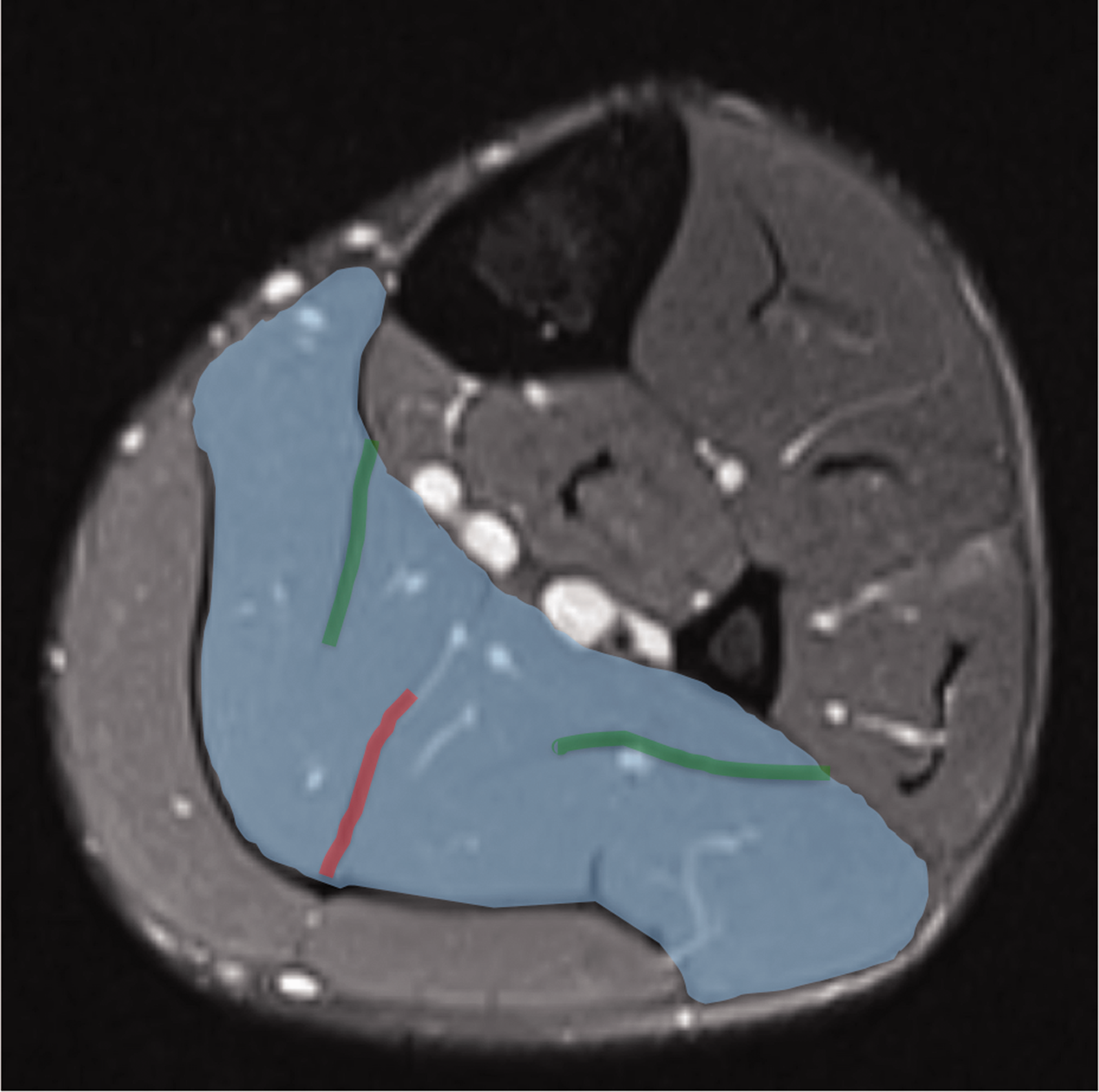
Axial STIR image through the mid soleus. Soleus muscle belly (blue); central intramuscular tendon (red); medial and lateral intramuscular aponeuroses (green). STIR, short tau inversion recovery.
These aponeuroses are considered intramuscular tendons both anatomically and functionally, which is reflected in their associated pathology. Distally within the soleus, a long intramuscular central tendon forms, initially from the deep epimyseal surface, before migrating centrally and condensing at the superficial surface. It then becomes confluent with the overlying gastrocnemius tendon to form the Achilles tendon.
The Achilles tendon is made up of three distinct fascicle bundles (the medial gastrocnemius, the lateral gastrocnemius, and the soleus), forming a twisted tendon unit (7), to distribute the varied force profiles of each muscle unit homogeneously throughout the structure (8).
Plantaris
Plantaris has a small proximal muscle belly and a long thin tendon. The muscle originates from the posterolateral distal femur, superomedial to the lateral head of gastrocnemius. It courses inferomedially across the popliteal fossa with the myotendinous junction around the level of the tibial origin of soleus (9). The tendon then courses distally between the medial belly of gastrocnemius and soleus. The distal insertion is variable; it is closely associated with the anteromedial aspect of the Achilles tendon and can blend with the tendon itself or remain independent with a separate insertion on the calcaneus (it can be seen to remain intact in cases of Achilles tendon rupture) (3).
Calf imaging
Magnetic resonance imaging (MRI) is the investigation of choice for comprehensive investigation of calf muscle injuries. Ultrasound remains a useful tool but more in its capacity to target intervention than in its diagnostic capabilities. It has limitations in accurate assessment of the soleus, particularly as the muscle is deep, multipennate, and a highly vascular structure which is often hypertrophic in athletes (10).
As well as detailing the anatomical structures involved and therefore accurately assessing the extent of the injury sustained, MRI has a further role in subsequent monitoring of the athlete during rehabilitation. A baseline MRI provides direct comparison aiding assessment of injury healing and ultimately resolution and recovery. Follow-up studies are useful in assessing for the presence of scar tissue formation; thick fibrous scar formation can alter the compliance of tissues at the interface between the scar and adjacent muscle/connective tissue and change the mechanics during contraction, thus predisposing an athlete to local re-injury (11,12).
MRI protocol
The MRI protocol for calf injury will vary according to practitioner preference. The authors’ preference is to acquire small field of view images of the calf (knee to ankle) using a combination of short tau inversion recovery (STIR) and proton density fat saturation (PD fat sat) sequences in the axial, sagittal, and coronal planes. A T1-weighted image (usually in the axial plane) is also chosen to detail the anatomy and can be helpful in detailing the degree of intratendinous injury (13). A radiological marker (e.g. oil capsule) should be applied to the skin to mark the site and level of symptoms.
MRI muscle injury reporting and classification systems
Radiological assessment of any muscle/tendon injury will require detailing of the topographical location, measurement of the cross-sectional and longitudinal area of involvement, and the degree of myofibril and tendinous distortion (loss of pennation angle, fiber disruption/discontinuity and fiber retraction, and loss of tension). Most of this information can be gleaned from a detailed examination of the images acquired in the axial plane (indeed these are the “go-to” images). For the assessment of parameters, looking at tension loss and retraction of the muscle/tendon complex and the longitudinal extent of the injury the images that are acquired in the coronal or sagittal plane may be more beneficial (Fig. 2).
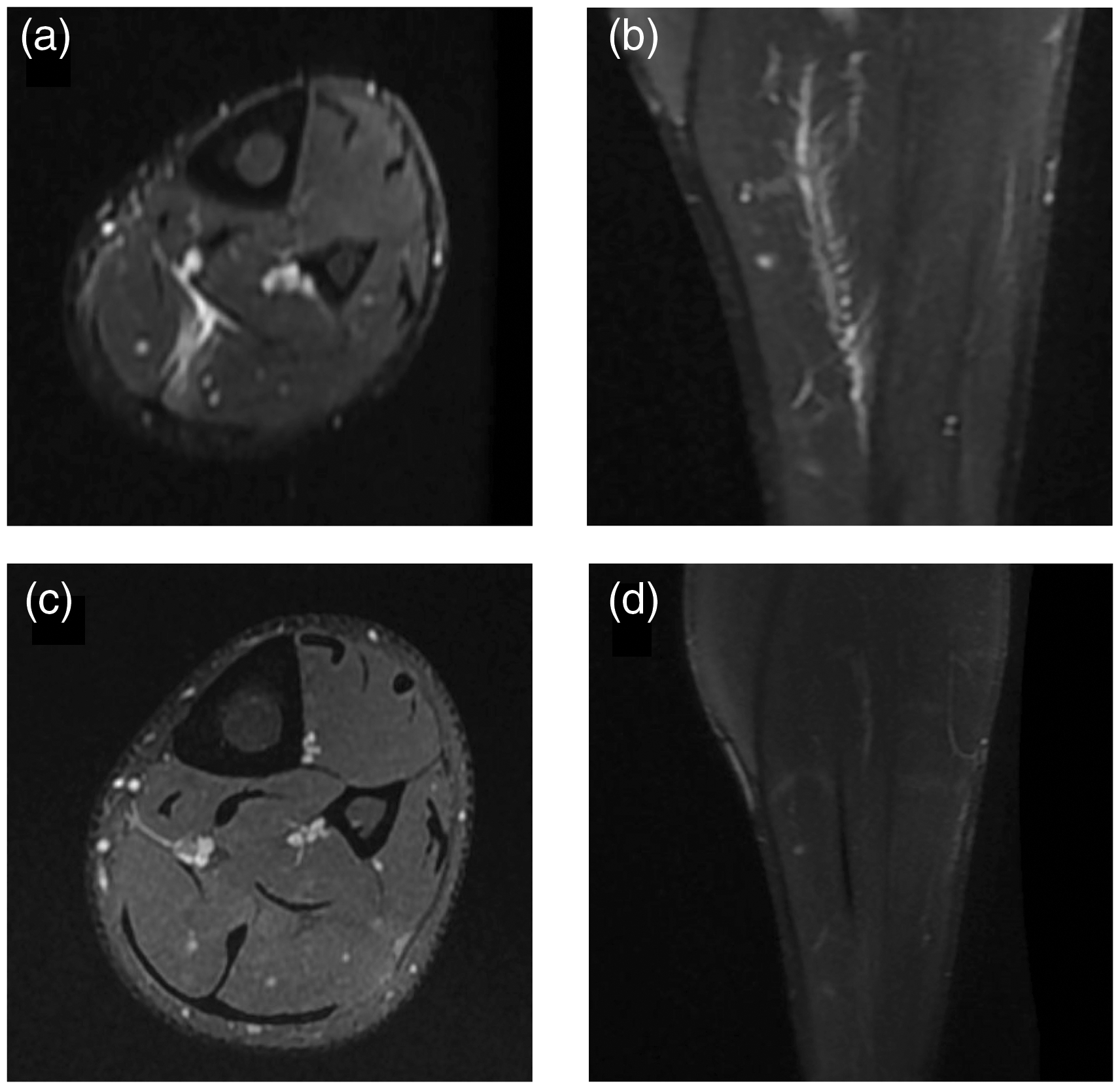
Central tendon injury soleus in a 25-year-old professional football player who developed calf tightness after a game with increasing pain on loading. No clear history of injury. (a, b) Initial injury; (c, d) 5 months later. (a) Axial STIR shows defect in the anterior portion of the central tendon with surrounding edema. BAMIC type c, Prakash Grade 3. (b) Coronal STIR demonstrating a wavy tendon in keeping with loss of tension. (c) Five months after initial injury, axial PD fat sat: the anterior portion of the central tendon has healed with mature scar formation with no appreciable defect. (d) Five months after initial injury, the corresponding coronal STIR image shows a healed, albeit thickened, tendon, which now has straight margins without loss of tension. The return-to-play time was 24 days after the initial injury with the player undergoing treatment with injection of platelet-rich plasma. BAMIC, British Athletic Muscle Injury Classification; PD fat sat, proton density fat saturation; STIR, short tau inversion recovery.
The topographical location of injury and the degree of involvement of the aforementioned connective tissue elements is thought to be key in predicting the time for recovery and return to play for the athlete (14). Indeed, injuries that extend into the tendon have already been shown to have extended times to return to full-time training and increased rates of re-injury in elite track and field athletes (15).
Another complicating factor that can have an impact on return-to-play time is the formation of intra- or intermuscular hematoma and is therefore an important component of the radiological report. A common site for intermuscular hematoma is between the medial head of gastrocnemius and soleus muscles (16), with injuries involving either of their apposing fascia implicated in its pathogenesis.
Reference to any scar formation or ectopic calcification subsequent to a previous injury is also important because of the implications on re-injury.
A grading system for calf muscle injuries has recently been proposed by Prakash et al. (17). This is based on the detection of myofibril distortion and integrity of the connective tissue elements in injuries of the calf muscle. Their grading system is simple to use but has yet to be validated. The “grade 0” classification is somewhat counterintuitive as, to the uninitiated, a grade 0 injury may be misinterpreted as “no injury identified” when in fact it represents a low-grade injury. The British Athletic Muscle Injury Classification (BAMIC) system has had its validity assessed with respect to hamstring muscle injury (18,19). While the system may not be fully applicable to calf muscle injury, the a, b, and c suffixes of the BAMIC classification system, representing myofascial, myotendinous, and intratendinous injury, respectively, can be applied to any muscle/tendon group. The a, b, c system is also clearly understood and already widely used by those working in the sporting community because of the correlation with increasing return-to-play times and re-injury rates in c injuries. Clearly, further work in this is required.
Injury mechanisms and MRI findings
When considering calf muscle injury mechanisms, it is important to have an appreciation of muscle function. Owing to the difference in origins of the gastrocnemius and soleus above and below the knee, respectively, there is variation in muscle contribution to plantar flexion depending on joint positioning. The soleus, gastrocnemeii, and plantaris are significant contributors to force absorption and generation during the various phases of foot plant during the running cycle.
On foot strike, the soleus acts eccentrically, controlling knee flexion via the mediation of tibial inclination (in relation to the ground) and through its action at the ankle and fixed foot. Juxtaposed is the action of the gastrocnemius, flexing the knee via its femoral attachment and controlling the position of the lower shank with the foot on the floor (20).
As the stance phase of the gait cycle begins, the soleus will provide propulsive forces of up to eight times body weight (21), working strongly at slow to moderate running speeds and providing up to 50% of the total vertical displacement forces (22).
As the stance phase progresses, the action of the gastrocnemius produces forward propulsive forces of up to three times the body weight (21), dependent on running speed and knee angle (increased knee flexion reducing gastrocnemius plantarflexion activity) (23–25).
It is therefore suggested that the soleus and gastrocnemius act functionally in isolation while sharing a common insertional structure: the Achilles tendon.
Each muscle of the calf group can be injured in isolation, but given their functional and anatomical relationships, they can be injured concomitantly; some studies report coexisting gastrocnemius and soleus strains in 17% of calf strains (Fig. 3) (26). This pays testament to the value of MRI in investigating the true origin of the injury as well as its use in prognostication and guiding rehabilitation/intervention. This is emphasized in the literature where studies have demonstrated that injuries in the region of the central tendon of soleus have longer return-to-play times (2), similar to other intramuscular tendinous injuries elsewhere in the body, for example the central tendon of the indirect head of rectus femoris (27). As well as proffering a classification, Prakash et al. (17) highlighted the importance of injuries that extend into the tendinous/aponeurotic structures with regards to their implications on return to play: Grade 0 and 1 injuries (no aponeurotic/tendinous involvement) are demonstrated to have a mean time to return to play of 8 and 17 days, respectively; Grade 2 and 3 tears, which involve these structures in increasing severity, have a time of 25 and 48 days, respectively (17).
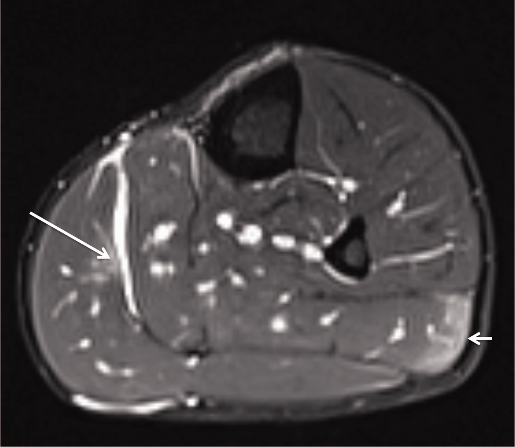
Axial STIR image. Epimyseal fibre injuries of medial gastrocnemius (long arrow; BAMIC type a; Prakash Grade 2) and lateral soleus (short arrow; BAMIC type a; Prakash Grade 0). Subsequent haemorrhage into the potential space between the medial gastrocnemius and soleus. BAMIC, British Athletic Muscle Injury Classification; STIR, short tau inversion recovery.
The distal myofascia of the medial head of the gastrocnemius is one of the most frequently encountered calf injuries in sport, often referred to as “tennis leg.” Although more complex injury patterns can occur, a commonly demonstrated morphology is that of an “L-pattern” tear (Fig. 4). A transverse component extends across the anterior fascia in the mediolateral plane before propagating proximally in a vertical linear manner. The vertical “split tear” component is thought to propagate due to retraction and separation of the initial transverse tear from the remainder of the intact fascia (28).
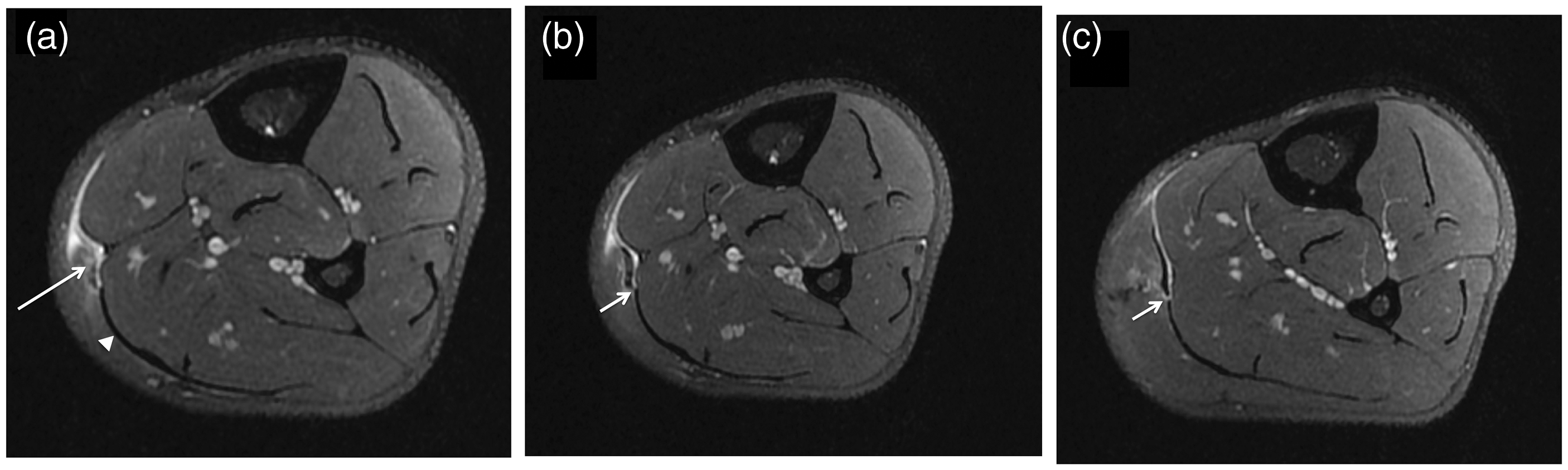
28 year old professional rugby player. Axial Proton Density fat-sat image demonstrating “L-pattern” tear of the distal myofascia of the medial head of gastrocnemius injury. BAMIC type c; Prakash Grade 3. Return-to-play 84 days; the player was injected with platelet rich plasma. (a) The medial portion of the anterior fascia is ill-defined (long arrow) representing a transverse tear. There is associated muscle oedema. Laterally, the anterior fascia is intact (arrowhead). (b) Axial image just cranial to A. The medial portion of the anterior fascia now appears intact. At its lateral extent however there is a clear defect in the fascia (short arrow). Blood tracks between the gastrocnemius anterior fascia and the underlying posterior fascia of soleus. (c) Axial image further cranial than B. The defect seen in B propagates superiorly representing the vertical split component of the tear. In this case the vertical tear extended cranially by 10cm. BAMIC, British Athletic Muscle Injury Classification; PD fat sat, proton density fat saturation.
Recently, Pedret et al. (14) have proposed a new classification system based around dynamic ultrasound assessment of this specific area (focusing on the junction between the aponeurosis/fascia of medial gastrocnemius and that of soleus before they combine to form the Achilles tendon). Although ultrasound can be useful in the assessment of medial gastrocnemius injuries, it remains a limited modality for assessment of the soleus, stressing the importance of more comprehensive evaluation with MRI. Their study also investigated the relationship between the different grades of injury and their respective associations with the formation of hematomas.
If injury of the medial gastrocnemius extends to involve the anterior fascia, hemorrhage can track into the potential space between this and the posterior fascia of soleus. This can complicate these injuries and has implications on a prolonged recovery time. Pedret et al. (14) suggest that intermuscular hematoma formation is less implicated with injuries isolated to the myofascial fibers (forming an intramuscular hematoma instead) but form when rupture of the gastrocnemius fascia/aponeurosis allows hemorrhage to dissect into the intermuscular plain between the apposing fascia.
In the context of such intermuscular hematoma formation, careful scrutiny is required to assess the plantaris tendon that courses through this space (Figs. 5 and 6). There may be biomechanical reasons that predispose the plantaris to injury in elite track and field athletes. It is a plantar flexor and therefore will be important in running and sprinting athletes, who require large plantar flexor forces, throughout the full range of plantarflexion. Athletes in other sports and recreational athletes may not require the same range or maximal plantarflexion recruitment. It has a long thin tendon that should confer stiffness and elastic energy return resulting in performance improvement in sprinting and fast-paced running (Fig. 7) (29).
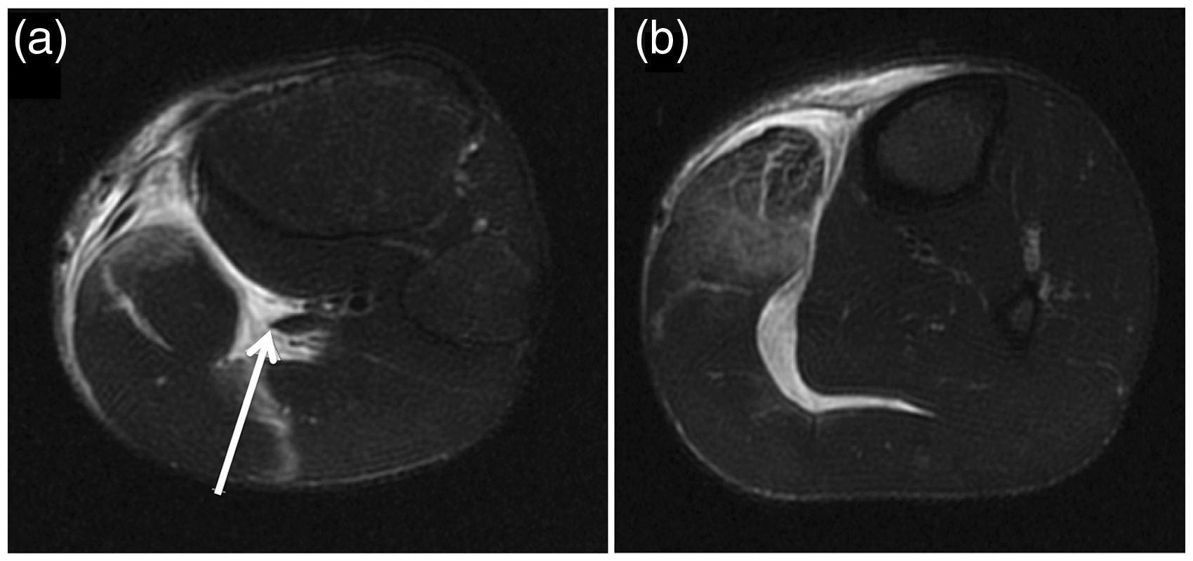
Axial Proton Density fat-sat images showing concomitant plantaris rupture and myofascial tear of the medial head of gastrocnemius. In (a) the plantaris myotendinous junction is clearly demonstrated (arrow). In (b) the plantaris tendon is not visualised in keeping with a rupture (BAMIC type 4c; Prakash Grade 3). There is a clear tear involving the muscle and anterior fascia of the medial head of gastrocnemius (BAMIC type a; Prakash Grade 2). Fluid and haemorrhage tracks in between the gastrocnemius and the underlying soleus. BAMIC, British Athletic Muscle Injury Classification; PD fat sat, proton density fat saturation.
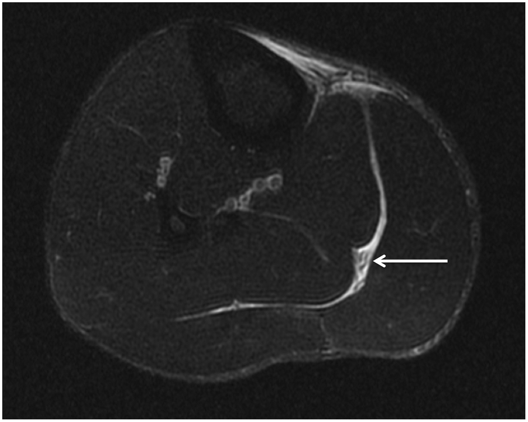
Axial STIR image at the level of the mid-calf. Hemorrhage expanding the space between the medial gastrocnemius and the soleus but no culprit injury to either the fascial surface or muscle on examination of the whole series. Irregular focal linear areas of low signal in the region of the expected plantaris tendon consistent with isolated plantaris tendon rupture (arrow). BAMIC type 4c, Prakash Grade 3. BAMIC, British Athletic Muscle Injury Classification; STIR, short tau inversion recovery.
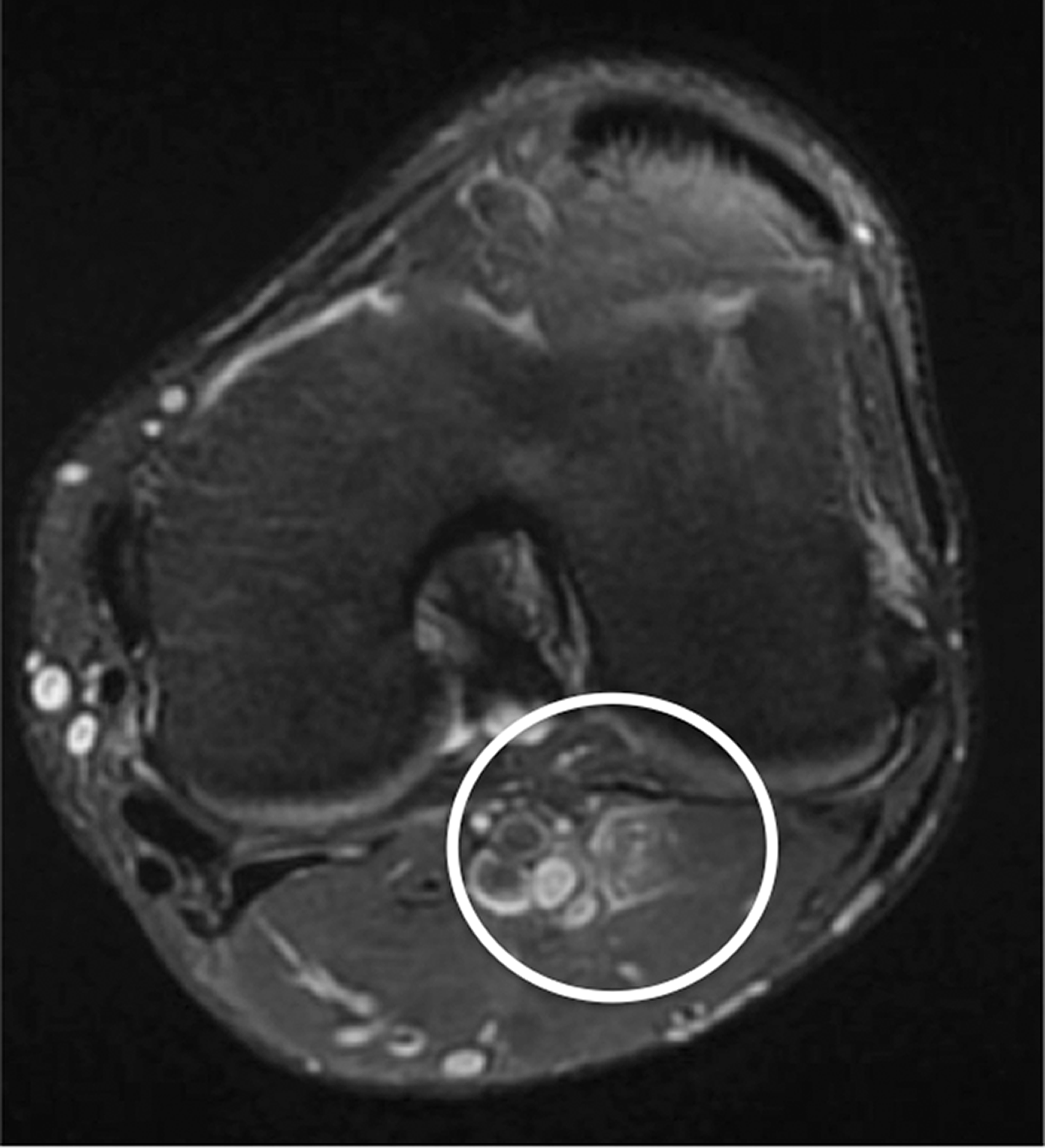
Axial STIR image. Subtle oedema within the plantaris muscle belly extending to the proximal myotendinous junction. BAMIC type b; Prakash Grade 0. BAMIC, British Athletic Muscle Injury Classification; STIR, short tau inversion recovery.
Soleus injuries have been described in five main sites: the anterior and posterior myofascial fibers (Fig. 8), the myotendinous junctions at the central tendon (Figs. 2 and 9), and the medial and lateral aponeuroses (Figs. 10–12) (6). Myotendinous injuries are more common, particularly involving the proximal medial intramuscular aponeurosis, which is clinically significant as injuries associated with the intramuscular tendons have considerable potential implications on rehabilitation, intervention, and return to play (Fig. 13).
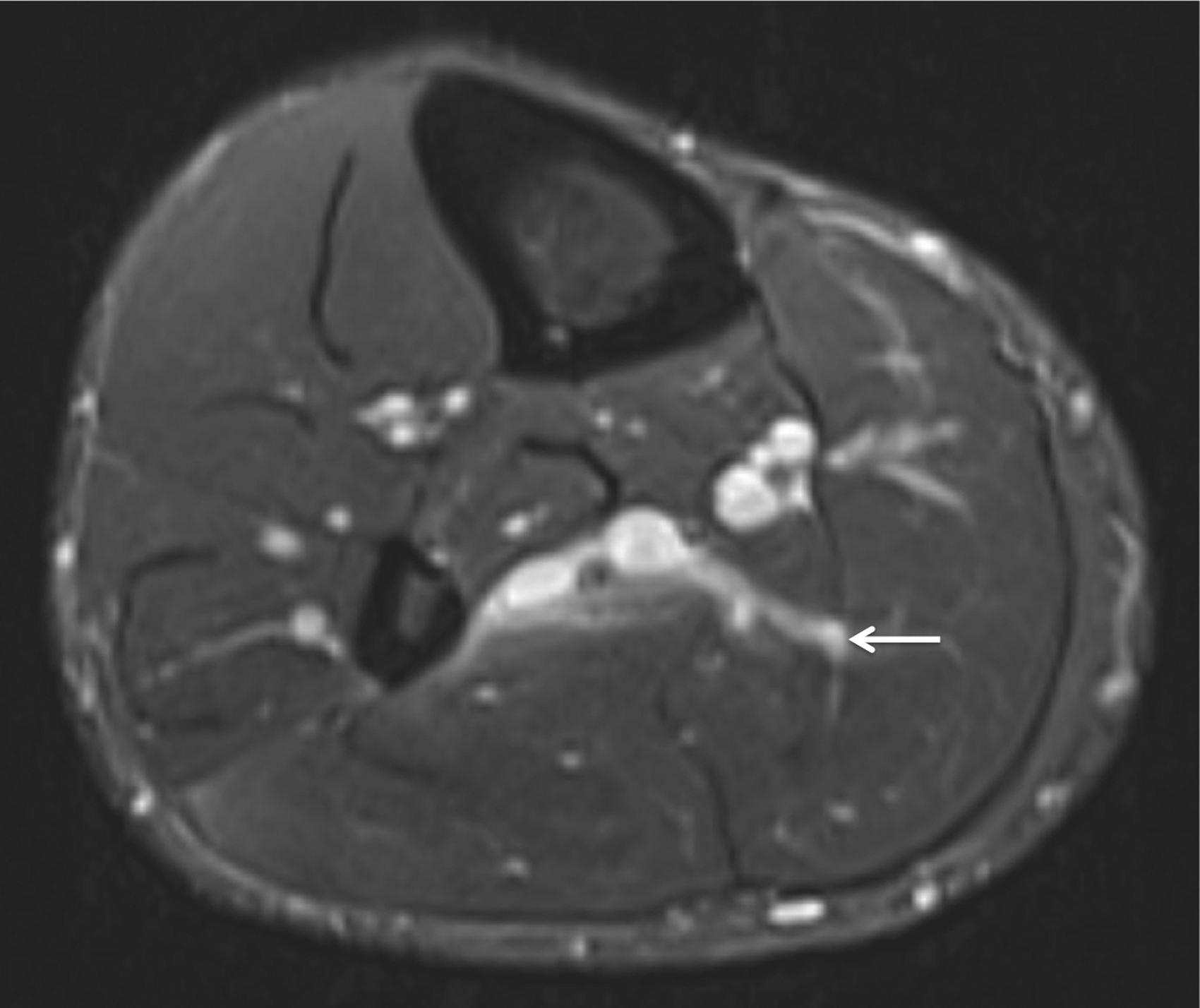
Axial STIR image. High signal at the anterior epimyseal surface of the soleus with extension into the medial intramuscular aponeurosis upgrading the injury to a BAMIC type c, Prakash Grade 2. BAMIC, British Athletic Muscle Injury Classification; STIR, short tau inversion recovery.

Axial STIR image at the level of the distal calf. High signal alongside the length of the central intramuscular tendon consistent with myotendinous fiber injury. The central tendon maintains uniform linear low signal with no loss of tension. BAMIC type b, Prakash Grade 1. BAMIC, British Athletic Muscle Injury Classification; STIR, short tau inversion recovery.
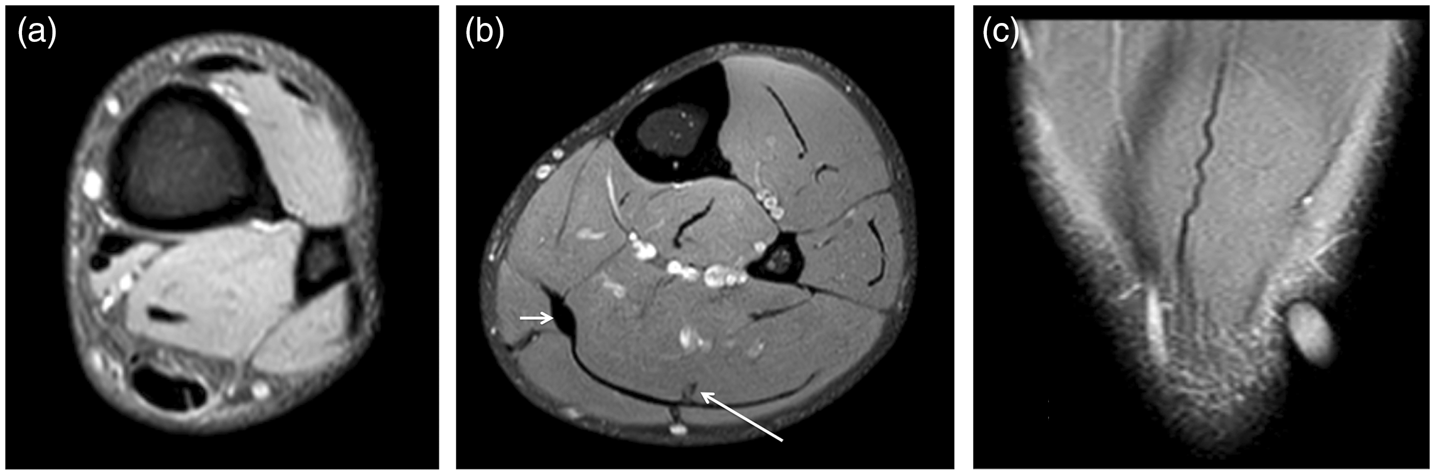
(a, b) Axial PD fat sat images of the distal calf and (c) coronal STIR image of the posterior soleus. New calf injury in the same player as Fig. 4. (a) Longitudinal split tear of the Achilles tendon. This extends cranially to involve the central tendon of soleus (long arrow in b) which demonstrates clear loss of tension (c; an oil marker is demonstrated on the skin surface indicating the level of symptoms as reported by the athlete). BAMIC type c, Prakash Grade 3. Healing with thickened scar of the previous anterior fascial tear of medial gastrocnemius (short arrow in b). BAMIC, British Athletic Muscle Injury Classification; PD fat sat, proton density fat saturation; STIR, short tau inversion recovery.
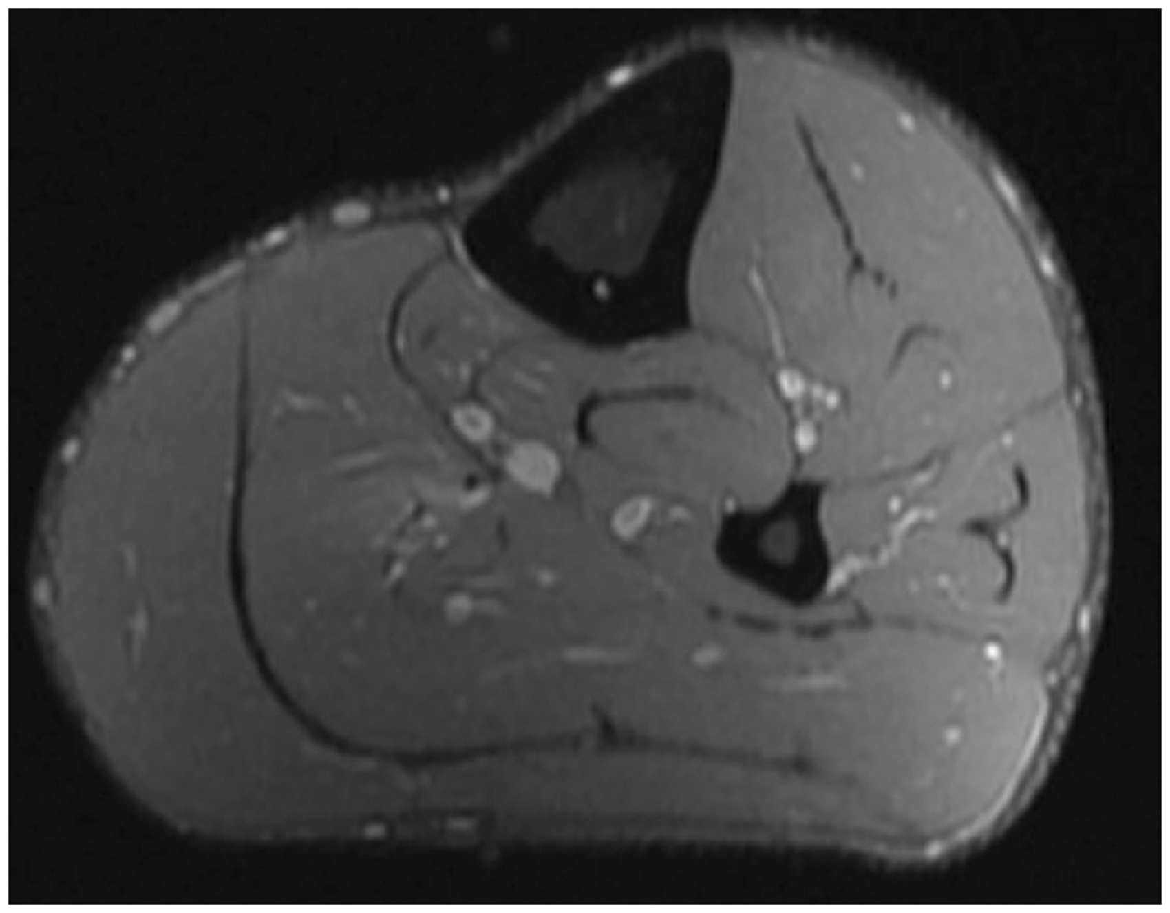
Axial PD fat sat image. High signal around the posterior and distal medial intramuscular aponeurosis of the soleus with focal discontinuity of the tendon. BAMIC type c, Prakash Grade 2. BAMIC, British Athletic Muscle Injury Classification; PD fat sat, proton density fat saturation.
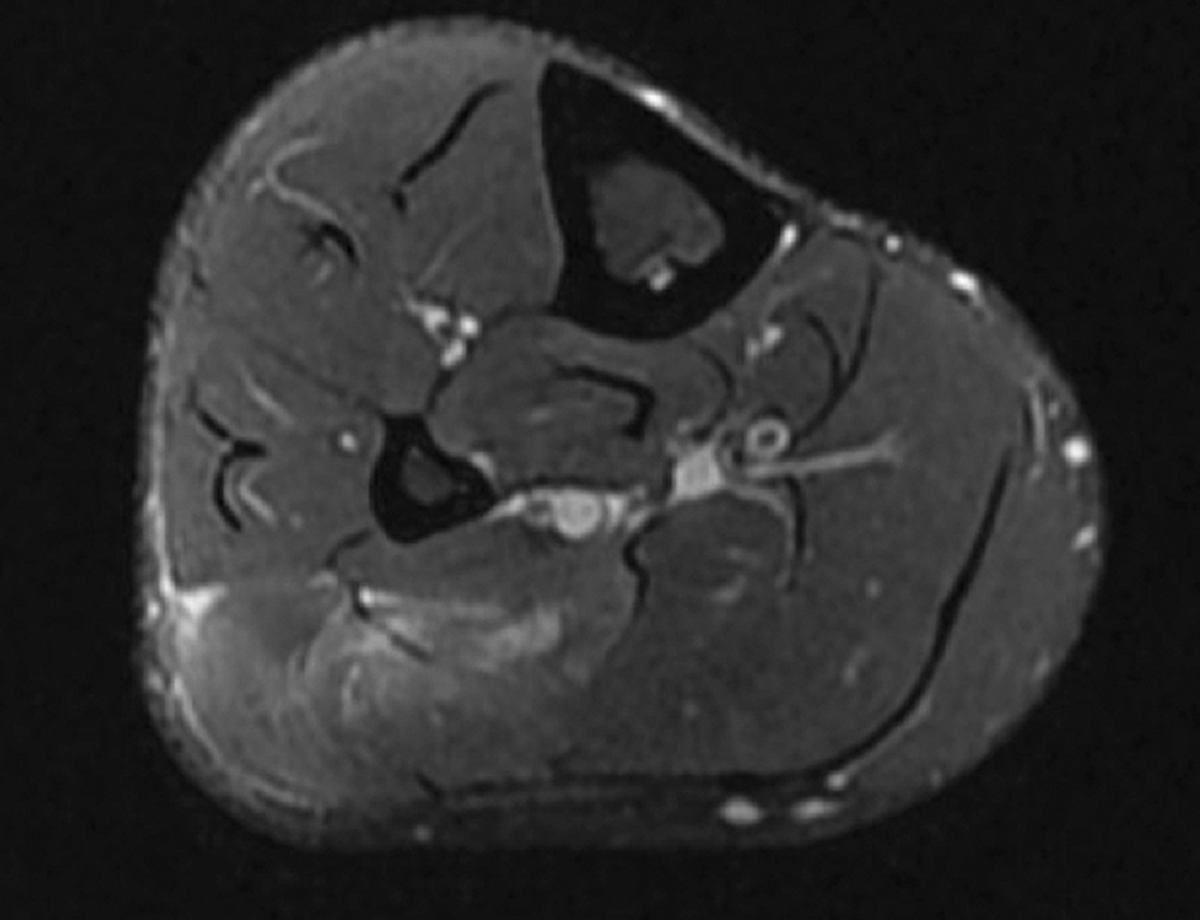
Lateral intramuscular aponeurosis of soleus injury in a professional football player. Axial Proton Density fat-sat image. There is focal disruption of the lateral intramuscular aponeurosis of soleus. BAMIC type c; Prakash Grade 2. Return-to-play 35 days; the player was injected with platelet rich plasma. BAMIC, British Athletic Muscle Injury Classification; PD fat sat, proton density fat saturation.

Axial PD fat sat image. Painful scarring of the lateral intramuscular aponeurosis of the soleus (close to the fibular attachment) in a professional rugby player. The scar denotes previous injury to the aponeurosis. The player complained of persistent deep lateral calf pain with increasing running intensity. PD fat sat, proton density fat saturation.
Conclusion
Calf injuries are common in elite athletes. The gastrocnemius, soleus, and plantaris complete the muscle group and can be injured either individually or in combination. Patterns of injury are determined by fundamental functional and anatomical differences between the muscles. Importantly, injuries involving tendinous or aponeurotic components are more predictive of injury severity as they have been proven to carry greater clinical implications with regards to rehabilitation, potential intervention, and length of time to return to play. The subsequent formation of scar tissue increases the susceptibility to re-injury due to alterations in compliance at the scar to muscle/connective tissue interface and mechanics during muscle contraction.
Examination with MRI permits more accurate measurement of the extent of structural defects and subsequent anatomical distortions, which in turn allows for more accurate assessment of injury severity. Furthermore, it evaluates for potential hematoma formation within the intermuscular planes of the calf, for example between the medial gastrocnemius muscle belly and soleus, the presence of which can be a significant complicating factor with associated prolonged recovery times.
Therefore, knowledge of the underlying anatomy and subsequent accurate interpretation of the findings carry significant clinical ramifications. As a result, MRI is vital in the comprehensive assessment of calf injuries.
Footnotes
Authors’ Note
Jonathan Williams is currently affiliated with Bristol Bears Rugby, Bristol, UK.
Declaration of conflicting interests
The author(s) declared no potential conflicts of interest with respect to the research, authorship, and/or publication of this article.
Funding
The author(s) received no financial support for the research, authorship, and/or publication of this article.
