



Abstract
Background
Pregnancy-related pain may be associated with sacroiliac joint (SIJ) changes, detectable by magnetic resonance imaging (MRI).
Purpose
To analyze the prevalence and course of SIJ MRI and clinical findings in women referred with low back pain and relate these to pregnancy.
Material and Methods
A retrospective follow-up study from a longitudinally collected cohort comprising 328 women.
Results
Women reporting debut of pain in relation to a pregnancy (PP group) tended to have a higher baseline prevalence of all investigated MRI findings, cumulated positive SIJ tests, and a potential fulfilment of the spondyloarthritis diagnosis compared to remainders. The prevalence of subchondral bone marrow edema (BME), any SIJ MRI finding, and potential fulfilment of the spondyloarthritis diagnosis were significantly higher in the PP group compared to women who had not been pregnant. In the total study group, the prevalence of ≥1 MRI finding increased over the four-year study period from 34% to 47% (P<0.001), driven by increasing prevalence of BME (25% to 32%; P=0.008) and fatty marrow deposition (FMD) (20% to 25%; P=0.020). In addition, the BME volume score increased. Over time, the PP group had persisting high prevalence of buttock pain and total MRI findings and their FMD volume score increased, but there were no between-group differences in MRI variables at follow-up.
Conclusion
Overall, the prevalence of MRI findings increased over time. Although the PP group had different clinical and SIJ MRI characteristics cross-sectional at baseline compared to remainders, longitudinal analyses revealed that these diminished over time.
Introduction
Back pain referable to the sacroiliac joints (SIJ) has been reported in 15%–30% of patients with low back pain (LBP), often occurring in relation to pregnancy, trauma, arthritis, and degenerative changes (1–5). Significant LBP and buttock pain in relation to pregnancy are reported with a prevalence of approximately 20% (6,7) and about 5% of all women who have given birth report chronic disabling pain induced in pregnancy and persisting several years after childbirth (8–10).
Postpartum SIJ findings have been studied by magnetic resonance imaging (MRI) (11–18), but it remains unclear if these findings are solely a result of pregnancies or whether women with explicit pain in relation to pregnancies may have different MRI findings.
Studies have demonstrated that postpartum SIJ MRI findings may simulate changes occurring in axial spondyloarthritis (SpA), a chronic inflammatory disease, where subchondral bone marrow edema (BME), sclerosis, erosions, and fatty marrow deposition (FMD) at the SIJs are characteristic findings (19). If such changes occur together with pain, there is a risk of establishing a false-positive diagnosis of SpA.
To improve the diagnostics of pregnancy-related pain conditions including differentiation from SpA, further knowledge regarding abnormal SIJ MRI findings in women in relation to pregnancies is needed.
In a large cohort, the Spines of Southern Denmark (SSD), consisting of 1037 patients referred with LBP, SIJ MRI changes were significantly more frequent in women than in men, mainly due to significantly increased frequencies of subchondral sclerosis and FMD. Subchondral BME and erosions were also more frequent in women, though not statistically significant (20). The reason for this gender difference may be due to mechanical strain on the SIJ in pregnancies, but this has not been explored.
Thus, the aims of the present study were to analyze the prevalence and course of SIJ MRI findings and concomitant clinical features during a four-year follow-up period in women of the SSD cohort and relate these findings to pregnancies and pain elicited by pregnancy.
Material and Methods
At baseline, the SSD cohort consisted of 1037 patients (660 women; age range = 18–40 years) referred to the Spine Centre of Southern Denmark from general practice with LBP persisting for 2–12 months and insufficient clinical response to conservative treatment. A detailed description of the baseline cohort has been published elsewhere (20,21). In brief, the cohort was recruited between March 2011 and October 2013, and all participants were invited by letter to participate in a follow-up study four years later. A total of 604 patients (58%) were included in the follow-up study; of them, 328 were women and they constitute the present study sample (Fig. 1).
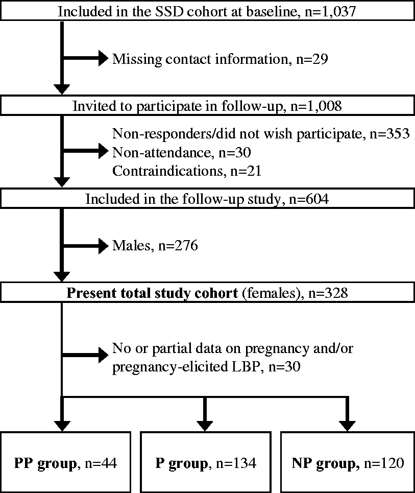
Flow diagram of patients included and excluded from baseline to follow-up and the formation of the three study groups. *the Spines of Southern Denmark. LBP, low back pain; NP, women who had never been pregnant; P, women that had been pregnant but did not report pain debut in relation to a pregnancy; PP, women reporting pain debut in relation to a pregnancy.
At baseline and follow-up, all included patients underwent MRI of the SIJs and the entire spine. Participants filled in a questionnaire including items on back and leg pain intensity (22), buttock pain, activity limitation (23), general health (24), and characteristics of inflammatory back pain (25). At baseline, participants answered a question regarding pain initiation in connection with a pregnancy, which at follow-up was supplemented by a question on history of previous pregnancies. The following three groups were formed: PP = women reporting debut of pain in relation to a pregnancy (n = 44); P = women who had been pregnant but did not report debut of pain in relation to a pregnancy (n = 132); and NP = women who had never been pregnant (n = 120).
At baseline, a clinical examination including the presence or absence of clinical features encompassed in the Assessment of SpondyloArthritis international Society (ASAS) criteria for SpA (26) and three SIJ tests (Gaenslen’s test, thigh trust’s test, and long dorsal ligament test) were performed in addition to blood sampling (human leucocyte antigen subtype B27 [HLA-B27] and high sensitivity C-reactive protein [hs-CRP].
A “potential fulfilment of SpA diagnosis” was defined as fulfilment of the ASAS definition of MRI sacroiliitis at baseline and one or more clinical or serological SpA features at baseline (the imaging arm of the ASAS criteria for SpA) (26).
The study was approved by the Regional Scientific Ethics Committee for Southern Denmark (reference number S-20102000-58), the Danish Data Protection Agency, and was conducted in accordance with the Declaration of Helsinki II and subsequent revisions. Approval from the institutional review board was granted. Before inclusion, patients gave written informed consent for research use and publication of their deidentified data.
MRI procedure
The scan protocols have been published previously (20). In brief, MRI of the SIJ and entire spine was performed with a 1.5-T MRI System (Philips Achieva, Best, The Netherlands) using a spine coil. Only data related to the SIJ were used in the present study. The following sequences were obtained at baseline and follow-up: semi-coronal T1-weighted and T1-fat saturated sequences; and semi-axial T2-weighted short tau inversion recovery (STIR) sequences. The same MRI scanner and MRI protocol were used at baseline and follow-up. Three experienced musculoskeletal radiologists evaluated the MRI images at baseline (AGJ, AZ and NE) and two performed the follow-up evaluations (AGJ and AZ). All readings were blinded to clinical information except patient’s age and gender; follow-up MRI evaluations were blinded to the findings at baseline.
MRI variables used in the data analyses
The following MRI findings were assessed: subchondral BME; FMD; sclerosis; and erosions. According to a previously described method of evaluation of the SIJs (27), each joint was subdivided into four osseous locations: the cartilaginous and ligamentous part of the iliac and the sacral bones, respectively (eight regions in total). In each SIJ region, the presence of each finding was divided into four grades (0 = absent, 1 = slight: <25% of the subchondral/subcortical bone area, 2 = moderate: 25 ≥ 50% of the area, and 3 = severe: ≥50% of the area). All lesions were only scored if a minimum of two lesions were seen on a single SIJ slice, or one lesion in at least two consecutive slices. In addition, sclerosis was only scored if extending ≥5 mm below the joint facet.
The maximal total sum score (volume score) for sclerosis and erosions was 24. For both BME and FMD, a depth score of 1 was added if the BME/FMD extended ≥ 1 cm below the joint surface and covered ≥ 1cm2. The maximal total BME and FMD sum + depth score (volume score) was thus 32.
Agreement within and between observers of the MRI evaluations has been tested in a previous study (28). All SIJ changes had kappa values > 0.6 for inter- and intra-observer agreement, except erosions and FMD depth score with inter-observer kappa values of 0.57 and 0.52, respectively (28).
Statistical analysis
Questionnaires and results of clinical and MRI evaluations were entered directly into a web-based registry (SpineData) (29) and analyzed using the STATA 15.0 (StataCorp, College Station, TX, USA).
Demographic and clinical characteristics were reported either as proportions for binary variables; continuous and ordinal variables were reported as medians with interquartile ranges (IQR). MRI variables were tabulated both as proportions (sum scores > 0) and average values of sum scores and sum + depth scores.
Comparison from baseline to follow-up within paired groups was tested using McNemar’s exact test for binary variables and the Wilcoxon signed rank test for ordinal and continuous variables. Cross-sectional comparison of baseline and follow-up characteristics between unpaired groups were tested using a two-way proportion test for binary variables and the Mann–Whitney rank sum test for ordinal and continuous variables.
P values < 0.05 were considered statistically significant.
Results
Baseline analyses
The flow chart from baseline to follow-up and reasons for dropout are shown in Fig. 1. Details on dropouts from baseline to follow-up are published elsewhere (30).
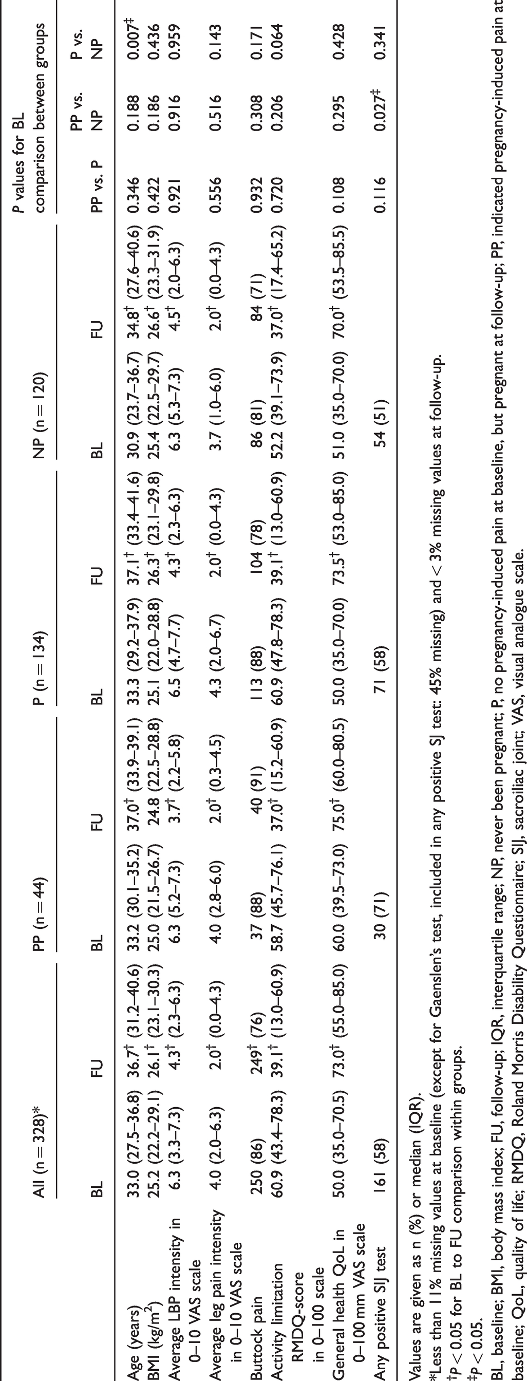
Table 1 shows the demographic and clinical characteristics at baseline and follow-up in the total cohort and the three subgroups: PP; P; and NP. There was no statistically significant difference at baseline in age, body mass index (BMI), average LBP and leg pain, buttock pain, activity limitation, and general health between the three groups, except that the NP group was significantly younger compared with the P group (P = 0.007).
Clinical characteristics.
Values are given as n (%) or median (IQR).
*Less than 11% missing values at baseline (except for Gaenslen’s test, included in any positive SJ test: 45% missing) and < 3% missing values at follow-up.
†P < 0.05 for BL to FU comparison within groups.
‡P < 0.05.
BL, baseline; BMI, body mass index; FU, follow-up; IQR, interquartile range; NP, never been pregnant; P, no pregnancy-induced pain at baseline, but pregnant at follow-up; PP, indicated pregnancy-induced pain at baseline; QoL, quality of life; RMDQ, Roland Morris Disability Questionnaire; SIJ, sacroiliac joint; VAS, visual analogue scale.
A least one positive SIJ test occurred in 58% of all study participants. Prevalence was 71%, 58%, and 51% in the PP, P, and NP groups, respectively, but there was only a statistically significant difference between the PP and NP groups (P = 0.027).
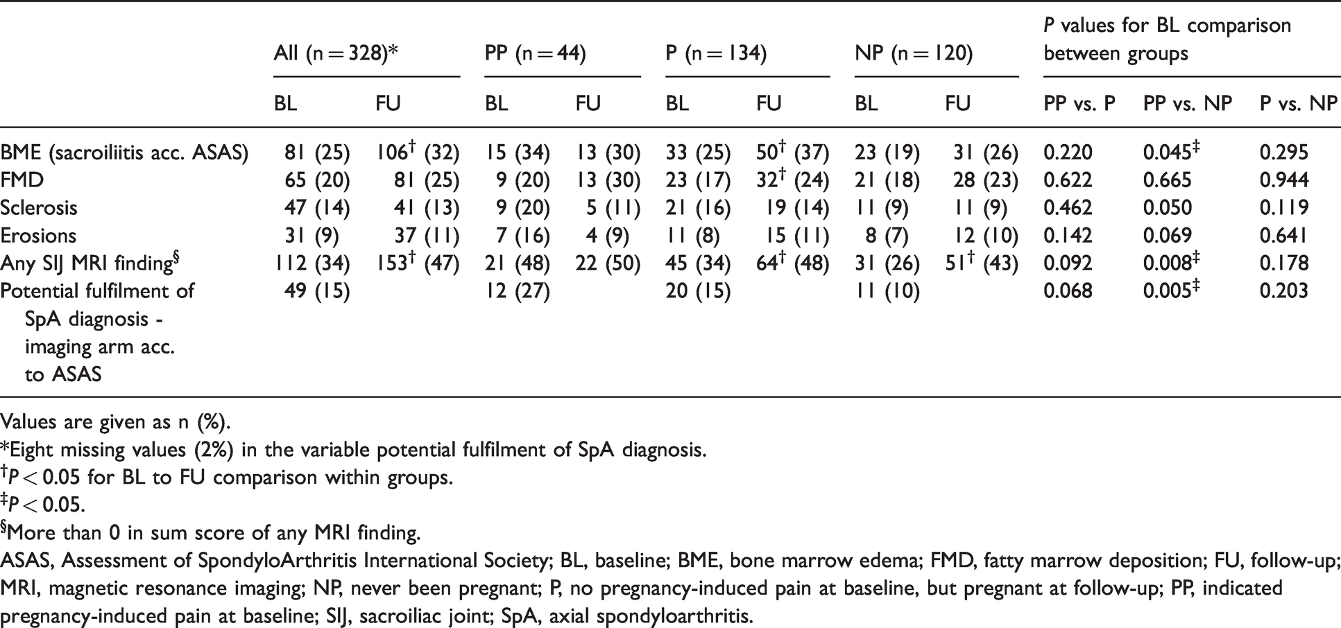
Table 2 shows baseline prevalence of MRI findings and the potential fulfilment of the SpA diagnosis. As visualized in Fig. 2, the PP group compared with the two other groups, had a tendency towards higher baseline prevalence of all MRI variables, any SIJ MRI findings, cumulative positive SIJ tests, and potential fulfilment of a SpA diagnosis according to ASAS imaging arm. The difference in baseline prevalence of BME (sacroiliitis according to ASAS), any SIJ MRI finding, and the potential fulfilment of the SpA diagnosis were significantly higher in the PP group compared with the NP group (P = 0.045, P = 0.008, and P = 0.005, respectively). No other results were statistically significant.
Prevalence of SIJ MRI findings and potential diagnosis of SpA.
Values are given as n (%).
*Eight missing values (2%) in the variable potential fulfilment of SpA diagnosis.
†P < 0.05 for BL to FU comparison within groups.
‡P < 0.05.
§More than 0 in sum score of any MRI finding.
ASAS, Assessment of SpondyloArthritis International Society; BL, baseline; BME, bone marrow edema; FMD, fatty marrow deposition; FU, follow-up; MRI, magnetic resonance imaging; NP, never been pregnant; P, no pregnancy-induced pain at baseline, but pregnant at follow-up; PP, indicated pregnancy-induced pain at baseline; SIJ, sacroiliac joint; SpA, axial spondyloarthritis.
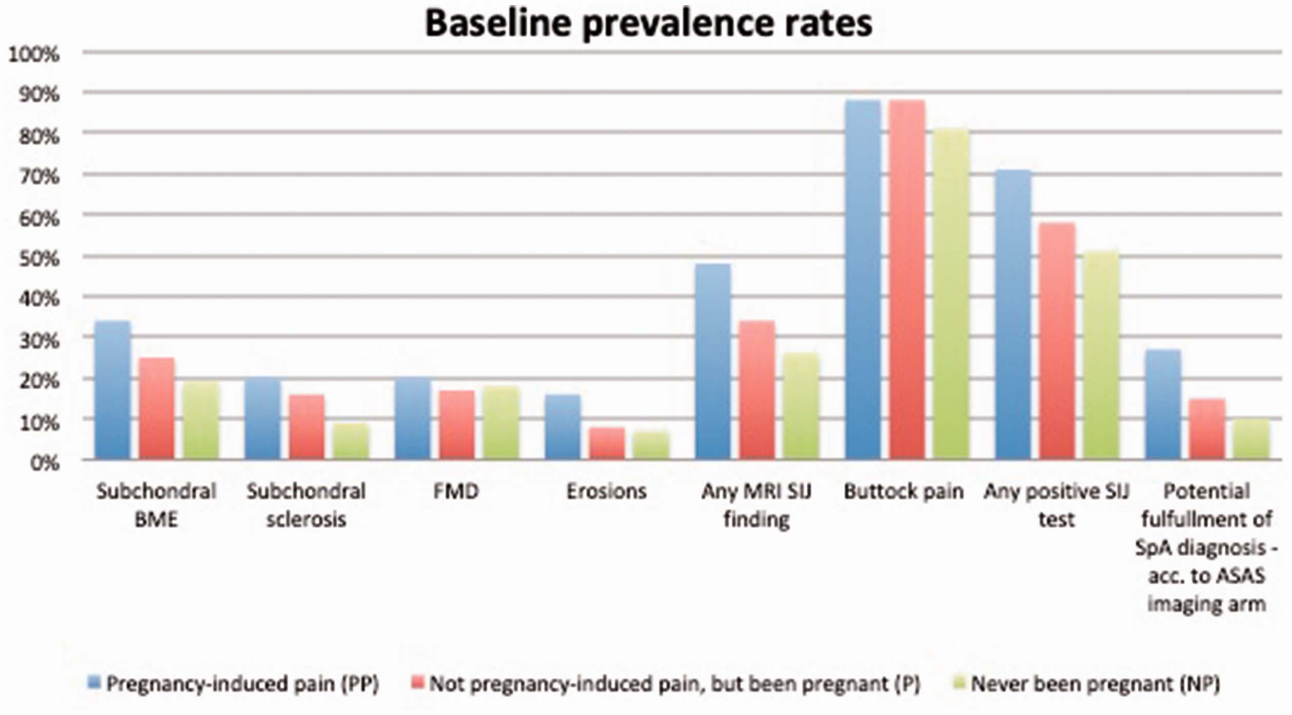
Between-group baseline comparison of prevalence of MRI findings, potential diagnosis of SpA, and clinical variables. ASAS, Assessment of SpondyloArthritis International Society; BME, bome marrow edema; FMD, fatty marrow deposition; SIJ, sacroiliac joint; SpA: axial spondyloarthritis.
Longitudinal and follow-up analyses
In the total study cohort, the following statistically significant changes from baseline to follow-up were observed regarding the clinical parameters (Table 1): increased BMI, decrease in average LBP and leg pain intensity, as well as the proportion of patients reporting buttock pain, along with improved general health and activity limitation.
The same results were evident in the three subgroups, except the prevalence of buttock pain and median BMI that remained unchanged in the PP group (BMI: P = 0.270; buttock pain: P = 1.000) compared to a statistically significant increase of median BMI (P < 0.001 in both the P and NP groups) and a tendency of decreasing prevalence of buttock pain in the remaining groups (P group: P = 0.050; NP group: P = 0.098). At follow-up, the PP group had a statistically significantly higher prevalence of buttock pain compared to the NP group (P = 0.007) and the P value between the PP and P group approached statistical significance (P = 0.052). The PP group also had a statistically lower BMI at follow-up compared to the NP group (P = 0.036). There were no other between-group differences in the clinical parameters at follow-up (data not shown).
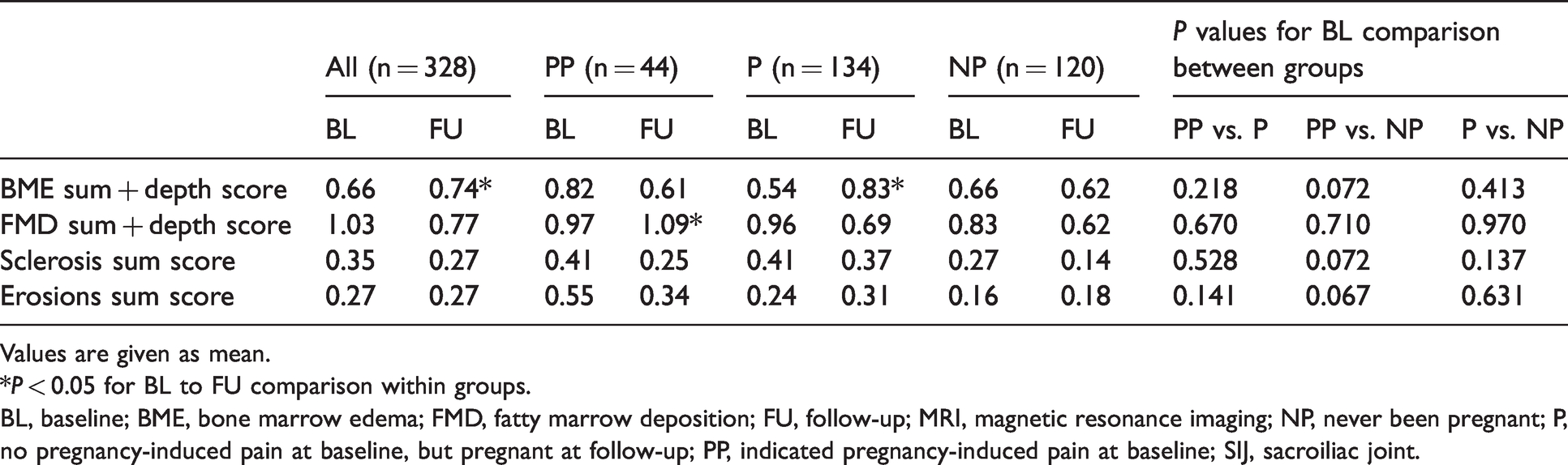
Over time, the total study cohort had a statistically significant increase in any SIJ MRI finding driven by an increased prevalence of BME (sacroiliitis according to ASAS) from 25% to 32% (P = 0.008) and of FMD from 20% to 25% (P = 0.020). The average BME sum + depth score also increased (0.66 to 0.74; P = 0.024), whereas the FMD sum + depth score remained unchanged (1.03 to 0.77; P = 0.604) (Table 3). The erosion and sclerosis prevalence rates and sum scores remained unchanged.
SIJ MRI: morphological analyses.
Values are given as mean.
*P < 0.05 for BL to FU comparison within groups.
BL, baseline; BME, bone marrow edema; FMD, fatty marrow deposition; FU, follow-up; MRI, magnetic resonance imaging; NP, never been pregnant; P, no pregnancy-induced pain at baseline, but pregnant at follow-up; PP, indicated pregnancy-induced pain at baseline; SIJ, sacroiliac joint.
In the PP group, the MRI prevalence rates remained statistically unchanged, whereas a statistically significant increase in the FMD sum + depth score was observed (Table 3). The P and NP groups changed in accordance with the overall group, but only the increase in “any SIJ MRI finding” in the NP group was statistically significant. There was no statistically significant difference between groups at follow-up when comparing prevalence, sum, and sum + depth scores (data not shown). Fig. 3 illustrates the evolution of the MR findings in a patient from the PP group.
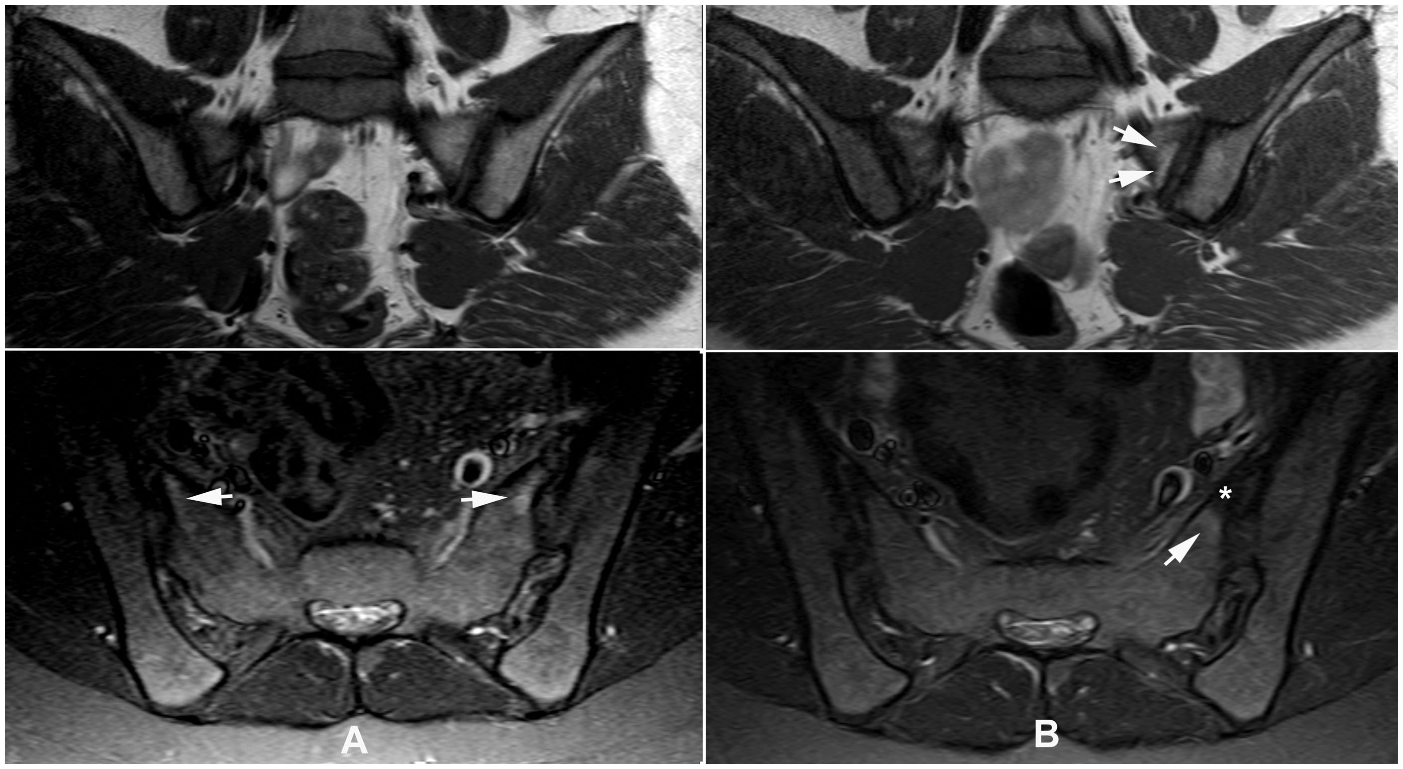
MRI of the sacroiliac joints in a 24-year-old woman reporting onset of LBP in relation to pregnancy. (a) Baseline MRI, semi-coronal T1 (upper image) and semi-axial STIR show bilateral subchondral edematous changes (BME) (arrows). (b) Four years later, semi-coronal T1 (upper image) and semi-axial STIR show disappearance of edema on the right side and regression on the left side, where there is persistence of slight edema (arrow) peripherally to a low intensity area on the STIR image compatible with fat deposition (FMD) (*). The fat deposition is more clearly visualized on the T1-weighted image (arrows). BME, bone marrow edema; FMD, fatty marrow deposition; LBP, low back pain; MRI, magnetic resonance imaging; STIR, short tau inversion recovery.
There was no exact number of patients with a final diagnosis of SpA. However, at the end of the follow-up period, 15 (5%) in the total study sample (25 missing values) received biological therapy; four of them were women in the PP group (9% of the PP group).
Anatomical distribution
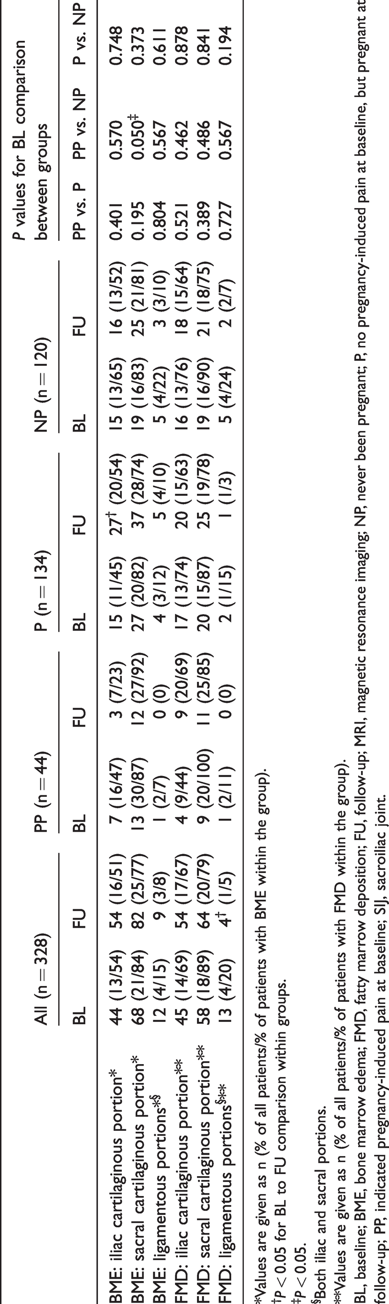
Tabulating the BME and FMD lesions at baseline in the iliac/sacral cartilaginous and ligamentous locations (Table 4) demonstrated that all groups had the highest BME and FMD prevalence in the sacral cartilaginous regions. It is worth noting that BME and FMD were also observed in the ligamentous portions of the SIJs. Over time, the highest prevalence remained located to the sacral cartilaginous portions, but a statistically significant increase of BME in iliac cartilaginous portions in the P group was observed (P = 0.036).
SIJ MRI: anatomical distribution of BME and FMD.
*Values are given as n (% of all patients/% of patients with BME within the group).
†P < 0.05 for BL to FU comparison within groups.
‡P < 0.05.
§Both iliac and sacral portions.
**Values are given as n (% of all patients/% of patients with FMD within the group).
BL, baseline; BME, bone marrow edema; FMD, fatty marrow deposition; FU, follow-up; MRI, magnetic resonance imaging; NP, never been pregnant; P, no pregnancy-induced pain at baseline, but pregnant at follow-up; PP, indicated pregnancy-induced pain at baseline; SIJ, sacroiliac joint.
Discussion
During the four-year study period, the total study group of 328 women referred with LBP reported a decrease in all pain-related questions as well as an improved activity level and general health; this is expected four years after referral. Surprisingly, this reported relief of symptoms was not reflected in the SIJ MRI findings at follow-up. On the contrary, the prevalence of women with at least one finding increased significantly; in particular, a significantly increased prevalence was seen for BME and FMD. However, in the morphological analysis, only the BME sum + depth score increased significantly (Table 3). The explanation for the increased prevalence in MRI findings but relief of symptoms is unknown but may partly be explained by age- or strain-related degeneration supported by the significant increase in BMI, which has also been suggested and demonstrated in previous studies (20,21,31–33).
At baseline and during the study period, women reporting pregnancy-induced LBP (the PP group) differed compared to the remaining study participants. Clinically, the PP group did not experience an increase in BMI and their high prevalence of buttock pain persisted; additionally, they had a tendency towards a higher baseline prevalence of cumulated positive SIJ tests. Concerning MRI, they had a tendency to a higher baseline prevalence of all measured lesions, including the potential fulfilment of the SpA diagnosis, compared to the other participants. Interestingly, the observed differences regarding MRI prevalence and volume scores along with the clinical parameters seemed to diminish during the study period with no statistically significant differences between groups at follow-up, except a higher follow-up prevalence of buttock pain and a lower BMI between the PP and NP group.
Previous studies have investigated SIJ MRI findings in relation to pregnancy and the postpartum period (11–18), but only a study from Renson et al. (18) was longitudinal. The detected BME prevalence was in the range of 21%–80% (13–18), with reports of higher prevalence in the early compared to the later postpartum period (17,18). The majority of previous studies have not succeeded in finding a detectable relation between pelvic pain and/or LBP in relation to pregnancy and presence of abnormal MRI findings (13,15,18). However, Hoballah et al. (17) reported that the presence of postpartum BME was associated with a history of LBP or pelvic pain and Seven et al. (16) found higher BME volume scores in women reporting pregnancy-related pain compared to women without pregnancy-related pain, but it was not reported whether this difference was statistically significant.
In the PP group, 34% had BME at baseline compared with 25% in the P group; at follow-up, the numbers were 30% and 37%, respectively. These results are comparable with the previously reported prevalence of pregnancy-related positive MRI according to ASAS, in the range of 12%–64% (14,17,18), as our definition of BME was similar. Our finding of a high baseline prevalence of the potential fulfilment of the SpA diagnosis in the PP group supports that clinicians should be cautious applying a SpA diagnosis to women reporting pain in relation to pregnancy, which may mimic SpA.
The persistent high prevalence of buttock pain in the PP group and the simultaneously unchanged prevalence of BME accompanied by an increasing FMD sum + depth score (Fig. 3) may indicate that the persisting high prevalence of buttock pain could be explained by ongoing subchondral inflammation in accordance with the observation of inflammatory BME areas transforming into FMD over time (34).
Other reasons for persisting buttock pain could be musculotendinous conditions or the occurrence of pain causing anatomical variations such as accessory SIJs and a lumbosacral transitional vertebra (35).
The finding of sacral predominance of BME and FMD (Table 4) is in contrast to the reported predominance in the lower iliac regions in two previous studies (13,14). This may partly be due to the use of semi-axial STIR in the current study compared to semi-coronal slice orientation in previous studies. A sacral location of BME may indicate predominantly strain-related changes. Thus,
We also demonstrated MRI findings in the ligamentous portions of the SIJs (Table 4). Only one Chinese study has previously reported the presence of such findings in postpartum women in a retrospective cross-sectional study of 27 women with symptomatic osteitis condensans ilii, often elicited by pregnancy (37).
With the current clinical and MRI results, we believe that there is a true difference between the three groups at baseline. However, the differences between groups seemed to vanish over time except for persisting buttock pain and unchanged BMI in the PP group compared to the remaining groups. The reason for this is unknown. Studies including histopathological characteristics of the SIJ in both symptomatic and non-symptomatic postpartum women would be of great interest, as the underlining cause of the presence of subchondral BME remains unclear; is it solely related to mechanical stress of the SIJs or is it partly a true inflammation?
With a study sample of 328 women and 656 MRI scans, the present study is, to our knowledge, the largest longitudinal study of pregnancy-related SIJ MRI findings, which is a major strength. However, the retrospective study design has limitations. We might have seen more differentiation between groups with further statistically significant results if we had had data on the full obstetric history, including knowledge of time delay between these events and the MRI. Furthermore, we did not have data on pregnancies and births during the follow-up period. Another limitation may be ascribed to the relatively heterogeneous cohort of patients with LBP including an unknown, but probably low, number of patients with SpA.
In conclusion, we have demonstrated that women reporting pregnancy-induced pain had different clinical and SIJ MRI characteristics at baseline compared to the remaining study participants; however, at four-year follow-up the differences diminished.
Footnotes
Acknowledgments
The authors thank Dr. Niels Egund for his contribution to the interpretation of MRI baseline findings.
Declaration of conflicting interests
The author(s) declared no potential conflicts of interest with respect to the research, authorship, and/or publication of this article.
Funding
The author(s) received no financial support for the research, authorship, and/or publication of this article.
