



Abstract
Background
Effect of decreased injection flow rate of contrast agent at the same iodine dose and delivery rate on aortic enhancement has not been clearly elucidated.
Purpose
To evaluate the effect of decreased injection flow rate of contrast agent on aortic peak enhancement in a dynamic flow phantom and on aortic enhancement in clinical dynamic 80-kVp computed tomography (CT) with contrast dose reduction.
Material and Methods
In the dynamic flow phantom experiment, the effect of a decreased injection flow rate at the same total iodine dose and delivery rate on simulated aortic peak enhancement was evaluated. In the clinical retrospective study, we searched 312 patients with renal dysfunction who underwent an 80-kVp abdominal dynamic CT with 40% reduction of contrast agent from a standard 120-kVp protocol and measured the aortic enhancement at the level of the hepatic hilum. Independent predictors for aortic enhancement were determined by multiple linear regression analysis, and after adjustment of significant predictors, independent variables for acquiring optimal aortic enhancement, ≥300 HU, were determined by multiple logistic regression analysis.
Results
In the phantom experiment, decreased flow rate showed a significant but small descent effect (6%–9%) on simulated aortic peak enhancement. In the multiple linear regression analysis, only age was an independent predictor of aortic enhancement; there was no independent predictor for optimal age-adjusted aortic enhancement of ≥300 HU.
Conclusions
Decreased injection flow rate had a small influence on aortic enhancement in vitro but had no significant effect on the aortic enhancement in clinical dynamic 80-kVp CT.
Introduction
Although the risk of acute kidney injury after intravenous injection of an iodinated contrast agent during computed tomography (CT) imaging has remained a topic of debate (1), administration of the lowest dose of contrast agent while maintaining diagnostic image quality is recommended (2). The potential risk of amplifying double-stranded DNA damage and radiation absorption by tissues due to the presence of an iodinated contrast agent has been proposed in previous studies (3,4), and the level of that risk also depends on the administered dose of contrast agent (5).
One of the effective ways to reduce the amount of iodinated contrast agent in clinical CT is the use of a low tube voltage, such as 80–100 kV peak (kVp) relative to the standard 120 kVp, because a low tube voltage peak moves the peak energy spectra of CT close to the iodine k-edge, which causes greater X-ray attenuation by iodine and improves contrast enhancement. However, a drawback of using a low tube voltage is increased image noise and image degradation because conventional filtered back projection (FBP) reconstruction is based on some mathematical assumptions of the CT system that differ from the actual CT acquisition situations, for which iterative reconstruction or deep-learning-based reconstruction has been introduced (6,7). Using CT of 80–100 kVp, the contrast agent dose can be effectively reduced by 38%–60% in coronary CT angiography (8,9), 25% in pulmonary CT angiography (10), 43% in cerebral CT angiography (11), and 40%–50% in abdominal dynamic CT (12–14) while maintaining image quality.
In general, a fixed injection duration of 25–35 s is more important than an increasing injection rate for maintaining the optimal time window for aortic peak enhancement because the optimal injection duration provides a “right shoulder” on the time density curve (TDC), while an insufficient injection duration leads to a “sloping shoulder” (15,16). Thus, effective reduction of the contrast agent in low tube voltage CT with a fixed injection duration leads to a low injection rate, which would be prominent in patients with a low body weight (BW). On the basis of previous evidence of a positive linear correlation between an increased injection flow rate and aortic peak enhancement in the range of 3–5 mL/s in a phantom experiment (15) and a low injection flow rate (1.9 mL/s) showing a negative effect on the aortic peak enhancement in comparison with that of high flow rate in an animal experimental study (17), we hypothesized that a low injection rate of <2 mL/s and close to 1 mL/s in abdominal dynamic 80-kVp CT with contrast dose reduction has a substantial negative effect on aortic enhancement.
Thus, the aim of the present study was to evaluate the effect of decreased injection flow rate on aortic peak enhancement by a dynamic flow phantom and on the aortic enhancement in clinical dynamic abdominal 80-kVp CT with contrast agent dose reduction.
Material and Methods
This phantom and retrospective clinical study was approved by our institutional review board, and the requirement for informed patient consent was waived.
Dynamic-flow phantom experiment
A dynamic-flow phantom was prepared (Fig. 1), and the contrast agent was injected by a power injector into an acrylic cistern and ejected to an acrylic tube simulating the aorta (internal diameter = 17 mm), which was embedded in an acrylic column containing water, by a pump at a pulsating flow of 60 beats per minute and a flow rate of 4.8 L/min, representing the cardiac output of a person with 50 kg BW. The contrast agent was accumulated in an acrylic tank filled with 2 L of water, which represented tissue storage blood, and then returned to a simulated inferior vena cava. A single-level CT scan was performed at the center of the acrylic column; the CT scan was started with the contrast injection and repeated 30 times at 2-s intervals (Fig. 1). CT scans were performed using a 320-row multidetector CT scanner with a fixed tube current of 100 mA and slice thickness of 1 mm. Iohexol (300 mgI/mL; GE Healthcare Japan, Hino, Japan) was used as the contrast agent in all experiments and injected at a fixed injection duration of 30 s; the acquired image was reconstructed by FBP. All experiments were repeated five times. A radiologist placed a region of interest (ROI) at the center of the acrylic tube while avoiding the acrylic tube wall on CT images and created the TDC, in which aortic peak enhancement was determined as the peak of TDC.
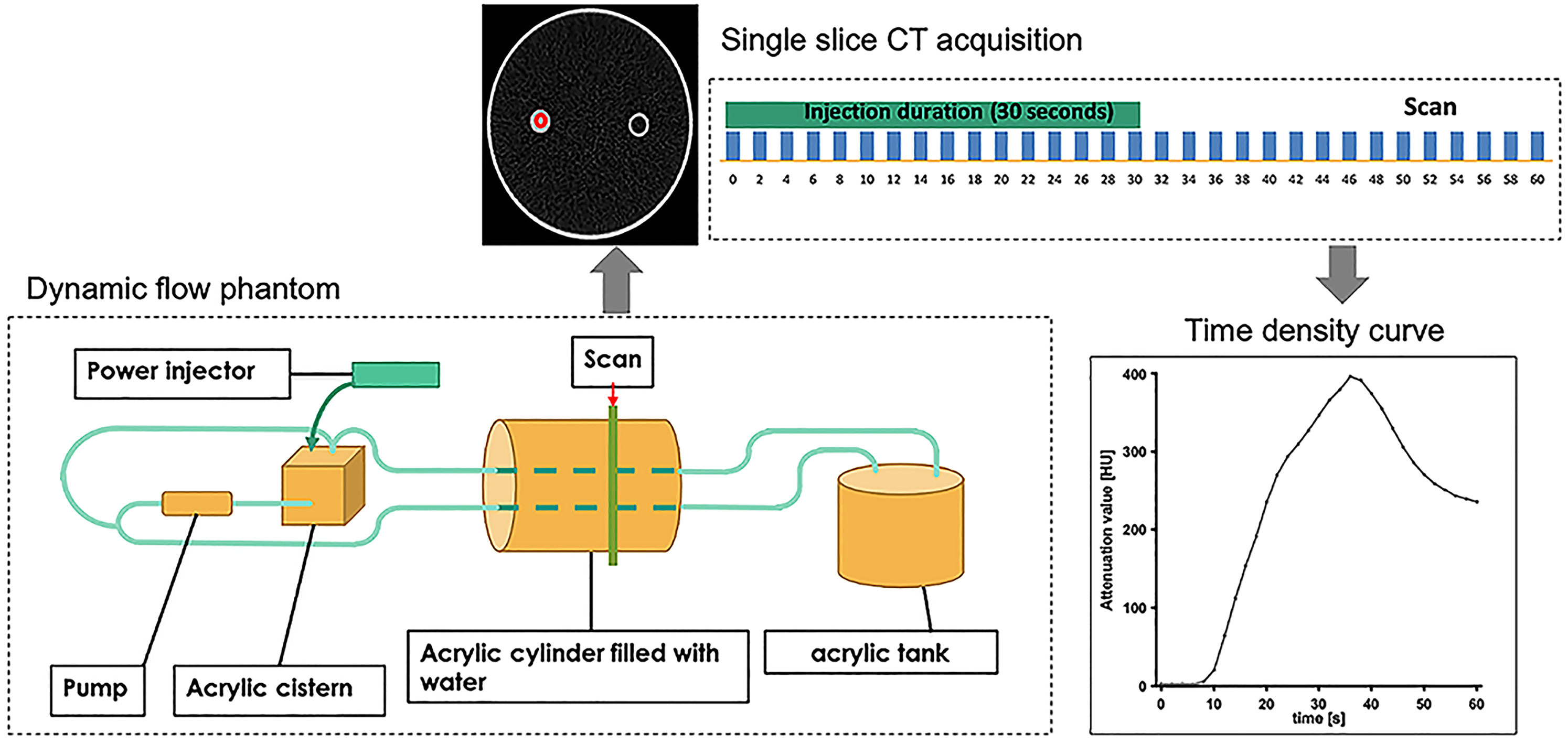
Dynamic flow phantom experiment. The contrast agent was injected by a power injector into an acrylic cistern and ejected by a pump to an acrylic tube simulating the aorta embedded in an acrylic column containing water. The contrast agent was accumulated in an acrylic tank filled with 2 L of water and then returned to a simulated inferior vena cava. A single-level CT scan was started at the center of the acrylic column at the same time as the contrast injection and repeated 30 times at 2-s intervals. A TDC was created, in which the aortic peak enhancement was determined as the peak of the TDC. CT, computed tomography; TDC, time density curve.
To evaluate the reduction in flow rate at the same total iodine dose and iodine delivery rate, two protocols were performed. In the first protocol, aortic peak enhancement at a total iodine dose of 90 mL injected at a flow rate of 3.0 mL/s and scanned at 120 kVp was compared with that of a total iodine dose of 54 mL (40% reduction) injected at a flow rate of 1.8 mL/s and scanned at 80 kVp, where the expected total iodine dose was considered to be equal between the two conditions (14,18) (protocol 1). In the second protocol, a total iodine dose of 30 mL was injected at a flow rate of 1 mL/s (no dilution), 2 mL/s with twofold saline dilution (total volume = 60 mL), or 3 mL/s with threefold saline dilution (total volume = 90 mL) and scanned at 120 kVp, and the findings at all three flow rates were compared (protocol 2).
Study population
To evaluate the effects of decreased injection flow rate of the contrast agent on aortic enhancement in vivo, we conducted a retrospective search for upper abdominal dynamic CT examinations performed with a reduced contrast agent dose in our radiology archive. From January 2014 to December 2016, a total of 317 patients with estimated glomerular filtration rate <45 mL/min/1.73 mm2 had received a 40% lower contrast agent dose from the reference dose of 600 mgI/kg and were scanned at 80-kVp CT in combination with an iterative reconstruction algorithm because of renal dysfunction (14). This strategy was applied only to patients with BW <70 kg because of the image degradation and increased radiation exposure in 80-kVp CT acquisition performed for patients with a large body mass in our clinical experience. Five patients were excluded from the cohort because of severe image degradation with high image noise, which impeded image analysis. Indications for upper abdominal dynamic CT were heterogeneous and included surveillance or examination of hepatic tumors, evaluation of the gallbladder, bile duct, pancreas, and renal diseases. During the study period, one of three iodine concentrations of 240, 300, and 370 mgI/mL (Iohexol or Iopamidol) were used in a weekly rotation. The contrast agent was injected over a fixed duration of 30 s without a saline chaser after non-contrast CT acquisition, and the arterial, portal venous, and delayed phases were acquired after 40 s, 70 s, and 180 s of the start of injection, respectively. Dynamic helical CT scans were performed using a 320-raw multidetector CT scanner (Aquilion-One; Cannon Medical Systems, Otawara, Japan) with a detector configuration of 64 × 0.5 mm, automated tube current adjustment, field of view of 350 × 300 mm, 13 Hounsfield units (HU) of preset image noise, and slice thickness of 5 mm with adaptive iterative dose reconstruction 3D-mild (AIDR 3D; Cannon Medical Systems, Otawara, Japan).
A ROI with an area of 100–150 mm2 was placed on the aorta at the level of the porta hepatis of the liver on both non-contrast and arterial phase images by a radiologist, and the difference in the aortic attenuation values between the two phases was determined as the aortic enhancement.
The patient's age, sex, BW, height, total iodine dose, injection flow rate, iodine concentration, and radiation exposure (CT dose index [CTDIvol]) were retrieved from the radiation information system.
Statistical analysis
Aortic peak enhancement in the phantom experiment was presented as mean and standard deviation (SD) and compared between the two conditions in protocol 1 by an unpaired t-test and among the three conditions in protocol 2 by an unpaired one-way analysis of variance and post-hoc Tukey's test. In the clinical study, a multiple linear regression analysis was performed to identify independent predictors of aortic enhancement, where the patient's age, sex, BW, height, total iodine dose, injection flow rate, iodine concentration, CTDIvol, and CT attenuation value of the aorta on non-enhanced images were included. Because only age was confirmed as an independent predictor, we adjusted the aortic enhancement in relation to age and determined the independent variables for acquiring optimal aortic enhancement by a multiple regression analysis, with values ≥300 HU considered as optimal aortic enhancement (16,19). In the sub-analysis, age-adjusted aortic enhancement ≥300 HU and <300 HU were plotted against injection flow rate and BW to figure out its distributions, where cutoff values for acquiring aortic enhancement ≥300 HU of injection flow rate and BW were calculated by receiver operating characteristic (ROC) analysis. Statistical analysis was performed using Prism 9 (GraphPad Software, San Diego, CA, USA).
Results
Dynamic flow phantom experiment
In protocol 1, aortic peak enhancement was observed 36 s after start of contrast agent injection with a total iodine dose of 90 mL, flow rate of 3.0 mL/s, and a 120-kVp acquisition, and at 38 s with a total iodine dose of 54 mL, flow rate of 1.8 mL/s, and 80-kVp acquisition. The TDC of both settings is shown in Fig. 2a. The mean aortic peak enhancement at 3.0 mL/s was 396 ± 0 HU and significantly higher than that at 1.8 mL/s (372 ± 0 HU; P = 0.031). In protocol 2, aortic peak enhancement was observed 36 s after start of contrast agent injection in with a total iodine dose of 30 mL and a flow rate of 1.0 mL/s, at 36 s with a total dose of 60 mL and a flow rate of 2.0 mL/s, and at 34 s with a total dose of 90 mL and a flow rate of 3.0 mL/s (Fig. 2b). The mean aortic peak enhancement was 144 ± 0 HU at 1.0 mL/s, 150 ± 0 HU at 2.0 mL/s, and 158 ± 0 HU at 3.0 mL/s, with significant differences among the three settings (P = 0.004); in the post-hoc analysis, the difference in aortic peak enhancement between 1.0 and 3.0 mL/s was significant (P = 0.003), while those between 1.0 and 2.0 mL/s and between 2.0 and 3.0 mL/s were not statistically significant (P = 0.127 and 0.077, respectively).

TDC of the dynamic flow phantom experiments. (a) In the first protocol, TDCs of a total iodine dose of 90 mL with a flow rate of 3.0 mL/s and 120-kVp acquisition and a total iodine dose of 54 mL (40% reduction) with a flow rate of 1.8 mL/s and 80-kVp acquisition are shown, where the expected total iodine dose is equal between the two conditions. The difference in the aortic peak enhancement between the two settings is significant (P = 0.031). (b) In the second protocol, a total iodine dose of 30 mL is injected at a flow rate of 1 mL/s (no dilution), at 2 mL/s by twofold saline dilution (total volume = 60 mL), and at 3 mL/s by threefold saline dilution (total volume = 90 mL) and scanned at 120 kVp. The difference in the aortic peak enhancement among the three settings is significant, and the difference between 1.0 mL/s and 3.0 mL/s was significant (P = 0.003) in the post-hoc analysis, while other comparisons were not significant. Symbols represent the mean value of five repeated measurements. TDC, time density curve.
Clinical study
After excluding five patients, 312 patients were included in the retrospective clinical study; 240 mgI/mL was used in 99 patients, 300 mgI/mL in 128 patients, and 370 mgI/mL in 85 patients. Patient demographics, total iodine dose, injection flow rate, iodine concentration, CT dose index, and CT attenuation measurements of the aorta are summarized in Table 1. The mean aortic enhancement in all patients were 400 ± 93 HU. Multiple linear regression analysis revealed that only age was an independent predictor of aortic enhancement (P = 0.021) (Table 1). Therefore, we defined age-adjusted aortic enhancement as follows:

Patients’ demographics, aortic CT attenuation measurements, and results of multiple regression analysis for predicting aortic enhancement.

Values are given as n or mean ± SD. Parameter estimates and their 95% CIs are given in multiple linear regression analysis for predicting aortic enhancement and ORs and their 95% CIs are given in multiple logistic regression analysis for predicting age-adjusted aortic enhancement >300 HU.
*The age-adjusted aortic enhancement is defined as follows:
CI, confidence interval; CT, computed tomography; HU, Hounsfield unit; OR, odds ratio.
The mean age-adjusted aortic enhancement in all patients was 400 ± 91 HU. In the multiple logistic regression analysis, there was no independent variables for acquiring ≥300 HU of age-adjusted aortic enhancement (Table 1). Fig. 3 shows the distributions of injection flow rate and BW in patients with age-adjusted aortic enhancement <300 HU (blue circle) and ≥300 HU (red square), where the three lines represent each of the three iodine concentrations (240, 300, and 370 mgI/mL); in the ROC analysis, the cutoff values for acquiring ≥300 HU of age-adjusted aortic enhancement was 1.5 mL/s for flow rate and 37 kg for BW (0.577 and 0.625 of the area under the ROC curve, respectively); the difference in the age-adjusted aortic enhancement between flow rates of <1.5 mL/s (359 ± 85 HU; n = 28) and ≥1.5 mL/s (403 ± 91 HU; n = 284) was significant (P = 0.014) and that between BW of <37 kg (318 ± 70 HU; n = 13) and ≥37 kg (403 ± 90 HU; n = 299) was also significant (P < 0.001) but with a substantial overlap in both variables. The difference in age-adjusted aortic enhancement between the three iodine concentrations was not significant (P = 0.329).

Plot of distributions of flow rate and body weight in patients with age-adjusted aortic enhancement <300 HU (blue circle) and ≥300 HU (red square), where the three lines represent the three iodine concentrations (240, 300, and 370 mgI/mL). Vertical and horizontal dotted lines represent thresholds calculated by receiver operating characteristic analysis. HU, Hounsfield unit.
Discussion
In the era of abdominal dynamic low-voltage CT, the amount of contrast agent used can be effectively reduced by using current reconstruction technologies, resulting in a lower injection rate or injection duration, where a fixed injection duration of 25–35 s (usually 30 s) is more important than increasing the injection rate for maintaining the optimal time window for aortic peak enhancement. In terms of aortic peak enhancement, the contrast dose can be reduced by approximately 20% in the 100-kVp acquisition and 40%–50% in the 80-kVp acquisition in comparison with a standard 120-kVp CT. Thus, a decreased injection rate of <2.0 mL/s and close to 1.0 mL/s, which is rarely seen in standard 120-kVp dynamic CT, is inevitable in 80-kVp dynamic CT.
In our dynamic flow phantom experiment, the simulated aortic peak enhancement decreased by 6% with a flow rate reduction from 3.0 mL/s to 1.8 mL/s with the same expected iodine dose (protocol 1) and by 9% with a flow rate reduction from 3.0 mL/s to 1.0 mL/s with the same iodine dose (protocol 2). As seen in Fig. 2, a decreased flow rate leads to a more sloping TDC with decreased peak enhancement and delayed peak time. The decrease in aortic peak enhancement was small; however, the effects of the low flow rate on the shape of the TDC and aortic peak enhancement require consideration.
Meanwhile, in the clinical study, only age was an independent predictor of aortic enhancement, while flow rate and iodine concentration were not. The positive effect of age on aortic enhancement has not been confirmed in a previous study (20); however, the paradoxical effects of cardiac output on arterial enhancement have been proposed (21,22), and we speculate that the lower cardiac output in older patients may have been responsible for the positive effect of age on aortic enhancement in our study. Iodine concentrations of 300–370 mgI/mL had no effect on the degree of contrast enhancement in coronary CT angiography and pulmonary CT angiography (23,24), which is consistent with our clinical results. In contrast, a low flow rate of 1.9 mL/s with high iodine concentration demonstrated significantly lower aortic peak enhancement than those observed at flow rates of 2.5 and 5.0 mL/s with an identical iodine dose and iodine delivery rate in the animal experiment (17). In this study, the distributions of flow rate and BW between patients with ≥300 HU and <300 HU mostly overlapped and we found no significant effect of injection flow rate, BW, and iodine concentrations on the aortic enhancement; the discrepancy between the phantom experiment and clinical evaluation would be partly because of an insufficient statistical power from a small number of patients with flow rate and BW below the thresholds, for which further evaluation with a large number of patients is needed. When considering a potential negative effect of extremely low BW and injection flow rate on aortic enhancement on abdominal dynamic CT, where the flow rate could be adjusted while the patient's BW cannot be changed, an easy way to increase the flow rate in a fixed-injection method is to use a contrast agent with a low iodine concentration because it bulks out the total amount of contrast agent (17). In addition to a simple fixed-injection method, other approaches such as an optimized multi-bolus injection protocol (25) and the use of a saline chaser (26) are optional.
The present study has some limitations. First, the BW of patients in our study was smaller than those of patients in western countries, where an injection flow rate <1.5 mL/s is rarely used. Second, other confounding factors associated with aortic enhancement, including cardiac output (20), were not evaluated in our study. Finally, the range of iodine concentrations in the phantom experiment (100–300 mgI/mL) was different from that in the clinical study (240–370 mgI/mL), which could lead to discrepancies in the effects of flow rate and iodine concentrations between phantom and clinical studies.
In conclusion, a decreased contrast agent injection flow rate resulted in a significant but small descent effect on the aortic peak enhancement in the dynamic flow phantom experiment and had no effect on aortic enhancement in clinical abdominal dynamic 80-kVp CT with contrast-dose reduction.
Footnotes
Acknowledgements
We thank Masanori Kondo and Koji Yuba, employees of Nemoto Kyorindo Co., Ltd., for their help with the dynamic flow phantom preparation.
Declaration of conflicting interests
The author(s) declared no potential conflicts of interest with respect to the research, authorship, and/or publication of this article.
Funding
The author(s) received no financial support for the research, authorship and/or publication of this article.
