



Abstract
Background
Magnetic resonance imaging (MRI) findings after subcutaneous extravasation of gadolinium-based contrast agent (GBCA) have not been investigated in detail.
Purpose
To present the MRI findings of iatrogenic extravasation and to evaluate the characteristic findings.
Material and Methods
In this retrospective study of 16,039 patients with cancer, 11 patients had significant extravasation of macrocyclic GBCA, and 7 of the 11 had MRI of the injection site. Characteristic MRI findings as well as symptoms and changes over time were evaluated.
Results
The forearms or antecubital fossa felt cold in all seven cases, and 3 (43%) patients felt pain at the injection sites. Fat-suppressed T1-weighted images showed a mosaic pattern of the extravasate with mixed high and low signal in the subcutaneous tissue in 7 (100%) cases. Contrast enhancement of the fascia toward the proximal portion was observed in 3 (43%) cases. There was no subfascial deep extension. On T2-weighted images, GBCAs were observed as low-signal fluid collection on the fascia, with linear spread to the dermis and subcutaneous tissue. Four patients underwent daily MRI scans, all of whom had their contrast disappeared within three days.
Conclusion
Macrocyclic GBCA disappears from subcutaneous tissue quickly after extravasation and is unlikely to cause serious sequelae.
Introduction
Extravasations are less common during gadolinium-based contrast agent (GBCA) injection for magnetic resonance imaging (MRI) than during iodine-based contrast injection for computed tomography (CT), but caution should be exercised when using automatic power injectors and in elderly patients (1–4). GBCAs have the advantage over iodine-based contrast agents in that they can be administered at lower flow rates and lower doses to achieve contrast effects (1). Since there have been few reports of MRI findings of iatrogenic extravasation of GBCA (5), knowing the MRI findings of peripheral subcutaneous extravasation is important in evaluating subsequent sequelae. The aim of the present study was to present the MRI findings of iatrogenic extravasation of GBCA and to evaluate the characteristic findings.
Material and Methods
This retrospective study was approved by the institutional review board (RO20190503), and all procedures performed in this study were in accordance with the ethical standards of the institutional and national research committee and with the 1964 Helsinki declaration and its later amendments or comparable ethical standards. Written informed consent was obtained from all patients for use of clinical data in research.
Patients
Patients with significant iatrogenic extravasation were retrospectively selected among patients with cancer who underwent contrast-enhanced MRI for radiotherapy treatment planning or tumor evaluation at our institution between October 2011 and August 2021. The definition of significant extravasation is as follows: (i) MRI after contrast injection shows no visual contrast enhancement effect; and (ii) on visual examination and palpation of the injection site, all of the GBCA and saline is considered to have extravasated. The extravasation volumes in this study were small compared to those that can occur with contrast-enhanced CT. The treatment protocol was topical corticosteroids. However, if the patient's hand showed paralysis or ischemia, the patient was sent to the emergency department where emergency physicians treated them.
Contrast media injection protocol
Contrast-enhanced MRI was performed using a dual-head automatic power injector (Sonic Shot7, Nemoto, Tokyo, Japan). A peripheral i.v. cannula (20 or 22 gauge) was inserted in the antecubital or forearm area for contrast administration. For outpatients, a fresh cannula was inserted in the radiology department immediately before imaging. For inpatients, a new cannula was inserted in the ward 1–3 h before imaging, the cannula was filled with heparin, and the patient remained in bed until the time of MRI. Macrocyclic GBCA was administered at a flow rate of 4 mL/s, followed by 30 mL of saline at the same flow rate. Since perfusion images are routinely used for radiotherapy treatment planning and evaluation of treatment effects, flow rates are set higher than those of typical contrast-enhanced MRI. The maximum pressure was limited to 150 pounds per square inch (PSI). The choice of GBCA was adjusted monthly to equal frequency of use based on the amount paid to the manufacturer for contractual reasons. Gadoxetate disodium (Primovist, Eovist, Bayer, Osaka, Japan) was excluded in this study due to different injection protocols.
MRI assessment
The injection site was the antecubital fossa for three patients and the forearm for four patients. Three-dimensional fat-suppressed T1-weighted (T1) volumetric interpolated breath-hold exam (VIBE) and T2-weighted (T2) turbo spin echo (TSE) or half-Fourier single-shot turbo spin-echo (HASTE) images of the antecubital fossa or the forearm were obtained with a 3.0-T MRI scanner (MAGNETOM Skyra; Munich, Germany). Outpatients (n = 2) underwent MRI only on the day of extravasation, while inpatients (n = 5) underwent daily MRI until the GBCA disappeared. It was allowed to discontinue MRI, depending on the patient's condition. Characteristic MRI findings were evaluated independently by two certified radiologists and consensus was reached through discussions.
Results
From October 2011 to August 2021, 16,309 contrast-enhanced MRI studies were performed in patients with cancer for radiotherapy treatment planning or tumor evaluation at our institution. Iatrogenic significant extravasation was observed in 11 (0.07%) patients, and MRI of the injection site was performed in 7 (64%) patients (6 men, 1 woman; median age = 69 years; age range = 67–74 years) because treatment of the extravasation was not prioritized. All patients had stage IV advanced malignancies: rectal cancer; pancreatic cancer; prostate cancer; hepatocellular carcinoma; ureteral carcinoma; non-small cell lung cancer; and malignant lymphoma. The MRI sites were the spine (n = 2), pelvis (n = 2), pancreas (n = 1), neck (n = 1), and kidney (n = 1). All patients had received at least one first-line systemic chemotherapy before MRI. Patient characteristics, symptoms, and MRI findings are shown in Table 1. Since no linear GBCA was used during the study period, all contrast media extravasated were macrocyclic GBCAs: gadoteridol (ProHance, n = 3); gadoterate meglumine (Magnescope, n = 2); and gadobutrol (Gadovist, n = 2). During contrast and saline administration, the injector pressure never reached the threshold (150 PSI) in all seven patients. The forearm or antecubital fossa felt cold in all cases, and 3 (43%) patients felt pain at the injection site. All symptoms were relieved within three days, and no late complications were observed. No patients showed paralysis or ischemia of the hands or were sent to the emergency room for treatment by an emergency physician.
Patient characteristics, symptoms, and MRI findings.

FE, fascial enhancement; FS-T1WI, fat-suppressed T1-weighted image; Gad., gadobutrol (Gadovist); GBCA, gadolinium-based contrast agent; Mag., gadoterate meglumine (Magnescope); Pro., gadoteridol (ProHance); T2WI, T2-weighted image.
MRI scans were taken within 15 min after the extravasation was identified. Fat-suppressed T1W VIBE images were obtained in all seven patients, while T2W images were available in four patients. Daily MRI scans were performed on four patients until the GBCA disappeared. Fat-suppressed T1W VIBE images showed a mosaic pattern of the extravasate with mixed high and low signal in the forearm subcutaneous tissue in all cases (Fig. 1) In addition, contrast enhancement of the fascia extending toward the proximal portion was observed in 3 (43%) cases (Fig. 1). There was no subfascial deep extension. Four patients had daily MRI scans. Labeling the day of extravasation as day 0, the GBCA was no longer visible on MRI performed on day 1 in two patients, day 2 in one patient, and day 3 in one patient (Fig. 2). On T2W images, GBCA was observed as low-signal fluid collection on the fascia, with linear spread to the dermis and subcutaneous tissue (Fig. 3).
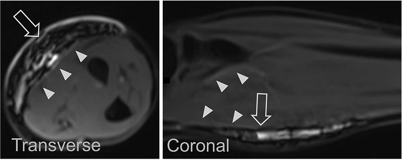
Three-dimensional fat-suppressed T1-weighted volumetric interpolated breath-hold exam images showed a mosaic pattern of the extravasate with mixed high and low signal in the forearm subcutaneous tissue (arrows). Contrast enhancement of the fascia extending toward the proximal portion was observed (arrowheads).
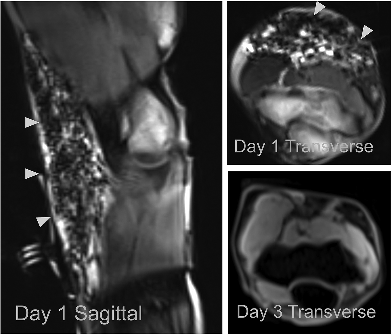
Ten minutes after extravasation, T1-weighted volumetric interpolated breath-hold exam images showed mosaic pattern of the extravasate with a mixture of high and low signals (arrowheads). Poor fat suppression is due to poor shimming and incorrect choice of the excitation center frequency. Three days after extravasation (day 3), the subcutaneous contrast agent disappeared completely.
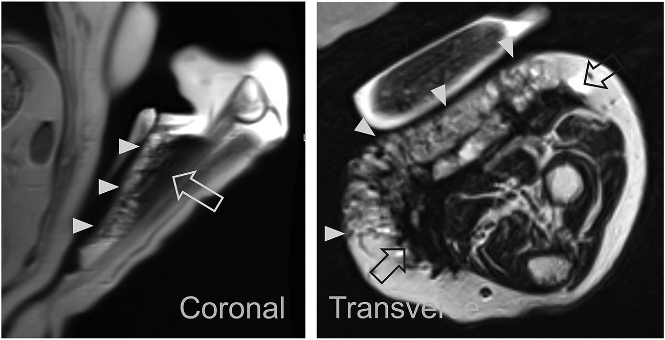
T2-weighted sagittal half-Fourier single-shot turbo spin-echo image and transverse turbo spin echo image showed low-signal fluid collection on the fascia (arrow), extending linearly to the skin surface (arrowhead).
Discussion
The results of this study showed a mosaic pattern of the extravasate with mixed high and low signal on fat-suppressed T1W images. The low-signal component is inferred to be saline or high-concentration GBCA, while the high-signal component is GBCA diluted with saline, blood, or tissue fluid. Contrast enhancement of the fascia is thought to be caused by GBCA seeping into the perifascial capillaries, lymphatic vessels, and the multilayered structure of deep fascia. On the other hand, the low-signal fluid collection on the fascia on T2W images reflects the spin-spin interactions of the high concentration of GBCA. These MRI findings are indicative of the location and distribution of the GBCA and are useful in assessing the clinical correlation with symptoms such as pain and paresthesia.
This study has several strengths. First, the location and distribution of high concentrations of GBCA, and the disappearance of the GBCA within three days was objectively assessed by non-invasive MRI. This may be an important clue for assessing the causal relationship with the extravasation when peripheral neuropathy is complicated after the disappearance of the GBCA. Second, this is the first report of MRI evaluation of changes over time of extravasated GBCA. The risk of GBCA deposition in peripheral nerves has been shown in rat experiments (6), but the deposition of GBCA in peripheral nerves after subcutaneous extravasation has never been studied in humans. Based on the results of this study, the risk of peripheral nerve deposition and associated neuropathy is unlikely because GBCA disappears within three days after extravasation.
The present study has some limitations. First, all patients in this study were patients with cancer. Furthermore, all patients with extravasation were aged >60 years and had received at least one first-line systemic chemotherapy before MRI. Given that the macrocyclic GBCA disappeared quickly even in patients with cancer with impaired tissue repair capacity and impaired clearance, it can be speculated that administration of the macrocyclic GBCA is safer in healthier and younger patients. Second, pathological evaluation of the subcutaneous tissue in which extravasation occurred was not performed due to invasiveness and ethical considerations. Although very small amounts of GBCA may have been deposited, the immediate disappearance of GBCA from the subcutaneous tissue suggests that large amounts of GBCA are unlikely to be deposited and cause serious damage. Third, MRI of the injection sites could not be performed in 4 (36%) patients because treatment of the extravasation was prioritized. That is, there is a bias towards imaging those with less immediately severe extravasations. Fourth, the overall incidence of extravasation, although low at 0.07%, may be relatively high in this cohort not only because of age and prior chemotherapy but also because the rate of 4 mL/s used for perfusion studies is faster than otherwise generally used for MRI scans. If the flow rate had been slower, the incidence of extravasation could have been reduced.
In conclusion, this article gives the first description of MRI findings of GBCA extravasation. Macrocyclic GBCA disappears from the subcutaneous tissue quickly after extravasation and is unlikely to cause serious sequelae.
Footnotes
Declaration of conflicting interests
The author(s) declared no potential conflicts of interest with respect to the research, authorship, and/or publication of this article.
Funding
The author(s) received no financial support for the research, authorship, and/or publication of this article.
