



Abstract
Background
Breath-hold volumetric interpolated breath-hold examination (BH-VIBE) of multiphase contrast-enhanced liver magnetic resonance imaging (MPCE-LMRI) requires good cooperative individuals to comply with multiple breath-holds.
Purpose
To develop a free-breathing modified VIBE (FB-mVIBE) as a substitute of BH-VIBE in MPCE-LMRI.
Material and Methods
We modified VIBE with a high acceleration factor (2 × 2) and four averages to produce the mVIBE scan. A total of 90 individuals (40 men; mean age = 54.6 ± 10.0 years) who had received MPCE-LMRI as part of a voluntary health check-up for oncology survey were enrolled. Each participant was scanned in four phases (pre-contrast, arterial phase, venous phase, and delay phase), and each phase had two sequential scans. To encounter the timing effect of contrast enhancement, three scan orders were designed: BH-VIBE and FB-mVIBE (group A, n = 30); BH-VIBE and FB-VIBE (group B, n = 30); and FB-mVIBE and BH-VIBE (group C, n = 30). The comparisons included the objective measurements and 25 visual-score by two abdominal radiologists independently.
Results
Consistency between raters was observed for all three sequences (intraclass correlation coefficient [ICC] = 0.741–0.829). For rater 1, the mean scores of FB-mVIBE (23.67 ± 1.32) were equal to those of BH-VIBE (23.83 ± 1.98) in groups C and B (P = 0.852). The mean scores of FB-mVIBE (22.07 ± 3.02), but significantly higher than those of FB-VIBE (14.7 ± 3.41) in groups A and B (P <0.001). Similar scores were found for rater 2. The objective measurement of FB-mVIBE were equal to or higher than BH-VIBE and markedly superior to FB-VIBE.
Conclusion
FB-mVIBE is a practical alternative to BH-VIBE for individuals who cannot cooperate with multiple breath-holds for MPCE-LMRI.
Keywords
Introduction
Assessment of arterial and venous phases of enhancement is essential for liver lesion detection and characterization (1). Multiphase contrast-enhanced liver magnetic resonance imaging (MPCE-LMRI) is vital for the evaluation of liver pathology (2–4). MPCE-LMRI typically uses a spoiled 3D gradient-echo variant sequence, such as the volume interpolation breath-hold sequence (VIBE) (1,5). MPCE-LMRI is usually defined as pre-contrast, arterial phase, portal venous phase, and delayed phase (6,7). However, multiple breath-holds (up to 15 s five times) in patients who are unable to adequately breath-hold often result in severe motion artifacts and uninterpretable images.
Advanced MRI sequences, such as a combination of compressed sensing, parallel imaging, and golden-angle radial sparse parallel (8–10), have been allowing for free-breathing (FB) scanning. Moreover, a combination of two techniques, such as TWIST-VIBE and Star-VIBE (11), can produce fast, high-quality, free-breathing liver dynamic contrast enhancement images. However, these products are only available on the very recently released scanner models and produce a large amount of images requiring dedicated postprocessing analysis; therefore, they may not be feasible for routine clinical examinations.
We are seeking a simple modification of the standard VIBE sequence to achieve free-breathing MPCE-LMRI on our 2011-installed scanner. We modified the acceleration and average algorithm without sequence editing, which we named the multi-average 3D-accelerated modified VIBE (mVIBE). The aim of the present study was to test whether the robustness of free-breathing mVIBE (FB-mVIBE) can achieve image quality comparable to that of breath-hold VIBE (BH-VIBE) for MPCE-LMRI. This technique may serve as an effective alternative for patients who cannot cooperate with multiple serial breath-holds, particularly for scanners without state-of-the-art sequences for free-breathing MPCE-LMRI.
Material and Methods
This study was reviewed by the institutional review board of our hospital (KSVGH21-CT13-02) and the requirement for written informed consent was waived.
We used a 3-T MR scanner (Magnetom Skyra; Siemens, Erlangen, Germany) with a VD13 Syngo MR-D13 operating system and an 18-channel body coil (3-T Tim body 18) for our study. To optimize the modification of VIBE to allow for a free-breathing scan, we used the parallel imaging technique (12) with CAIPIRINHA (Controlled Aliasing in Parallel Imaging Results in Higher Acceleration) (13,14). This acceleration effectively shortened the acquisition time. In addition, we revised the averaging mode of VIBE (e.g. number of excitations [NEX] = 4) to “long-term average mode,” which acquires the complete k-space lines within 1 NEX and then repeats the acquisition for the second NEX, and so on to 4 NEX. The default averaging mode is “short-term” average mode: a phase encoding step is acquired through all 4 NEX; then we move on to the next phase encoding steps, go through the 4 NEX, and so on, to completely fill in the k-space. Fig. 1 shows the results of a few pilot cases where we compared different scan parameters and found the optimal parameters. Details of BH-VIBE, FB-VIBE, and FB-mVIBE are shown in Table 1.

Comparison of different pulse sequences and scan methods. Non-contrast phase liver MRI of a 69-year-old woman weighing 48 kg. (a) a1. FB-mVIBE (long-term) series, 4 NEX; a2, 2 NEX; a3, 1 NEX. (b) FB-mVIBE 4 NEX (short-term). (c) BH-VIBE 1 NEX. (d) FB-VIBE 1NEX. lt, long-term; st, short-term (see the “Material and Methods” section).
Comparison of the scan parameters.

BH-VIBE, breath-hold VIBE; CAIPIRNHA, controlled aliasing in parallel imaging results in higher acceleration; FB-mVIBE, free-breathing modified VIBE; FB-VIBE, free-breathing VIBE; FOV, field of view; GRAPPA, GeneRalized autocalibrating partial parallel acquisition; NEX, number of excitations; TE, echo time; TR, rotation time; VIBE, volumetric interpolated breath-hold examination.
Between June 2020 and April 2021, we enrolled 90 volunteers (40 men) who received MPCE-LMRI as part of their health check-up whole-body MRI. The mean age of the participants was 55 years (age range = 33–72 years), with a mean body weight of 65.5 kg (range = 43–112 kg). The participants were randomly divided into three groups (as shown in Table 2), with each person receiving MPCE-LMRI once, composed of four phases: pre-contrast; arterial phase; venous phase; and delayed phase. Contrast enhancement was achieved using MultiHance (Bracco Diagnostics, Monroe, NJ, USA) at a dose of 0.1 mmol/kg, with the injection performed using a power injector at a rate of 1.5 mL/s. Each phase was scanned with two sequential sequences: BH-VIBE and FB-mVIBE (group A, n = 30); BH-VIBE and FB-VIBE (group B, n = 30); and FB-mVIBE and BH-VIBE (group C, n = 30). The details of the scan sequence order, phases, and scan timing of each group were described in Table 2. First, we performed group A and group B to examine the image quality of FB-mVIBE and FB-VIBE. Second, we added group C with a reverse sequence order of group A to balance the order effect of the BH-VIBE and FB-mVIBE.
Order table of the three scan sequences in the three groups.
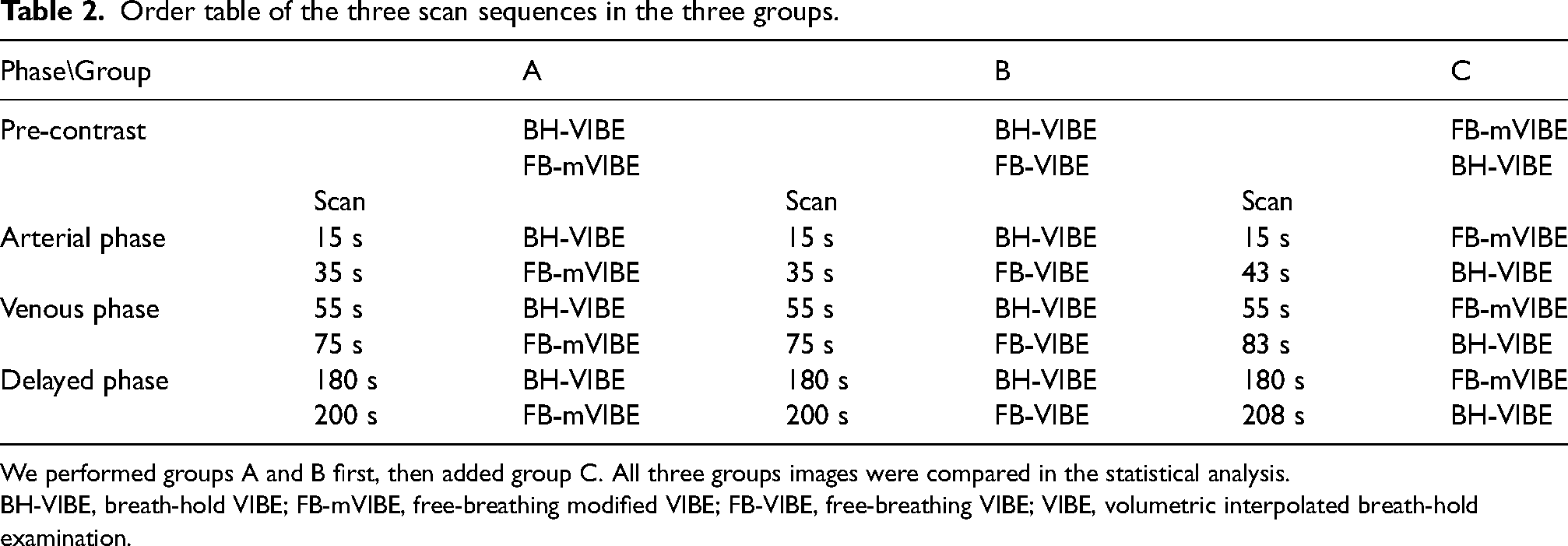
We performed groups A and B first, then added group C. All three groups images were compared in the statistical analysis.
BH-VIBE, breath-hold VIBE; FB-mVIBE, free-breathing modified VIBE; FB-VIBE, free-breathing VIBE; VIBE, volumetric interpolated breath-hold examination.
Two abdominal radiologists, with 25 years and 15 years of experience, respectively, independently assessed the images based on five quality indices: pre-contrast image quality; contrast-enhanced image quality; liver edge definition; liver vascular clarity; and respiratory motion artifacts. The grading scale ranged from 5 (good) to 1 (poor), with scores as shown in Table 3.
Definition of the five indices and scores of image quality.

For objective evaluation of image quality, we measured the signal-to-noise ratio (SNR) of the liver, aorta, and paraspinal muscle using an ROI of approximately 1 cm2 at a single slice of each phase. We calculated the contrast-to-noise ratio (CNR) for the liver versus muscle and aorta versus liver of each phase. The measurements were performed by a senior MR technologist with 15 years of experience (MHC). We used a two-sample t-test to compare the SNR and CNR between the two groups. To compare the visual scores between the two readers, we used the intraclass correlation (ICC). We used SPSS version 22.0 (IBM Corp., Armonk, NY, USA) for the analyses.
Results
Fig. 2 shows examples from groups A, B, and C in panels a1/a2, b1/b2, and c1/c2, respectively, for the pre-contrast phase. The arterial, venous, and delay phases of the same three cases are shown in Figs. 3, 4, and 5, respectively.

Paired comparison of non-contrast phase of the three sequences. Three example cases from groups A (a1, a2), B (b1, b2), and C (c1, c2), respectively. Clearly, the image quality of FB-mVIBE is comparable to BH-VIBE, and much superior to that of FB-VIBE.

Paired comparison of arterial phase of the three sequences. The arterial phase of the same three sample cases from Fig. 2 clearly showed the image quality of FB-mVIBE is comparable to BH-VIBE, and much superior to that of FB-VIBE. The reverse scan order of groups A and C is to compare the scan timing effect.
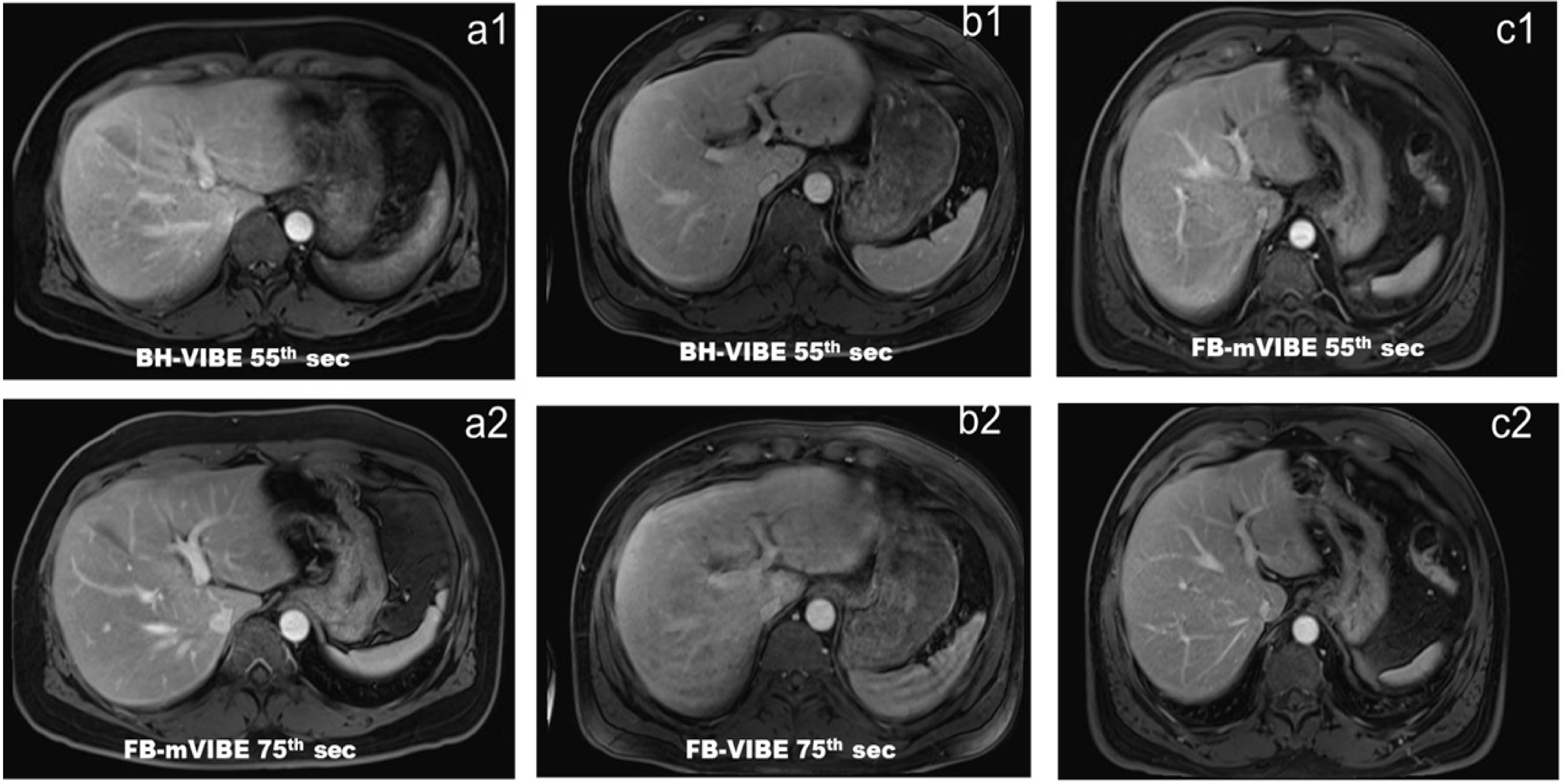
Paired comparison of the venous phase of the three sequences. Venous phase of the same three sample cases from Fig. 2 clearly showed the image quality of FB-mVIBE is comparable to BH-VIBE, and much superior to that of FB-VIBE. The reverse scan order of groups A and C is to compare the scan timing effect.
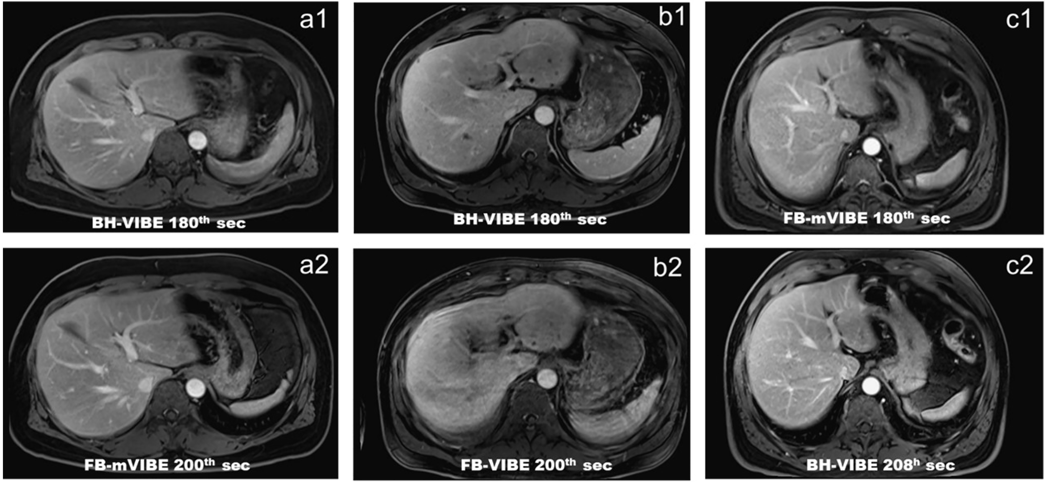
Paired comparison of the delay phase of the three sequences. Delayed phase of the same three sample cases from Fig. 2 clearly showed the image quality of FB-mVIBE is comparable to BH-VIBE, and much superior to that of FB-VIBE. The reverse scan order of groups A and C is to compare the scan timing effect.
The objective evaluation of image quality by SNR and CNR for the three sequences is shown in Tables 4 and 5. In Table 4, we compared the FB-mVIBE sequence of group A to the FB-VIBE sequence of group B. Both sequences had the same second timing for each phase (i.e. before, 35th s, 75th s, and 200th s after contrast injection). We found that most of the SNR and CNR values of FB-mVIBE were superior to those of BH-VIBE (9 out of 12 SNR and 5 out of 8 CNR parameters), while others were equal. In Table 5, we compared the FB-mVIBE sequence of group C to the BH-VIBE sequence of group B. Both sequences had the same first timing scan for each phase (i.e. before, 15th s, 55th s, and 180th s after contrast agent injection). We found that most of the SNR and CNR values of FB-mVIBE were equal to those of BH-VIBE (9 out of 12 SNR and 5 out of 8 CNR parameters), while others were better.
Quantitative comparison of image quality of FB-mVIBE and FB-VIBE.

Values are given as mean ± SD unless otherwise indicated.
P < 0.05.
CNR, contrast-to-noise ratio; FB-mVIBE, free-breathing modified VIBE; FB-VIBE, free-breathing VIBE; SNR, signal-to-noise ratio; VIBE, volumetric interpolated breath-hold examination.
Quantitative comparison of image quality of FB-mVIBE and BH-VIBE.
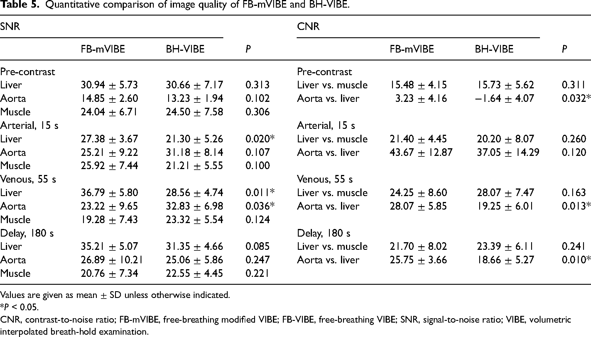
Values are given as mean ± SD unless otherwise indicated.
P < 0.05.
CNR, contrast-to-noise ratio; FB-mVIBE, free-breathing modified VIBE; FB-VIBE, free-breathing VIBE; SNR, signal-to-noise ratio; VIBE, volumetric interpolated breath-hold examination.
The subjective evaluation of the three sequences by two raters is shown in Table 6. Comparison of the visual scores by the two raters showed good consistency for all three sequences (ICC = 0.741–0.829). We found that, according to Rater 1, the visual score of FB-mVIBE was equal to that of BH-VIBE (P = 0.852) and higher than that of FB-VIBE (P <0.001). Similar findings were noted by Rater 2 (P = 0.721 and <0.001, respectively).
Visual scores of image quality of the three sequences, with a total score of 25 for each case.

Values are given as mean ± SD.
For further analysis of the scores of FB-mVIBE for the 90 volunteers, we found an inverse linear regression between body weight and scores (R = −0.33, P <0.001 for rater 1, and R = −0.348, P <0.001 for rater 2). Both raters scored the 45 cases of <63 kg (23.8 ± 1.6, 24.0 ± 1.0) higher than the 45 cases of >63 kg (22.7 ± 2.3, 23.2 ± 1.9) (P = 0.005 and 0.003, respectively). In addition, both raters found that the 50 women (23.8 ± 1.7, 23.8 ± 2.6) had higher scores than the 40 men (22.6 ± 2.3, 23.8 ± 1.7) (P = 0.002 and 0.025, respectively). However, there was no difference in scores related to age, and there was no single independent predictor of the scores by multivariate analysis.
We also found one case in group A that showed a hepatic tumor (Fig. 6), which was later identified as hepatocellular carcinoma. This participant was unable to cooperate and hold their breath during the examination, resulting in better image quality and lesion distinction with FB-mVIBE compared to BH-VIBE.
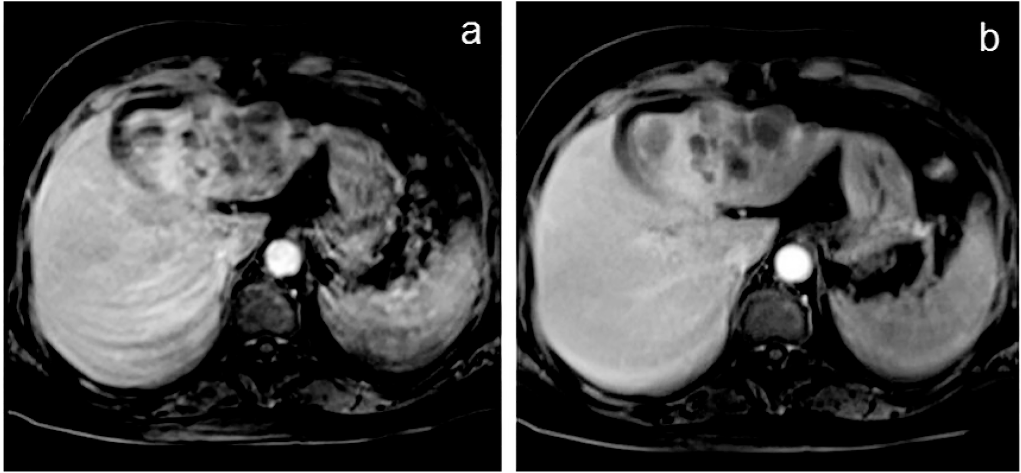
Venous phase of a participant with hepatic cellular carcinoma. There is remarkable motion artifact in FB-VIBE, obscuring the identification of the tumor characteristics.
Discussion
This study has several unique features. First, the modification of VIBE can be performed on the standard scan console. Second, we conducted a paired comparison of three sequences in a large number of individuals and demonstrated that FB-mVIBE was not inferior to BH-VIBE. To the best of our knowledge, this is the first study to offer a practical solution for individuals who are unable to hold their breath during the exam, while still producing images of similar high quality to MPCE-LMRI.
Time frame is critical for successful MPCE-LMRI (2–4). Our mVIBE used 3D acceleration, which effectively shortens the data acquisition time, allowing for 4 NEX in a period of 23 s and fitting within the standard time frame of MPCE-LMRI (13). Signal averaging helps reduce motion artifacts, and these advantages of FB-mVIBE effectively reduce tissue displacement caused by breathing, making it comparable to BH-VIBE images. The advantage of this multi-average 3D-accelerated modification is that it can be performed on our standard operation console, which, in our case, was installed 10 years ago.
To compare two sequences at the same timing of each phase, we designed BH-VIBE as the default first timing scan in groups A and B to ensure a diagnostic image quality in the optimal scan timing. Group C had the inverse order of group A to balance potential order bias in statistical analysis.
The behavior of contrast depends on various factors, such as the acquisition period and k-space trajectory (8–10). In the case of our mVIBE, which uses 3D acceleration, the data acquisition time is effectively shortened, allowing for 4 NEX within 23 s, which fits within the standard time frame of MPCE-LMRI (2–4). The average effect also helps reduce motion artifact, making the image quality comparable to BH-VIBE, despite tissue displacement caused by breathing. Moreover, the multi-average 3D-accelerated modification can be performed on standard operation consoles, which are easily available.
During our study, we compared two sequences at the same timing of each phase to ensure equal scan timing in comparison. Although the central k-spacing timing of mVIBE is longer than VIBE, we found no significant difference in contrast-enhancement patterns between the two sequences, even in the arterial phase. In fact, we observed that FB-mVIBE was superior to BH-VIBE in a volunteer who was uncooperative with breath-holding.
For FB-mVIBE, we did notice that female participants and those with lower body weight tended to have better image quality, likely due to the lower amount of soft tissue surrounding the liver. The use of abdominal compression bands may further suppress the range of abdominal respiratory motion and improve image quality.
Finally, we found that FB-VIBE was not acceptable for interpretation, which is often the case in clinical practice when patients are unable to hold their breath for extended periods. Therefore, our proposed FB-mVIBE provides a practical solution for such difficult scans (15).
The present study has some limitations. In this voluntary cohort, which underwent whole-body MRI as part of a preventive healthcare program, we did not find many cases with significant liver lesions. Therefore, we cannot determine the detectability of hepatic lesion detection using FB-mVIBE. Based on this preliminary result, we plan to conduct a clinical trial to test the diagnostic efficacy of FB-mVIBE for hepatocellular cancer using a follow-up MRI protocol in patients who cannot hold their breath.
In conclusion, compared to several advanced variants of the VIBE sequence, which are specially designed for free-breathing MPCE-LMRI (8–11), our proposed modification method of VIBE is relatively simple and available on scanners without the most updated models. FB-mVIBE showed comparable image quality to that of conventional BH-VIBE. For many people who are unable to cooperate in performing five consecutive 15-s breath-holds, our simple FB-mVIBE can be an option to obtain high-quality MPCE-LMRI.
Footnotes
Acknowledgments
We appreciated the participation of Siou-Fong Pan and research assistants, Jan-Yi Lee, Li-Ling Hung, and Chiung-Chih Hu for data management. We also appreciated Yi-Yu Shih for his consultation about MR technology.
Declaration of conflicting interests
The authors declared no potential conflicts of interest with respect to the research, authorship, and/or publication of this article.
Funding
The authors disclosed receipt of the following financial support for the research, authorship, and/or publication of this article: This study is supported by grants from VGHUST108-G3-3-2, VGHKS 110-119, and MOST 108-2314-B-010-019-MY3.
