



Abstract
Background
In acromegaly, the primary tumor is usually found during magnetic resonance imaging (MRI) of the pituitary gland. A remnant tumor after surgery is, however, harder to depict. When a tumor is missed, the remaining option is usually lifelong pharmacological treatment.
Purpose
To identify tumors by reassessment of all available MRI scans in pharmacologically treated patients, operated or not, and to compare our results with the routine MRI reports.
Material and Methods
Adult patients diagnosed with acromegaly and managed at a tertiary care center between 2005 and 2021 and currently on pharmacological treatment were included. MRI scans were evaluated in a standardized manner and classified independently by a radiologist and an endocrinologist into “certain,” “suspected,” or “no tumor.” In case of disagreement, consensus was achieved with a senior neuroradiologist. The results were compared using the clinical radiologists’ routine MRI reports.
Results
We identified certain and suspected tumors in 29/74 and 36/74 patients, respectively. No tumor was identified in nine patients. In five of these, no MRI contrast agent was given. Discrepancy between our results and the routine MRI reports was found in 31/74 patients (P = 0.01). In 22 patients, the routine reports described no tumor while we identified certain tumors in 2/22 patients and suspected tumors in 13/22 patients.
Conclusion
In most patients with pharmacologically treated acromegaly, we identified a certain or suspected pituitary tumor. These findings were more frequent compared to the routine MRI reports. Based on our results, patients will be considered for a change in long-term treatment modality.
Introduction
Acromegaly is a rare disease caused by overproduction of growth hormone (GH), nearly always due to a pituitary adenoma (1). The primary aims of neurosurgical intervention in acromegaly are cure of GH hypersecretion and treatment of tumor compression symptoms, if present (1,2). Residual GH hypersecretion is found in a substantial proportion of patients after surgery due to a remnant somatotroph adenoma (3). The primary tumor is often found on magnetic resonance imaging (MRI); however, a remnant tumor after surgery is often undetectable (4). Without a visualized target, it is challenging to proceed with surgery, re-surgery, or radiation therapy. Usually, the only option for these patients is lifelong, expensive pharmacological treatment that requires regular follow-up and is associated with potential side effects (5).
The Endocrine Society clinical practice guidelines on the management of acromegaly recommend repeat surgery to be considered in patients with a residual intrasellar tumor, but this option may be overlooked in clinical practice (1). Further, defined intra- and extrasellar tumor remnants may be accessible for radiotherapy (6). There are numerous studies on biochemical outcomes after primary pituitary surgery for acromegaly (7); however, data concerning tumor remnants after primary treatment for acromegaly are lacking.
Routine pituitary MRI during postoperative follow-up in patients with biochemical residual activity may be insufficient to identify small tumor remnants (4). This also applies to patients with small pituitary tumors that may be difficult to identify at diagnosis or during preoperative pharmacological treatment with somatostatin receptor ligands (SSRL). Optimized MRI protocols or functional imaging may improve the identification of residual or recurrent somatotroph adenomas (8,9). Further, optimal detection of tumor remnants requires clearly formulated radiology referrals with sufficient information in order to optimize MRI protocols and radiological assessment. Assessment of MRI of the pituitary gland may be difficult, and suboptimal MRI protocols and failure to detect a pituitary adenoma on MRI may lead to loss of treatment opportunities, since MRI reports are the basis for clinical decisions regarding targeted treatment options with potentially permanent cure, including radiotherapy and pituitary surgery. Clinical endocrinologists at our center usually follow up patients with acromegaly and rely on the information provided in the primary routine MRI reports. A quality study can detect missed tumors and could potentially offer patients definite treatment instead of long-term medical treatment.
The aims of the present study were to assess the proportion of patients with pituitary tumors that can be identified by a standardized reassessment of all available pituitary MRI scans of pharmacologically treated patients with active acromegaly and to identify discrepancies between the standardized reassessment and the primary routine MRI reports.
Material and Methods
Patients
The Department of Endocrinology at Oslo University Hospital is a tertiary referral center for patients with pituitary disease, covering approximately 3 million people. Eligible patients, operated or not, were identified from the internal pituitary quality registry. Patients diagnosed with acromegaly based on clinical and biochemical findings between 1 January 2005 and 30 June 2021, and currently on pharmacological treatment for acromegaly were included in the study cohort. Thus, included patients have GH excess requiring treatment and were expected to have a tumor in the sellar region. Patients who died before MRI assessment in November 2022 (n = 11), were diagnosed with McCune Albright syndrome (n = 1), were lost to follow-up due to relocation (n = 2), or had CT scans only (n = 1) were excluded.
Patients had follow-ups according to clinical practice, usually with annual visits assessing the clinical symptoms of acromegaly, medication, and biochemistry. MRI was performed routinely or as clinically indicated. Clinical data and MRI from visits until 30 June 2022 were included, resulting in at least one year of follow-up after diagnosis.
Baseline and last available markers of secretory activity (GH and insulin-like-growth factor 1 [IGF-1]) analyzed as clinical routine at the department of clinical chemistry were registered.
MRI
At our center, the standard protocol of pituitary MRI consists of T1 and T2 sequences. Gadolinium-based contrast agent (GBCA) is given in preoperative imaging but not always in late follow-up MRI (usually given at first postoperative scan [within 48 h] and at the three-month follow-up).
MRI was primarily described (“routine MRI report”) at the department of radiology at our center, collaborating care centers, or private radiology clinics.
For the systematic reassessment, the most recent MRI was used for tumor classification based on all available T1-weighted (T1W) and T2-weighted (T2W) images, the same images as the routine MRI reports. Moreover, all available previous scans were also reassessed to facilitate the identification and classification of tumors in the reassessment. Slice thickness and magnetic field strength (1.5 or 3 T) were registered. MRI quality was assessed subjectively as “good” or “poor,” based on a total impression with regard to spatial resolution, signal-to-noise ratio, whether GBCA was sufficiently applied, and presence of image artifacts. Size (micro- or macroadenoma [<10 or ≥10 mm] in the largest dimension) and cavernous sinus invasion (KNOSP-Steiner grade (10)) were assessed. MRI scans were assessed and classified independently by an endocrinologist (SA) and a radiologist (MKA). In case of disagreement, consensus was achieved by reassessment together with a board-certified radiologist (GR) with 17 years of experience in neuroradiology. Before the MRI assessment, the same endocrinologist and radiologist (SA, MKA) were trained by the senior radiologist (GR) in the reading of pituitary MRI and underwent training in the classification routine on MRI scans from 12 pilot patients who were not included in the study.
Tumor classification based on MRI reassessment
Results of MRI reassessment in all patients were classified into three distinctive categories: certain tumor; suspected tumor; and no tumor.
A certain tumor is a sellar or extra-sellar lesion with characteristics in T1-signal, T2-signal, and/or features of GBCA enhancement different from normal pituitary tissue. A tumor is typically enhanced with GBCA, usually to a lesser degree than the normal pituitary gland.
In postoperative MRI, a remnant tumor should be situated in the same localization as the primary tumor. It usually has the same intensity as the primary tumor and may grow over time (11).
It does not have the typical characteristics of scar tissue. Fibrous scar tissue is typically hyperintense in T2W imaging and may have prominent GBCA enhancement. Even though scar tissue may resemble a tumor, it is expected to gradually reduce in size over time. Nor should the tumor appear as a homogenous cyst or hemorrhage but may have cystic or hemorrhagic components (11).
A suspected tumor is a pituitary or parasellar lesion that does not meet the characteristics described above for a visible tumor and cannot be defined as “no tumor” described below. These include: (i) a lesion that is not situated where the primary tumor once was; (ii) a lesion that has a different intensity from the primary tumor; and/or (iii) a lesion that does not grow (11). Pharmacological treatment may change the intensity of the tumor (12).
No tumor findings have the following characteristics: (i) normal pituitary and extra-sellar findings; (ii) an empty sella and no para-sellar tumor components; or (iii) a cyst without any identifiable solid tumor component, usually situated in the midline.
If the patient has undergone surgery, postoperative findings such as scar tissue, fluid in an operation cavity, hemorrhage or fat and muscle fascia graft remnants may be present. A fat graft is hyperintense on T1W imaging, and usually degenerates gradually (11). Muscle with fascia may be slightly enhanced in its peripheral parts after GBCA on T1, whereas it may be hyperintense on T2W imaging. Fascia may be presented as a hypointense line (13). Further, T2W images may, in some cases, provide more information than T1W contrast images in the assessment of the postoperative sellar region (14).
Clinical routine MRI reports were compared to our consensus classifications. The routine reports were also classified as “certain,” “suspected,” or “no tumor” based on information in the reports.
Statistics
Descriptive statistics with medians and interquartile ranges (IQR) were used for age, follow-up time, IGF-1, and GH levels of the study cohort using Excel and RStudio. McNemar`s test was performed using RStudio for the 3 × 3 contingency table (Table 1), which compares the reassessment findings with the routine MRI reports.
Summary of the results of reassessment and primary MRI reports (n=73).
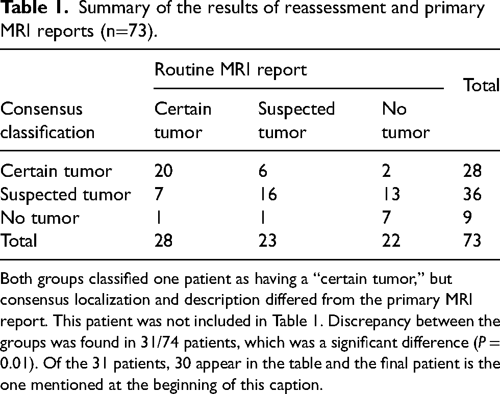
Both groups classified one patient as having a “certain tumor,” but consensus localization and description differed from the primary MRI report. This patient was not included in Table 1. Discrepancy between the groups was found in 31/74 patients, which was a significant difference (P = 0.01). Of the 31 patients, 30 appear in the table and the final patient is the one mentioned at the beginning of this caption.
Ethics
The study was approved by the hospital authority. As the study was a registry-based quality control study using clinical data, the requirement to obtain any informed consent was waived by the Regional Ethical Committee of the involved Health Region.
Results
The screening cohort consisted of 206 patients, of whom 74 fulfilled the inclusion criteria of the study: 62 patients had undergone pituitary surgery and 12 were on primary pharmacological treatment without previous surgery. The characteristics of the study cohort at baseline and follow-up are presented in Table 2.
Baseline and follow-up characteristics.
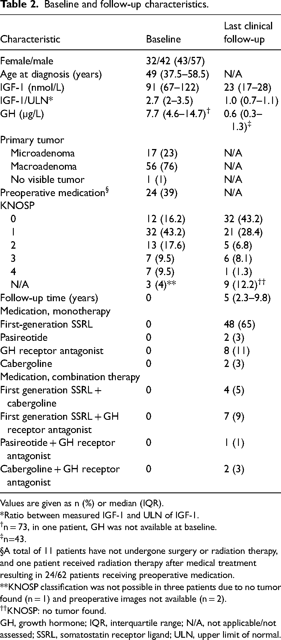
Values are given as n (%) or median (IQR).
*Ratio between measured IGF-1 and ULN of IGF-1.
n = 73, in one patient, GH was not available at baseline.
n=43.
A total of 11 patients have not undergone surgery or radiation therapy, and one patient received radiation therapy after medical treatment resulting in 24/62 patients receiving preoperative medication.
**KNOSP classification was not possible in three patients due to no tumor found (n = 1) and preoperative images not available (n = 2).
KNOSP: no tumor found.
GH, growth hormone; IQR, interquartile range; N/A, not applicable/not assessed; SSRL, somatostatin receptor ligand; ULN, upper limit of normal.
MRI
T1W and T2W images were available for all 74 patients, except for in one patient who did not have T2 images. The median slice thickness was 1 mm (IQR = 1–1.375 mm) in T1 and 3 mm (IQR 2–3 mm) in T2. The magnetic field strength was 1.5 T in 57 patients, 3.0 T in 16 patients, and unknown in one patient. In 23 patients, GBCA was not given at the last MRI scan. In five of these patients, no tumor was detected. The general MRI quality was deemed good in 61 patients and poor in 13 patients.
We identified certain tumors in 29/74 (39%) patients and suspected tumors in 36/74 (49%) patients. No tumor was found in 9/74 (12%) patients. In Table 3, types of intervention are summarized and stratified by their current KNOSP classification. Most patients with certain or suspected tumors were classified as KNOSP 0 or 1 (Table 3).
KNOSP classification after treatment based on the latest magnetic resonance imaging subdivided into type of intervention.

*Reintervention: undergone re-surgery and/or radiation therapy; one patient had undergone radiation therapy only.
In total, 12 (16%) patients had not undergone surgery due to the following reasons: radiation therapy without surgery (n = 1); no definite tumor (n = 4); awaiting debulking pituitary surgery after primary pharmacological treatment by the time of MRI reassessment (n = 4, all KNOSP 3 or 4; follow-up time was approximately one year); patient preference and/or tumor deemed not suitable for surgery (n = 3).
Table 1 summarizes the results from the MRI reassessment and routine MRI reports. Any discrepancy between the groups was found in 31/74 (42%) patients (P = 0.01). An example of MRI scans where the discrepancy occurred is presented in Fig. 1.
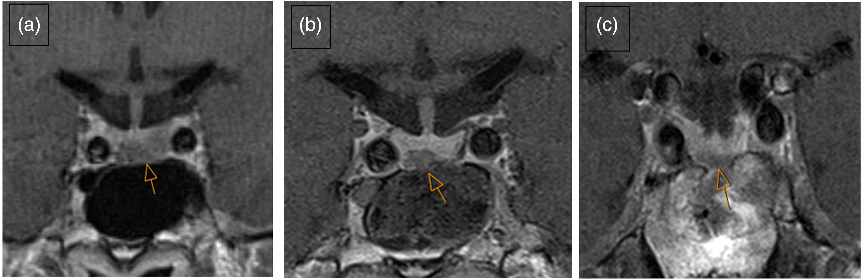
Example of a patient and MRI scans in T1 + GBCA series where there was discrepancy between the consensus classification and the routine MRI report. (a) Tumor (arrow) before surgery: there was GBCA enhancement in the pituitary adenoma, which was hypointense compared to the pituitary gland. The tumor was isointense to the pituitary gland in T1 series without GBCA and at T2, thus difficult to visualize without GBCA. (b) First postoperative day: at T1 after contrast, the surgical cavity (arrow) corresponds well with the location of adenoma on preoperative MRI. (c) The lesion four years after surgery was classified as a certain tumor (arrow) by structured reassessment due to its location at the site of the primary tumor and since postoperative changes and scar tissue should have been dissolved or reduced in size (11). The routine MRI report described no remnant tumor or other findings. GBCA, gadolinium-based contrast agent; MRI, magnetic resonance imaging.
Discussion
In the present study of patients with pharmacologically treated acromegaly, we identified a certain or suspected pituitary tumor in most of the cases (88%). Only a few patients were classified as having no visible tumor. In patients with one prior surgical intervention, most had non-invasive tumor remnants or recurrences, thus potentially being available for future intervention. Further, there was a significant discrepancy between the primary MRI report and the standardized reassessment. We identified a certain or suspected tumor in more than half of the patients who were described to have no tumor in the primary reports.
Several factors may have contributed to the discrepancy between primary reports and the results of reassessment: (i) with the knowledge of persistent GH excess, a radiologist may be more alert to the presence of a pituitary tumor due to higher pre-test probability in patients with active disease. Thus, small tumors and tumor remnants are less likely to be overlooked or considered irrelevant to report. Information from the referring clinician on GH excess is therefore crucial; (ii) the primary reporting radiologist did not always have access to all the prior images, including preoperative MRI scans. This may impact the evaluation of the latest images as remnant tumors may be small and are usually located at the site of the primary tumor (11); (iii) time to evaluate serial MRI scans is limited in a routine setting. Although reassessment was time and resource demanding, we conclude it to be beneficial for both patients and clinics. In the long term, it may even be time- and resource-saving; (iv) pharmacological treatment with SSRL might reduce the tumor size (15), information all radiologists might not have been aware of. Several primary reports in the present study concluded that lesions cannot be a tumor as they decreased in volume, and typically rather concluded with scar tissue.
Many of the routine MRI scans were acquired with a magnetic field strength of 1.5 T without GBCA enhancement. These protocols are usually sufficient for the follow-up of non-functioning pituitary adenomas, but not always for small secreting pituitary lesions or tumor remnants (8,16). The rate of identified lesions might increase by application of dedicated imaging protocols for pituitary MRI at 3 T, which has higher spatial resolution and better signal-to-noise ratio, enabling detection of smaller lesions (17). Such dedicated MRI protocols may include high resolution T2W images (slice thickness of typically 1.5–2 mm) in three orthogonal planes, 3D T1W scans with isotropic voxels pre- and post-GBCA, and even dynamic imaging or delayed imaging, typically 30–40 min after contrast.
In addition, in the clinical routine follow-up of patients receiving long-term pharmacological therapy, surgery or radiation therapy may not have been considered as alternative treatment modalities, even though a tumor is known and described in the primary MRI report.
Studies assessing remnant or recurrent GH producing tumors are scarce, and we have not found systematic studies on reassessment of MRI in acromegaly. A recent study reported that 37/83 (45%) patients had residual tumors after primary transsphenoidal surgery, but further residual tumor characteristics were not reported (7). Other data on remnant tumors in acromegaly included mainly patients undergoing redo-surgery and thus represented highly selected patients (18,19). A recent study reported that 23.7% of operated patients had postoperatively persistent GH excess despite the absence of tumor remnants on MRI (4). This is a similar proportion as provided by the routine MRI reports in the present study. After reassessment, there were substantially fewer patients in the “no tumor” category. According to the Endocrine Society’s guidelines, patients with intra-sellar tumors after primary surgery should be considered for redo-surgery (1). About half of the patients selected to undergo repeat surgery achieved remission in previous studies with an even higher probability in non-invasive remnants (18,19). Thus, it is of interest to identify small, non-invasive tumors by reassessment of previous MRI. Optimized MRI or functional imaging, such as 11C-methionine PET, may further increase the rate of tumor identification (8,9).
The present study has some limitations. The primary MRI reports did not classify the findings systematically as certain, suspected, or no tumor. These classifications were extracted from the written information in the reports. However, we did not experience this to be difficult. Further, MRI scans from different scanners and protocols were analyzed retrospectively. To assess the real prevalence of MRI detectable lesions, identical, optimized MRI protocols would be necessary. Moreover, we do not have histological or clinical validation of the source of GH excess from the identified lesion and we did not assess whether the identified or suspected lesions were suitable for a targeted therapeutic approach, or if the patients wanted to undergo radiotherapy or neurosurgery. These matters should be addressed in future studies after shared decision-making with the patients. Possible management pathways include optimized imaging, continued medical treatment, trans-sphenoidal surgery, or radiation therapy after multidisciplinary team (MDT) assessment.
In conclusion, systematic reassessment of MRI can identify certain or suspected pituitary tumors in most patients with pharmacologically treated acromegaly. These findings were more frequent compared to the routine MRI reports. In addition, the collection of all previous MR images, sufficient time utilization to evaluate serial MR images, and optimized clinical referrals may increase detection rates of tumors. Based on the results of this study, MRI images will be assessed by a multidisciplinary team for the necessity of optimized imaging, and selected patients will be considered for a change in long-term treatment modality.
Footnotes
Declaration of conflicting interests
The authors declared no potential conflicts of interest with respect to the research, authorship, and/or publication of this article.
Funding
The authors disclosed receipt of the following financial support for the research, authorship, and/or publication of this article: Helse Sør-Øst RHF (Regional Health Department of Southeast Norway), project number 2020081.
