
Abstract
Background
Advances in molecular imaging strategies have had an effect on precise diagnosis and treatment. Research has been intensified to develop more effective and versatile radiopharmaceuticals to uplift diagnostic efficiency and, consequently, the treatment.
Purpose
To label the flutamide (FLUT) coupled with diethylenetriamine pentaacetate (DTPA) with technetium-99 m (99mTc) and to evaluate its binding efficiency with rhabdomyosarcoma (RMS) cancer cells.
Material and methods
Radiolabeling of FLUT with 185 MBq freshly eluted 99mTcO4−1 was carried out via DTPA bifunctional chelating agent using stannous chloride reducing agent at pH 5. The labeled compound was assessed for its purity using chromatography analysis, stability in saline and blood serum, AND charge using paper electrophoresis. Normal biodistribution was studied using a mouse model, while binding affinity with RMS cancer cells was studied using an internalization assay. The in vivo accumulation of RMS cancer cells in a rabbit model was monitored using a SPECT gamma camera.
Results
Radiolabeling reaction displayed a pharmaceutical yield of 97% and a stability assay showed >95% intact radiopharmaceutical up to 6 h in saline and blood serum. In vitro internalization studies showed the potential of [99mTc]DTPA-FLUT to enter into cancer cells. This biodistribution study showed rapid blood clearance and minimum uptake by body organs, and scintigraphy displayed the [99mTc]DTPA-FLUT uptake by lesion, induced by RMS cancer cell lines in rabbit.
Conclusion
Stable, newly developed [99mTc]DTPA-FLUT seeks its way to internalize into RMS cancer cells, indicating it could be a potential candidate for the diagnosis of RMS cancer.
Introduction
Rhabdomyosarcoma (RMS) is considered a primitive mesenchymal cell (PMC) malignancy comprising neoplastic primitive precursor cells that possess histological features associated with myogenic differentiation. It can arise anywhere in the body but is more likely to start in the head and neck, bladder, reproductive system (vagina, uterus, and testes), arms, or legs. RMS is a type of rare soft tissue malignancy; in most cases, it is associated with familial syndromes. Types of RMS include spindle/sclerosing (∼10%), pleomorphic (∼10%), alveolar (∼20%), and embryonal RMS (∼60%) (1,2). The literature reports that RMS is a common childhood and adolescent soft tissue sarcoma, i.e. childhood tumor (3%) and pediatric soft tissue sarcomas (50%) (3,–5). Different types of histologic subtypes of RMS, such as embryonal, alveolar, pleomorphic, and spindle/sclerosing, are associated with newly developed diagnostic, therapeutic, and prognostic implications (6). The pathological diagnosis of RMS is sometimes challenging due to its similarity in size, shape, and color of cell tumors of bone and soft tissue in childhood. There are no confirmed physical signs and symptoms of RMS, which mostly vary due to site of origin, age of patients, and stage of sarcoma. The patient’s initial evaluation with suspected RMS mainly aims to chalk out the anatomic boundaries of the sarcoma, which will help define the tumor stage, risk stratification, and therapeutic protocol (4). Computed tomography (CT) and magnetic resonance imaging (MRI) in association with bone scan, bilateral bone-marrow aspirate, and biopsy are the common initial diagnostic workup (7). Immune-histochemical, molecular-genetic, and/or ultra-structural techniques are considered to confirm the diagnosis of RMS (8). A nuclear medicine technique (NMT), based on radionuclide labeled bioactive molecules, helps in diagnosis, irrespective of morphological changes (9). Currently, the NMT in combination with CT or MRI explains the 3D morphology of diseased tissue, which helps treat the patient effectively (10). Therefore, the development of radiopharmaceuticals to diagnose and study a variety of diseases is a hot area of research.
FLUT belongs to a class of non-steroidal anti-androgen drugs with a broad-spectrum application for the treatment of prostate and bladder cancers (11). FLUT was approved by the U.S. Food and Drug Administration (FDA) in 1989 for the treatment of androgen-dependent prostate cancer due to its antagonism for androgen receptors (AR) (12). Ankley et al. reported the affinity of FLUT and its metabolite 2-hydroxyflutamide for mammalian ARs and its potential to block the expression of AR-mediated responses in vivo (13). Moreover, Koch et al. highlighted the effectiveness of FLUT as non-toxic putative aryl-hydrocarbon receptor (AhR) ligand with persistent anticancer efficiency and reported that FLUT can inhibit the growth of various cancer cells independent of AR expression (14). FLUT and 2-hydroxyflutamide were investigated after labeling with 99mTc using 99mTc(CO)3 and fac-99mTc[(OH2)3(CO)3+)] core for the diagnosis of prostate cancer (15,16). However, the labeling procedure is lengthy and needs much care to avoid radio-contamination. The aim of the present study was to design the protocol of radiolabeling of FLUT using the DTPA bifunctional chelating agent with 99mTc using mild reaction condition and to evaluate its binding affinity with RMS cancer cells using in vitro and in vivo biological assays for the diagnosis of RMS carcinoma.
Material and Methods
Chemical and reagents
All the chemicals used in this work were of analytical grade. Flutamide, sodium hydroxide, diethylenediamine pentaacetate acid (DTPA) anhydride, stannous chloride dihydrate (SnCl2.2H2O), acetonitrile, acetone, trifluoroacetic acid (TFA), DMSO, and ethanol were purchased from Sigma-Aldrich (Germany). All other chemicals were purchased from Alfa-Acer (Germany). Radioactivity was obtained from the Pakistan Institute of Nuclear Science and Technology (PINSTECH) Islamabad, Pakistan in the form of PakGen Moly-generator.
Cell line and animal models
The RMS cell line for the internalization study was kindly provided by the Photo-medicine Research Laboratory, Pakistan Institute of Engineering and Applied Science (PIEAS), Islamabad, Pakistan. Healthy Sprague–Dawley mice (mean weight = 150–200 g) for biodistribution and RMS cancer-induced New Zealand white rabbit (mean weight = 2.5 kg) for scintigraphy were obtained from the National Institute of Health (NIH) Islamabad, Pakistan.
Coupling of DTPA with FLUT
The coupling of DTPA with FLUT was carried out using the reported protocol (17). DTPA dianhydride (2.5 g, 14 mmol) was dissolved in 75 mL of dimethyl formamide followed by slow addition of FLUT (28 mmol) at 65°C. The reaction mixture was stirred for 15 h. The white precipitate was then extracted in diethyl ether (250 mL) followed by washing thrice. Evaporation of diethyl ether yielded DTPA-FLUT (4.24 g, 61%) as a white solid. 1H NMR (400 MHz, D2O): δ1.29 (6H, d, J = 7.6 Hz), 2.30 (4H, t, J = 7.5 Hz), 2.34 (4H, t, J = 7.5 Hz), 3.39 (4H, s), 3.56 (4H, s), 3.59 (2H, s), 3.98 (1H, m), 7.1 (1H, d, J = 7.4 Hz), 7.9 (1H, s), 8.1 (1H, d, J = 7.4); FTIR (ν, Cm−1): 2921, 1640, 1728, 1510, 1450, 1400, 1290, 1192.
Labeling of DTPA-FLUT with 99mTc
In order to optimize the best radiolabeling conditions to a fixed amount, 200 µg/mL of DTAP-FLUT was added to 5–40 µg of SnCl2.2H2O (with an increment of 10 µg in each set of the experiment), followed by adjusting the pH in the range of 3–8 using 0.1 M NaOH or HCl solution. Lastly, freshly eluted 185 MBq/mL Na99mTcO4 was added into the reaction vial, shaken, and heated for different lengths of time (10–15 min) at 50°C. The reaction mixture was then cooled to room temperature. Hydrolyzed radioactive impurity was removed by filtering the reaction mixture through 0.22 µm Millipore channel. To analyze the radiochemical purity of [99mTc]DTPA-FLUT), the mixture was subjected to ITLC and HPLC analysis. Fig. 1 shows the DTPA-FLUT and [99mTc]DTPA-FLUT synthesis.

Coupling of DTPA with FLUT and radiolabeling of DTPA-FLUT with 99mTc.
Instant thin layer chromatography
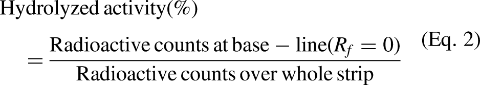
At the baseline of the ITLC strip (10 × 1 cm) an aliquot of 1 µL reaction solution was spotted. The strip was placed in a glass jar and pre-saturated with mobile phase to elute the sample components. Acetone was taken as the mobile phase to determine free 99mTcO4− while 0.5 N NaOH was chosen to determine the hydrolyzed activity. Upon completion of elution, each strip was dried and cut into 1-cm patches. The counts of each patch were recorded by placing into well-type NaI detector. From the knowledge of the free and hydrolyzed activity, the radiochemical yield of [99mTc]DTPA-FLUT was calculated using the following expressions:


High-performance liquid chromatography analysis
Reverse-phase high-performance liquid chromatography (RP-HPLC) equipped with 168-diode-array ultraviolet and sodium-iodide (NaI) flowthrough radiation detectors (NaI-FRD) were used to analyze non-labeled and labeled DOTA-FLUT, respectively. Two solvent systems were used, i.e. a weaker solvent (solvent A; water with 0.1% TFA) and a stronger solvent (solvent B; acetonitrile). The elution strength was increased with time starting from 95% solvent A for the first 5 min. The gradient was then increased to 80% solvent B over the next 10 min.
Electrophoresis
Electrophoresis of the labeled compound was performed to evaluate the charge over [99mTc]DTPA-FLUT complex. Whatman #1 paper (20 × 2 cm) was stretched over a plastic support and its ends were dipped into anode and cathode chambers filled (3/4 volume) with sodium phosphate buffer at pH 6.8. An aliquot of 2 µL of [99mTc]DTPA-FLUT was introduced at the middle of the strip. Electrophoresis was then carried out at 300 V potential for 1 h. A gamma counter was used to determine the radioactivity at the middle, right, and left ends of the paper.
[99mTc]DTPA-FLUT stability study
The in vitro stability of the radiopharmaceutical was evaluated in saline at pH 5.5 and freshly obtained blood serum at pH 7.35. First, 185 MBq [99mTc]DTPA-FLUT was mixed with 1 mL saline and incubated for up to 24 h at room temperature. For analyzing stability in serum, 1.8 mL serum was mixed with 185 MBq [99mTc]DTPA-FLUT in a glass vial followed by 24 h of incubation at 37°C. Both mixtures were analyzed using paper chromatography at predefined time points.
Biodistribution study
For the biodistribution study, 200 µL (111 MBq) [99mTc]DTPA-FLUT was injected through the tail vein of healthy Sprague–Dawley male mice (weight 25–30 g). At predefined time intervals, i.e. 0.25, 0.5, 1, and 3 h, the mice were anesthetized using chloroform and sacrificed to collect body organs. The blood sample was taken from cardiac puncture. The body organs, such as the brain, heart, lungs, liver, stomach, spleen, kidneys, intestine, bladder, femur, and carcass-A (car-A, inflamed thigh tissue) and carcass-B (Car-B, normal thigh tissues) were separated, washed twice with saline to remove residual activity, weighted, and counted for activity using well-type NaI detector. The distribution of the radiopharmaceutical throughout the body was calculated as the percentage of injected dose per gram organ (%ID/g). The biodistribution study in tumor-bearing mice was not conducted due to non-availability of immunocompromised nude mice.
Cell binding and internalization study
For the internalization experiment, RMS cells were seeded at a density of 1 × 10⁶ cells per well in a six well plates (Corning, New York, USA) and grown to 80%–90% confluent cells at 37°C in 5% CO2 incubator for 48h. On the day of the experiment, cells with confluence >80% were washed twice with ice cold internalization medium (RPMI-1640 supplemented with 1% (v/v) FBS), followed by incubation for another 1 h at 37°C in fresh growth medium (1 mL per well). [99mTc]DTPA-FLUT (3–5 pmol) in 150 µL PBS/1% BSA was added in each wells with and without cells, along with 150 µL PBS/1% BSA (for determining total binding). The wells without cells were served as controls to determine the total radioactivity added. The plate was incubated at 37°C and 5% CO2 for a further 10, 30, 45, 60, and 120 min. At each time point, the medium containing [99mTc]DTPA-FLUT was removed and internalization was halted by rinsing the cells twice with ice cold internalization medium. Thereafter, the cells were washed twice for 5 min in ice cold 50 mM glycine-HCl buffer (pH 2.8), followed by a rapid rinsing with the internalization medium. The resulting buffer washed supernatants in each case were pooled in test tubes and counted as surface-associated bound fractions. Furthermore, the cells were lysed with 1 M NaOH for 10–15 min and collected as the internalized fractions in another test tube, followed by washing with internalization medium in same test tubes. All fractions were counted in a NaI well-type gamma-counter (LKB-1282). The internalized [99mTc]DTPA-FLUT fractions were expressed as a percentage of the total cells associated activity (i.e. surface-associated plus internalized activity).
Scintigraphy study
For the scintigraphy study, subcutaneously induced RMS tumor rabbit model was placed on a flat surface under dual-headed gamma camera (Siemens, Berlin, Germany) integrated with high-resolution parallel-hole collimator and interconnected with dedicated online computer system. Initially, the rabbit was anesthetized using a single dose of diazepam injection (2 mg/kg body weight) followed by administration of [99mTc]DTPA-FLUT (185 MBq/250 µL saline) through marginal ear vein. Static images of the whole body were acquired at predefined time intervals (i.e. 15 min, 0.5 h, 1 h, and 4 h).
Results
Coupling of DTPA with FLUT and radiolabeling
The coupling reaction progress was monitored with TLC. Finally, the coupled product was extracted in diethyl-ether and analyzed with RP-HPLC, which showed a single peak at 6.39 min (Fig. 2a), indicated the purity of the DTPA-FLUT coupled product. The structure of DTPA-FLU was elucidated by 1H NMR and FT-IR spectroscopic techniques. The spectroscopic data (1H NMR and FT-IR) is given in the experimental part. Radiolabeling of DTPA-FLUT with 99mTcO4− at different sets of reaction conditions showed a different yield of radiochemical, which is summarized in Table 1. The maximum radiochemical yield (97%) was obtained when 200 µg DTPA-FLUT was mixed with 20 µg stannous chloride, and 185 MBq 99mTcO4− at room temperature and pH 5. The HPLC radio-chromatogram, as shown in Fig. 2b, is displaying one peak at 7.4 min.
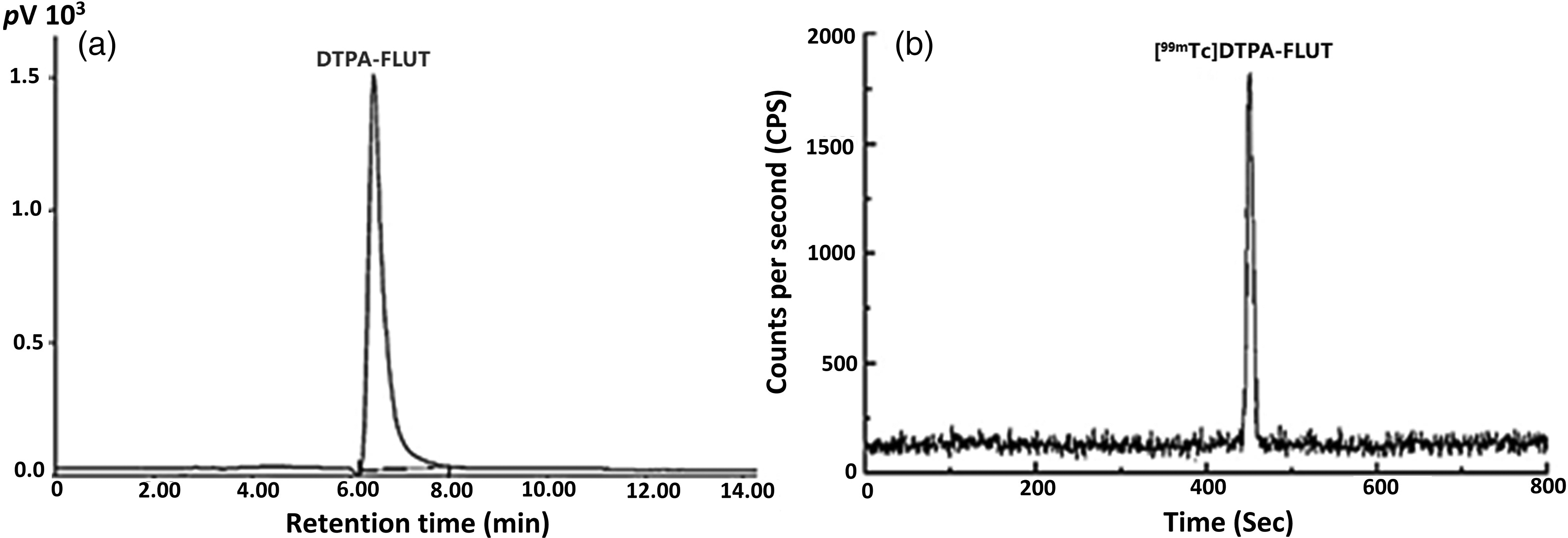
(a) RP-HPLC chromatograms of DTPA-FLUT and (b) radio-chromatogram of 99mTc labeled [DPTA]FLUT.
Radiochemical yield at various quality control parameters to validate the optimized labeling conditions for radiosynthesis of [99mTc]DTPA-FLUT (n = 3).
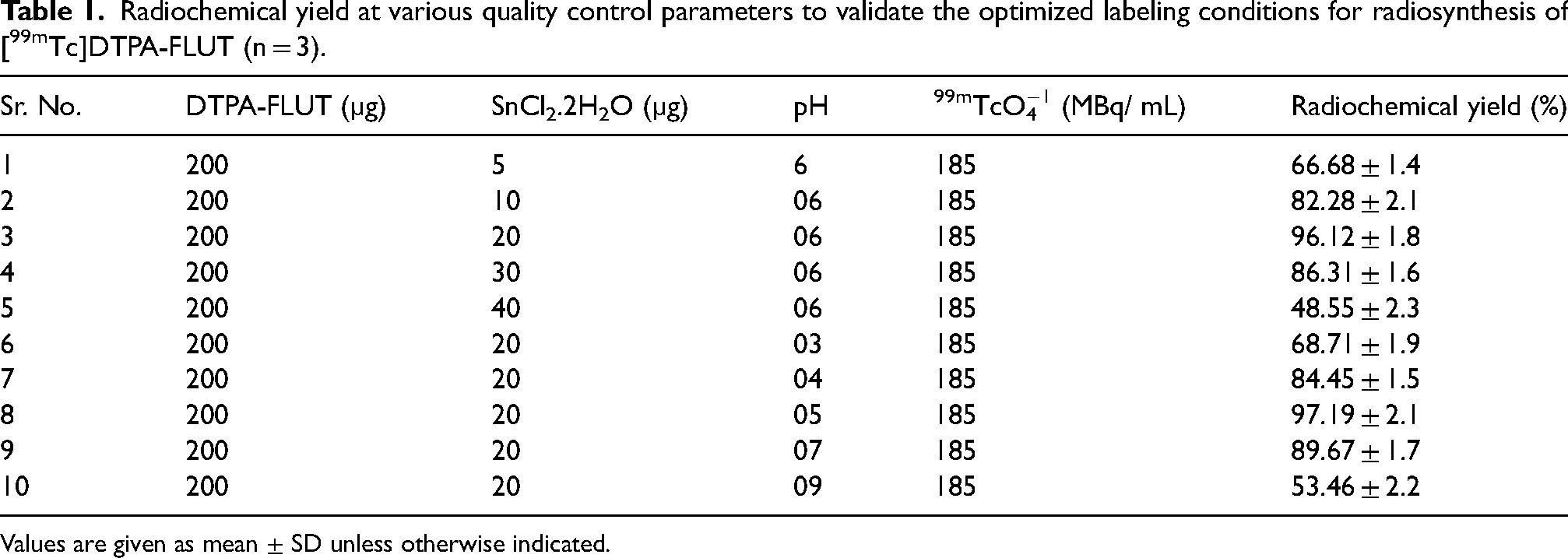
Values are given as mean ± SD unless otherwise indicated.
Physicochemical properties evaluation
Stability tests
A 6-h incubation of [99mTc]DTPA-FLUT in an equivalent volume of saline displayed 95.87% intact radiopharmaceuticals, while in serum it was recorded as 92.91%. The results of the radiopharmaceutical stability in both mediums up to 12 h time points are shown in Fig. 3.
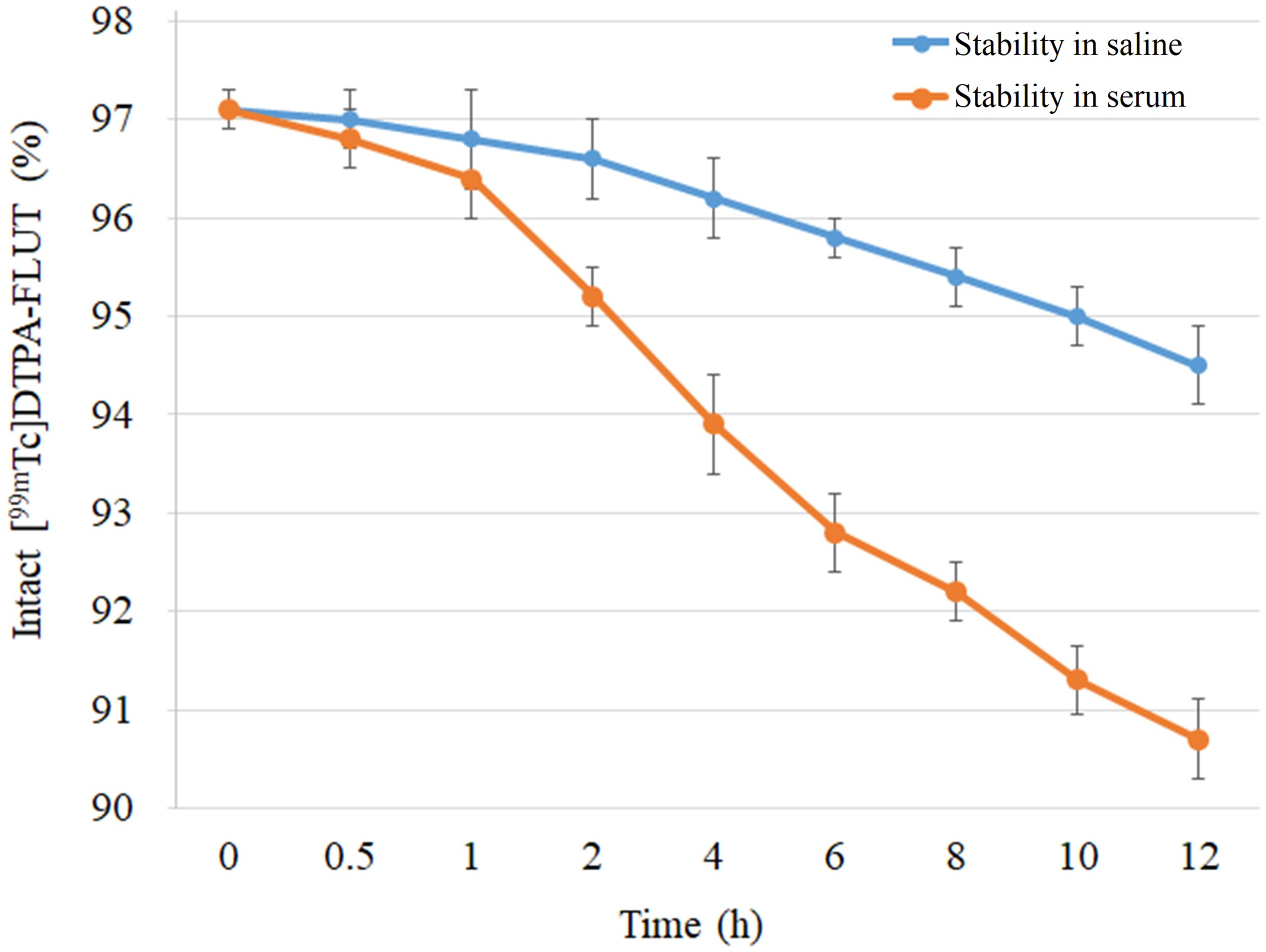
Stability of [99mTc]DTPA-FLUT in saline and serum medium.
Electrophoresis
The results of the electrophoresis analysis are shown in Fig. 4. Maximum counts were recorded at the point of radiopharmaceutical introduction, while a small number of radioactivity counts were detected near to the anode terminal.

Electrophoretogram of [99mTc]DTPA-FLUT.
Biodistribution study
Biodistribution of [99mTc]DTPA-FLUT was assessed in inflammation-induced Sprague–Dawley mice by following the guidelines issued by the animal ethical committee of our institute. The animal ethical committee of Government College University Faisalabad gave their approval to carry out biodistribution and scintigraphy studies using animal models (GCUF/ERC/ 4202). The tissue distribution pattern was represented as % ID/g. Fig. 5 shows the graphical representation of [99mTc]DTPA-FLUT biodistribution in different organs of mice.
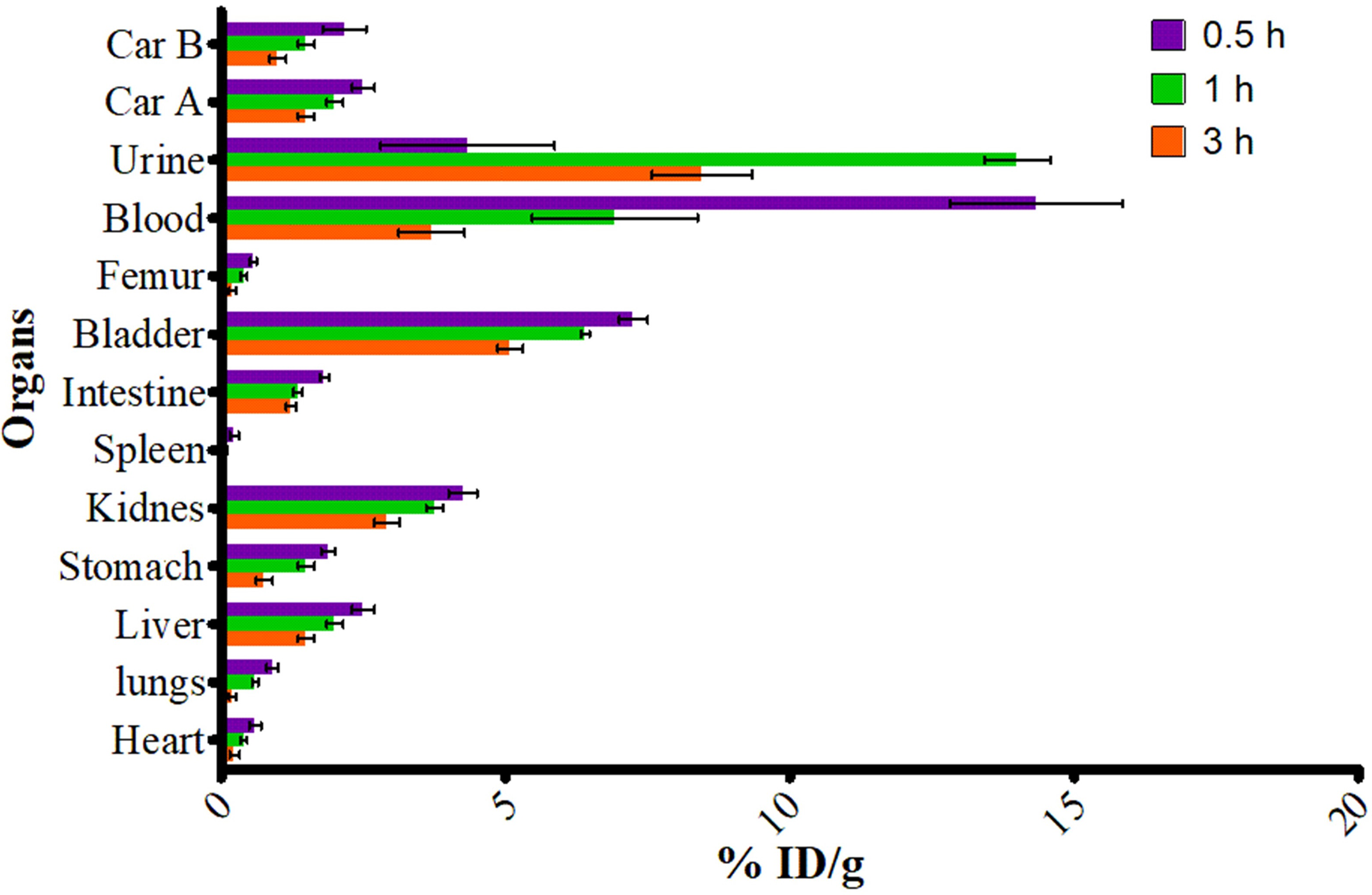
Biodistribution study of [99mTc]DTPA-FLUT in healthy mice models (mean ± SD).
Internalization study
To evaluate the [99mTc]DTPA-FLUT interaction with RMS cancer cells and its uptake, the cancer cell line was incubated with 3 pmol free [99mTcO4]− or [99mTc]DTPA-FLUT at various time intervals. Fig. 6a shows the internalized activity, surface-bound activity, and washed-out activity (fraction of externalized activity at the 30 min time point and onward) at different time intervals. Fig. 6b and c displays the morphology of RMS cell at start of incubation with radiopharmaceutical and at 2 h time points.
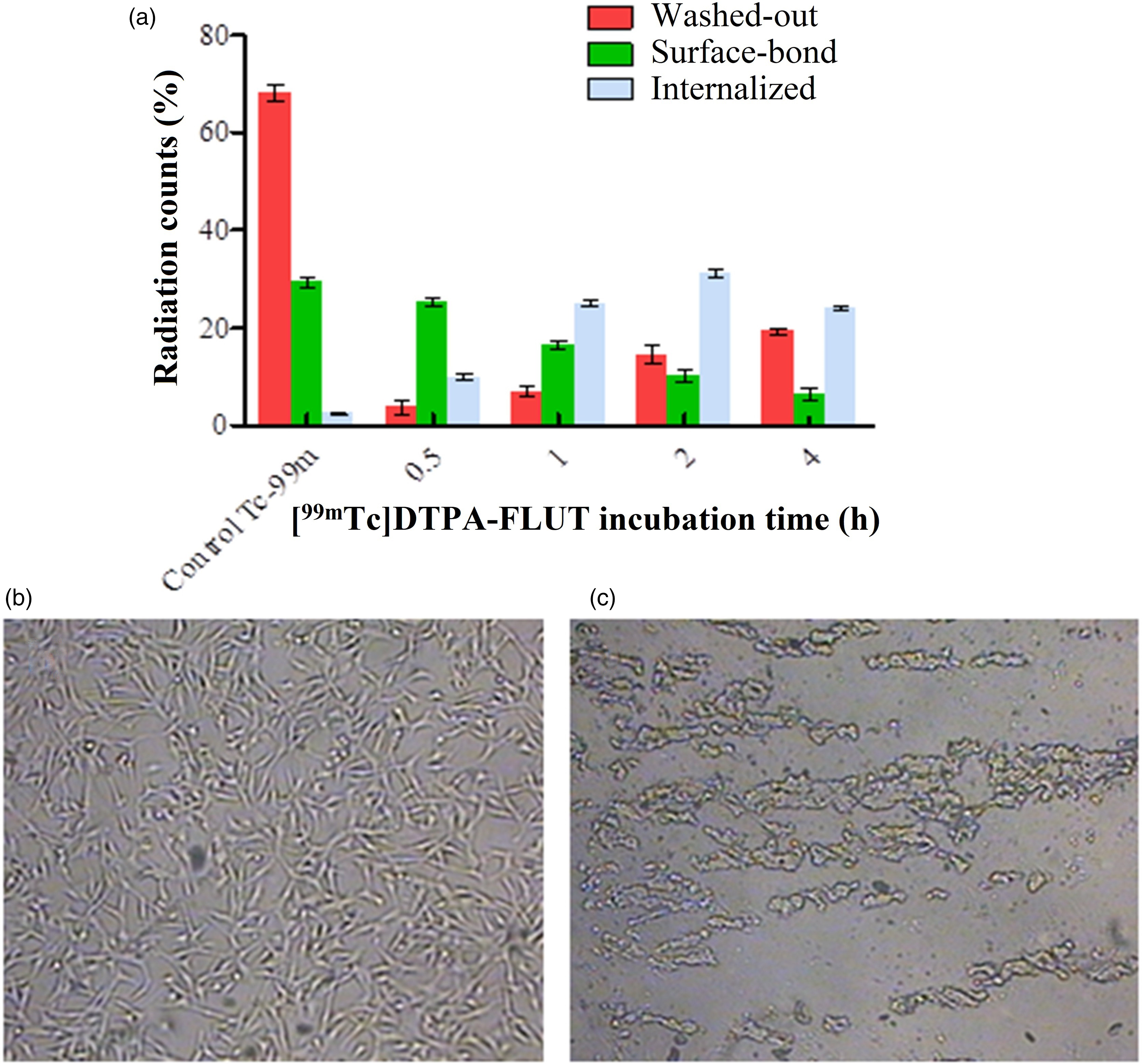
Representation of (a) internalized, surface-bound and washed-out (externalized) activity, and (b) the microscopic morphology of pre-incubated and (c) post-incubated RMS cells.
Scintigraphy study
Scintigraphy studies were conducted using a subcutaneous RMS cancer-induced rabbit model. The scintigraphy images were captured by administrating the 185 MBq/250 µL saline [99mTc]DTPA-FLUT through the marginal ear vein. The scintigraphy images were captured at the 15-min, 30-min, 1-h, and 4-h time points (Fig. 7).

Scintigraphy study of [99mTc]DTPA-FLUT at; (a) 15 min, (b) 30 min, (c) 1 h, and (d) 4 h after injection time points.
Discussion
DTPA is a well-reputed bifunctional chelating agent, commonly coupled with those biological active molecules that are unable to hold radionuclide directly. Multiple electron donor atoms (nitrogen and oxygen) over DTPA ensure a stable complex with reduced 99mTc. Before radiolabeling the purity of the coupled product was confirmed with RP-HPLC. DTPA-FLUT, [99mTcO4]− and a reducing agent were mixed at pH 5 to acquire [99mTc]DTPA-FLUT at room temperature with a high labeling yield. RP-HPLC hyphenated with NaI-FRD showed a single peak at 7.4 min, indicating high purity (Fig. 2b), which prevails the non-targeted accumulation and ensures the possibility of target-specific accumulation (7,18). Compared to many other clinical radiopharmaceuticals, the preparation of [99mTc]DTPA-FLUT does not require heating and purification, thus making it easier for clinical application. Paper electrophoresis displaying the complex is uncharged. Maximum radioactivity counts were detected over paper at the point of introduction while a small number of counts coming from free [99mTcO4]− were detected near the anode terminal. The uncharged radiopharmaceutical favors accumulation at cancer cells compared to positively charged radiopharmaceuticals; however, a negative charge over the radiopharmaceutical favors greater accumulation (19). The stability tests at pH 5 indicate that the [99mTc]DTPA-FLUT complex is sufficiently stable for the period required for a patient study. 99mTc-labeled radiopharmaceuticals are considered to be stable in saline and serum at least for 6 h (half-life of 99mTc) to avoid any background radiation, whichs reduce the imaging efficiency (20). In addition, the radiopharmaceutical stability in blood serum excludes the radiopharmaceutical serum-protein binding factor and degradation of complex. This potentially favors the availability of the radiopharmaceutical to accumulate in target tissue and minimum background counts (21). However, the non-pharmacological potential of radiopharmaceuticals and the absence of radiopharmaceutical acceptor chemical moieties at the disease’s cell surface are other key factors that lower the efficiency of the radiopharmaceuticals to target the disease tissue precisely (22).
The biodistribution study of [99mTc]DTPA-FLUT in inflammation-induced mice models revealed 4.67%ID/g of the injected dose of radioactivity was excreted through urine, 14.12%ID/g was detected in the blood pool, 2.46%ID/g in the liver, 4.29%ID/g in the kidneys, 1.97%ID/g in the stomach. and 1.81%ID/g in the intestine at 0.5 h after the injection. At the 3-h time point, the stomach showed 0.78%ID/g and the intestine showed 1.01%ID/g, which reveals the presence of a minute quantity of free 99mTc. A similar pattern of biodistribution was also reported, previously, using 99mTc-labeled epirubicin (an anti-cancer agent) (7). All the data was analysed statistically (23). The kidneys and urinary bladder exhibited an increased accumulation of [99mTc]DTPA-FLUT and showed a slow washout during the period of the study. Renal accumulation is most probably due to its role as excretory path and lipophilic nature of the radiopharmaceutical. Other organs, such as the heart, lungs, spleen, and femur, showed a negligible number of counts at the end of study period, as shown in Fig. 5. Car-A, at each time point in the study, showed slightly more uptake than Car-B, which might be due to the presence of cancer cells that were injected for tumor growth. Cancer cells were injected into the left thigh muscle (Car-A) to induce a tumor in non-immunocompromised mice but no tumor was grown; however, minute inflammation was noted. The biodistribution and pharmacokinetics of [99mTc]DTPA-FLUT in various organs of healthy mice were found to be consistent with previously reported data by different research groups.
An in vitro cell binding study displayed that [99mTc]DTPA-FLUT had the promising cell binding potential for RMS cells than that of free [99mTcO4]−, which showed no binding affinity in comparison. At the 30-min time point, 31% activity appeared to bind with cells and 2% was found to internalize into the cell matrix. The internalized activity gradually increased to 29% at the 2-h time point, which then decreased to 24% at the 4-h time point, which was due to the externalization of internalized activity. In another study, DTPA coupled cetuximab conjugated with 99mTc (99mTc-p-SCN-Bzl-DTPA cetuximab) and showed 19.55% internalized radiopharmaceutical into FaDu tumor cells; compared to this result, [99mTc]DTPA-FLUT showed greater internalization potential (24). According to the literature review, RMS cell membranes bear no receptor proteins for FLUT for binding but it is assumed that [99mTc]DTPA-FLUT may develop some cell membrane chemical interaction that triggers the endocytosis/internalization process. The microscopic morphology of pre-incubated (Fig. 5b) and post-incubated RMS cells (Fig. 5c) were also recorded to note the effects of the internalization and externalization processes. At the 2-h and 4-h post-incubation time points, the RMS cells showed deteriorated morphology (Fig. 5c), which might be due to the externalization of the radiopharmaceutical. FLUT, in particular, is used as a therapeutic drug for prostate and bladder carcinoma by blocking the action of endogenous and exogenous testosterone, and as an inhibitor of prostatic DNA synthesis. Therefore, the [99mTc]DTPA-FLUT could be tested for the imaging of prostatic and bladder carcinoma. The exact mechanism of internalization of [99mTc]DTPA-FLUT into RMS cancer cells is not known but the results refer to value added use in the imaging of RMS carcinoma as well. The assumption on the basis of the literature could be made that the yes-associated protein (YAP) might be involved in interacting with [99mTc]DTPA-FLUT over RMS cell membrane for the endocytosis process (25,26).
The scintigraphy study was conducted using a subcutaneous RMS cancer-induced rabbit model. Administration of [99mTc]DTPA-FLUT (185 MBq/250 µL saline) through the marginal ear vein followed by capturing the static images at 15 min, 30 min, 1 h, and 4 h showed mild uptake of lesion tissues. The liver and kidney showed relatively high uptake, which is due to metabolic excretory pathway and lipophilic nature of [99mTc]DTPA-FLUT (logP = 1.79). The SPECT dynamic imaging showed the rapid clearance of activity from the blood circulation. Most of the activity was excreted to the bladder, which was the final excretory path. The rapid clearing feature of [99mTc]DTPA-FLUT from the body organs and filtering from the body fluid can significantly reduce the radioactivity body burden (27). For the imaging lesion, the region of interest (ROI) was drawn followed by the calculation of uptake activity in the tumor and normal thigh muscle, and target-to-non–target ratio (T/NT). SPECT images (Fig. 7) showed 1.169 T/NT at 30 min and 1.428 T/NT at 1 h in the post-injection period. At 15 min, the imaging phase with 50% bar value showed the radiopharmaceutical uptake and low background value. The 30- and 60-min imaging phases taken at bar values of 25%–30% showed sufficient uptake by different organs and tumor cells. The final imaging phase (at 4 h) recorded with the maximum bar value showed weak activity counts in different organs that were very weak at 50% or low bar value, which indicates up to 4 h maximum amount of [99mTc]DTPA-FLUT clear from body. The T/NT value cited in literature for various radiopharmaceuticals is in good agreement with our results (9,28).
In conclusion, [99mTc]DTPA-FLUT could be considered for preclinical and clinical studies to collect more evidence for its suitability as an imaging agent for RMS carcinoma. However, further studies are also needed to evaluate the interaction of [99mTc]DTPA-FLUT with RMS cell membrane and mechanism of internalization.
Footnotes
Acknowledgment
The authors are grateful to Dr Muhammad Rafi, Pakistan Institute of Engineering and Applied Science (PIEAS), Islamabad, Pakistan, for providing the RMS cancer cell line; the National Institute of Health (NIH) Islamabad, Pakistan, for providing healthy Sprague–Dawley mice and New Zealand white rabbits for the biodistribution and scintigraphy study; NESCOM Hospital, Islamabad, for obtaining scintigraphy images. The animal ethical committee of Government College University Faisalabad gave approval to carry out biodistribution and scintigraphy studies using animal models.
Declaration of conflicting interests
The author(s) declared no potential conflicts of interest with respect to the research, authorship, and/or publication of this article.
Funding
The author(s) received no financial support for the research, authorship, and/or publication of this article