



Abstract
Background
SynthesiZed Improved Resolution and Concurrent nOise reductioN (ZIRCON) is a multi-kernel synthesis method that creates a single series of thin-slice computed tomography (CT) images displaying low noise and high spatial resolution, increasing reader efficiency and minimizing partial volume averaging.
Purpose
To compare the diagnostic performance of a single set of ZIRCON images to two routine clinical image series using conventional CT head and bone reconstruction kernels for diagnosing intracranial findings and fractures in patients with trauma or suspected acute neurologic deficit.
Material and Methods
In total, 50 patients underwent clinically indicated head CT in the ER (15 normal, 35 abnormal cases). A non-reader neuroradiologist established the reference standard. Three neuroradiologists reviewed two routine clinical series (head and bone kernels) and a single ZIRCON series, detecting intracranial findings or fractures and rating confidence (0–100). Sensitivity, specificity, and jackknife free-response receiver operating characteristic (JAFROC) figure of merit (FOM) were compared (limit of non-inferiority: −0.10).
Results
ZIRCON and conventional images demonstrated comparable performance for fractures (sensitivity: 51.5% vs. 54.5%; specificity: 40.2% vs. 34.2%) and intracranial findings (sensitivity: 88.2% vs. 91.4%; specificity: 77.2% vs. 73.7%).The estimated difference of JAFROC FOM demonstrated ZIRCON non-inferiority for acute pathologies overall (0.003 [95% CI=−0.051–0.057]) and fractures (0.048 [95% CI=−0.050–0.145]) but not for intracranial findings alone (−0.024 [95% CI=−0.100–0.052]).
Conclusion
Thin-slice, low noise, and high spatial resolution images can be created to display intracranial findings and fractures replacing multiple images series in head CT with similar performance. Future studies in more patients and further algorithmic development are warranted.
Introduction
Computed tomography (CT) is widely used for the diagnosis of various acute and chronic diseases in the head (1–3). Current practice in clinical radiology is to create (i.e. reconstruct) multiple image series from the same acquired data to separately visualize the brain and calvarium (4–6). The reconstruction process can utilize filtered back projection, iterative reconstruction, or deep-learning algorithms. During reconstruction, the spatial resolution and image noise of the resultant images can be adjusted using a parameter known as a kernel. Sharp reconstruction kernels are used to maximize spatial resolution in the bone at the cost of increased noise but display sharp margins at fracture boundaries; smooth kernels are used to visualize the brain and soft tissues to minimize image noise, so that small attenuation differences can be better appreciated, but at the cost of blurred anatomical details (7). As a result, no single set of CT images is “best” for all diagnostic purposes. Avoiding the inefficiency of reconstructing, transmitting, storing, presenting, and reading multiple image series might make better use of technical and personnel resources, particularly radiologist interpretation time.
The need for different reconstruction kernels for different diagnostic tasks is well understood. Earlier studies have reported combination multi-kernel techniques (8–10), but no method that combines the advantages of brain and bone images into a hybrid image series has been shown to produce clinically acceptable images in a reader study. SynthesiZed Improved Resolution and Concurrent nOise reductioN method (ZIRCON) is a convolutional neural network (CNN)-based denoising technique that creates a single thin slice CT image series that offers both low image noise and high spatial resolution (11,12). ZIRCON has been previously validated in abdominal CT (11).
This work addresses additional diagnostic challenges to ZIRCON present in head CT. Smooth reconstruction kernels for the head often incorporate additional processing to enhance the small contrast differences between gray and white matter (13,14). To account for this, two objective functions were added to ZIRCON to minimize noise in the brain while maintaining high spatial resolution in bone imaging tasks (12). We hypothesized that this modified ZIRCON approach could provide a single, thin-slice image series adequate for both brain and bone imaging without compromising diagnostic performance compared to multiple conventional image series. The aim of the present study was to compare the diagnostic performance of a single set of ZIRCON images to two routine clinical image series using conventional CT head and bone reconstruction kernels for diagnosing intracranial findings and fractures on unenhanced head CT.
Material and Methods
Patients and reference
Our Institutional Review Board approved this HIPAA-compliant study and waived the requirement for informed consent for this retrospective case-control study. All patients permitted the use of their clinical records for research purposes.
The inclusion criteria were as follows: (i) patient consent to use of clinical records for research purposes; and (ii) archived CT projection data from patients who underwent head CT scan for trauma or suspected acute neurological symptoms in our emergency department. CT scans and CT projection data of qualifying patients were archived daily by study personnel, and subsequently reviewed by a non-reader neuroradiologist (F.E.D., with 12 years of staff experience at Mayo Clinic after completing neuroradiology fellowship). The reference standard for intracranial findings and fractures was also established by this non-reader neuroradiologist, who examined clinical CT images (including oblique, coronal, and sagittal images), comparing the clinical CT report to imaging findings, as well as prior and subsequent CT or magnetic resonance imaging exams and all existing medical records (including mechanism of injury if relevant and physical exam), marking positive findings on clinical images in cases and recording absence of positive findings for controls. The exclusion criteria were as follows: (ⅰ) insufficient reference information; (ⅱ) obvious imaging findings (e.g. large subdural hematoma or vast lobar brain infarction); (ⅲ) notable metal or motion artifact; and (ⅳ) non-unique patients. Cases were excluded by the non-reader neuroradiologist, who created the reference standard for intracranial findings and fractures. Before the start of the reader study, the target number of patients for the study cohort, which was a convenience sample, was set at 50 by the study co-authors, to include 35 abnormal cases and 15 controls (to allow for evaluation of potential false-positive findings).
CT acquisition and reconstruction
Unenhanced spiral head CT examinations were performed using our clinical head trauma or acute stroke protocols. Both protocols utilize the following parameters: tube rotation time = 1 s; detector collimation = 192 × 0.6 mm; pitch = 0.6; 120 kV; 350 effective mAs (without tube current modulation); and CTDIvol = 49.7 mGy. For reader evaluation, reconstructed images included a bone series (slice thickness = 0.75 mm; sharp Siemens [Siemens Healthineers, Forchheim, Germany] Hr69 kernel, window-level L = 60 HU, W = 3700 HU), two brain series (slice thickness = 5 mm and 0.75 mm, head Siemens Hr40 kernel, both with brain window-level setting L = 40 HU, W = 80 HU), and a single axial ZIRCON series (slice thickness = 0.75 mm, with preset displays using bone and brain window settings).
SynthesiZed Improved Resolution and Concurrent nOise reductioN (ZIRCON)
ZIRCON is a convolutional neural network (CNN) method to create a single series of thin-slice CT images displaying both low noise and high spatial resolution. The original implementation of kernel synthesis using a U-Net architecture was developed for abdominal imaging (11). To account for the unique challenges of head imaging, ZIRCON features a task-based loss function with two terms to enable parameterized tuning of specific image quality parameters specific to neuroimaging (12). The algorithm was separately trained using 300,000 paired low- and routine-dose 64 × 64 voxel patches randomly sampled from 100 patient scans, with an additional 10 patient scans used for training validation and hyperparameter tuning. These 110 exams were not included in this reader study.
Reader interpretation
In the first two sessions, three neuroradiologists (A.A.N., T.J.P., and D.R.D.) with 4, 22, and 22 years of experience, blinded to clinical and image information, randomly reviewed either clinical or ZIRCON CT images, with each patient's images displayed only once per session. Reader evaluation was performed using a computer workstation, which displayed axial clinical or ZIRCON images.
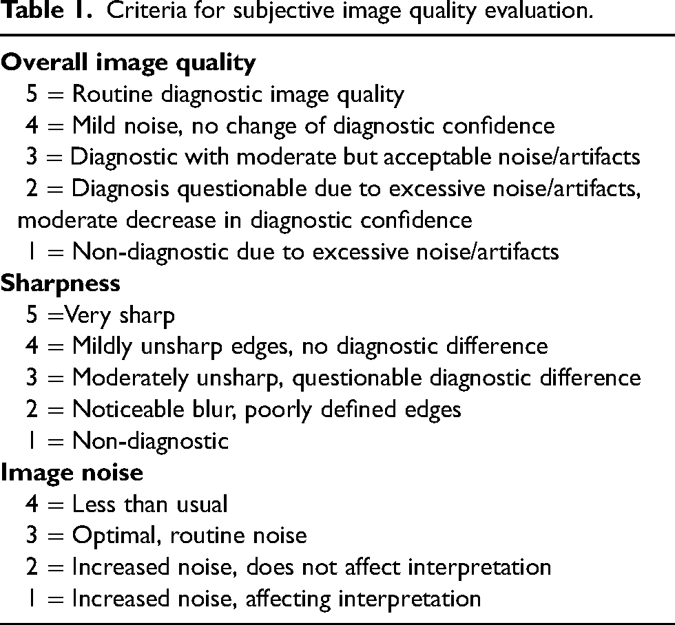
Readers were asked to mark acute intracranial findings, including subdural/epidural hematoma, subarachnoid hemorrhage, intraparenchymal hemorrhage, acute/subacute infarction, or intracranial mass, on images on thin-slice brain window images and then mark fractures on thin-slice bone window images. Window-level setting adjustments were permitted. Readers were instructed that they did not need to mark non-acute findings and extracranial acute pathology, such as a scalp hematoma. For each intracranial finding or fracture, readers recorded their confidence in diagnosis (scale of 0–100: 0 = definitely absent (i.e. no lesion), 50 = questionably present, 100 = definitely present), with knowledge that a confidence level of 25 would be used for the calculation of sensitivity and positive predictive value (PPV). In a third reading session, readers also scored overall image quality, sharpness, and image noise using a Likert scale (Table 1) (15). Reading sessions were separated by 3 weeks. Before the formal reading sessions, the readers were trained using five non-test set cases to learn how to use the workstation, with a training focus on marking imaging findings and assigning confidence scores.
Criteria for subjective image quality evaluation.
Matching of reader marking and references standard
A non-reader radiologist (A.I.) compared the readers’ markings to reference markings created by the non-reader neuroradiologist. If the reader correctly marked the lesion(s), a true positive was recorded. False-positive markings were also recorded. When readers failed to mark a reference lesion, a false negative was recorded. In case multiple marks were placed in the same continuous pathology, one mark was rated as a true positive, with additional marks not considered false positives. The senior neuroradiologist who determined the reference standard and excluded cases with obvious imaging findings examined false-negative interpretations to determine potential causes of interpretive error.
Statistical analysis
Results were analyzed for all acute findings, and separately for intracranial findings and fractures. For patient-level analyses, concordance or discordance with the reference was evaluated as follows: true positive if at least one true-positive marking was recorded in an abnormal case with a reference finding; false positive if at least one false-positive marking was recorded in a normal control; true negative if no marking was recorded in a normal control; and false negative if no true positive marking was recorded in an abnormal case. Sensitivity and specificity were calculated along with 95% confidence interval (CI). For lesion-level analyses, sensitivity and PPV with 95% CI were calculated. A generalized estimating equation (GEE) was used to provide the pooled estimate across the three readers for each imaging strategy.
Jackknife free-response receiver operating characteristic (JAFROC) figure of merit (FOM) was employed to compare reader performance using a non-inferiority study design (16). JAFROC FOM takes reader confidence and imaging findings into account and requires site-specific correlation of reader and reference markings. FOM values in JAFROC analysis are in the range of 0–1, similar to the area under the ROC curve. JAFROC FOM was calculated for each modality across readers. The difference in JAFROC FOM between routine and ZIRCON images was evaluated using forest plots, with a non-inferiority limit of −0.10 set before the start of the study. ZIRCON images were considered non-inferior to clinical routine images if the lower limit of the 95% CI of estimated difference between imaging modalities was greater than −0.10 (17–19).
For subjective image quality analysis, mean scores for overall image quality, sharpness, noise, and texture were compared between clinical routine and ZIRCON images using a linear mixed effects model; P <0.05 was considered to be a statistically significant difference. All statistical analysis was performed using R version 3.4.2 (R Foundation for Statistical Computing, Vienna, Austria).
Results
Patients
Between March 2020 and January 2021, 589 patients with head trauma or acute-onset neurological symptoms suspicious for acute intracranial pathology underwent a clinically indicated head CT in our emergency department. In total, 555 patients consented to retrospective use of their medical records for research and had CT projection data archived. Acute imaging findings were present in 88 patients. Of them, 53 patients with positive findings were excluded for the following reasons: (ⅰ) insufficient reference standard (n = 9); (ⅱ) obvious imaging findings (n = 29); (ⅲ) chronic imaging findings (n = 8); (ⅳ) notable artifact (n = 5); and (ⅴ) duplicated patient (n = 2). In the order of enrollment, 15 normal cases confirmed by the reference neuroradiologist were selected (Fig. 1).
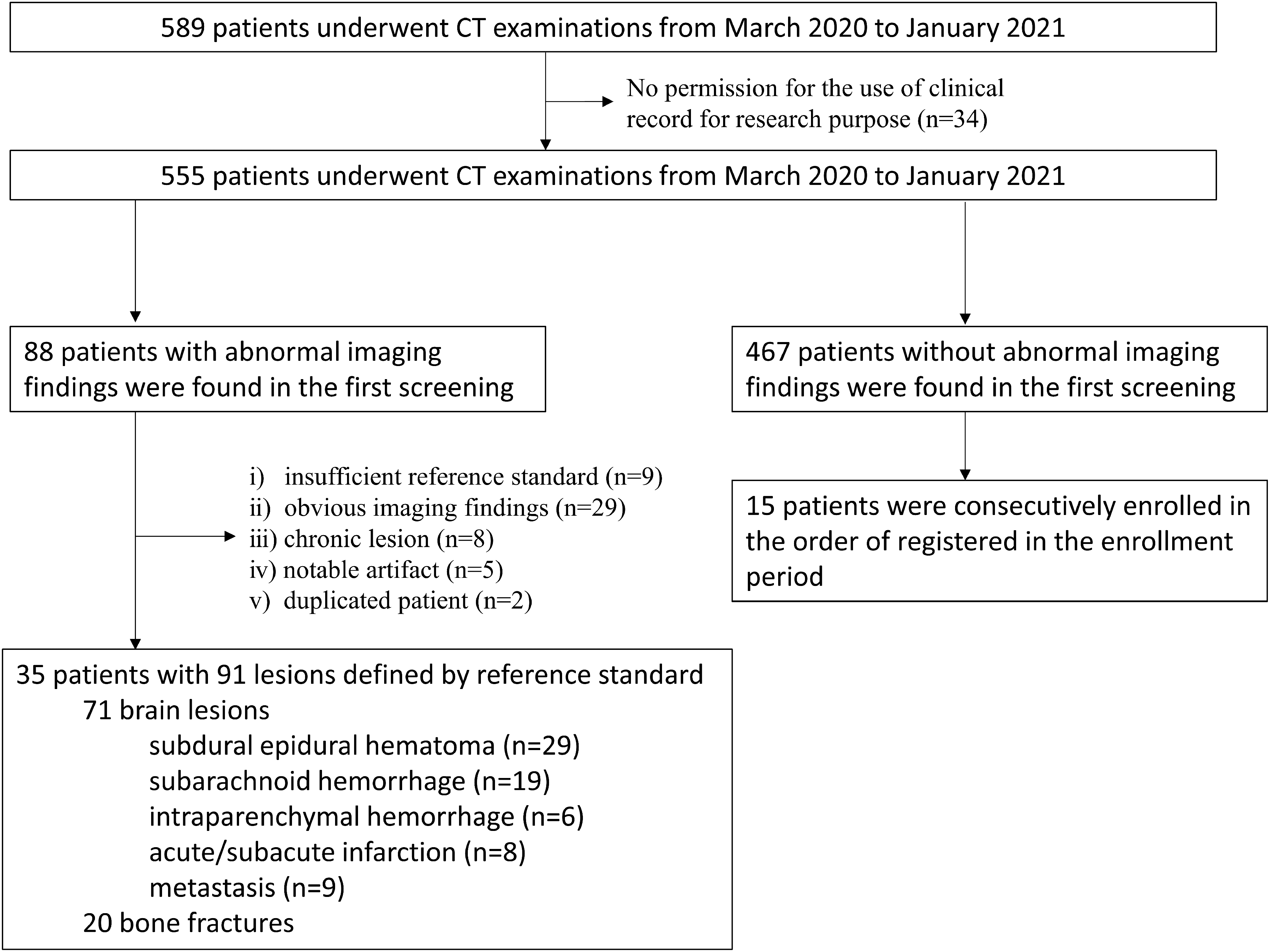
Patient screening and study population.
A total of 50 patients (20 women; mean age = 66.5 ± 18.9 years), including 35 abnormal patients and 15 normal patients, comprised the study cohort. These patients included 91 positive findings by the reference standard (71 intracranial findings in 31 patients; 20 fractures in 11 patients). Intracranial findings included subdural/epidural hematoma (n = 29), subarachnoid hemorrhage (n = 19), intraparenchymal hemorrhage (n = 6), acute or subacute infarction (n = 8), and metastases (n = 9).
Patient-level analyses
Table 2 shows the patient-level sensitivity and specificity for all acute findings, intracranial findings alone, and fractures, for each neuroradiologist reader and across readers using generalized estimate equations. For each reader and overall, there was a similar sensitivity and specificity for overall findings, intracranial findings, and fractures, with widely overlapping 95% CIs. Sensitivity and specificity for fractures were lower than for intracranial findings, regardless of whether ZIRCON or conventional images were viewed.
Patient-level sensitivity and specificity across neuroradiologist readers for intracranial findings and fractures on unenhanced head CT using two conventional reconstruction kernels (at two slice thicknesses) vs. a single ZIRCON reconstruction kernel on a per-patient level.

Values in parentheses are 95% CI.
CI, confidence interval; GEE, generalized estimate equations; PPV, positive predictive value; ZIRCON, SynthesiZed Improved Resolution and Concurrent nOise reduction.
Lesion-level and reader confidence analyses
Table 3 shows the sensitivity and positive predictive value for all acute findings, intracranial findings alone, and fractures, for each neuroradiologist reader and across readers using generalized estimate equations. For all acute imaging findings, as well as intracranial findings and fractures, pooled GEE sensitivity for ZIRCON and routine clinical images also demonstrated similar sensitivity and PPV (Figs. 2 and 3). A similar pattern was observed for each reader individually.

Early subacute brain infarction (low attenuation lesion). A 90-year-old woman presented with left upper extremity flaccid paresis, left gaze preference, left visual field deficit. Head CT was performed 2 days later. (a, b) The low-attenuation region involving gray and white matter in the right occipital lobe compatible with early subacute brain infarction is observed on (a) a 0.75 mm ZIRCON (arrows) as well as (b) a 5 mm brain setting clinical image (arrows). ZIRCON image more clearly depicts gray-white junction compared to clinical image despite the slightly altered texture of brain parenchyma. One and two readers correctly diagnosed the lesion on ZIRCON and clinical images, respectively. In the reader who correctly diagnosed both images, the confidence score compared to the clinical image was decreased on ZIRCON (from 90 to 70). This effect may be related to the relative unfamiliarity with the technique by the readers. CT, computed tomography; ZIRCON, SynthesiZed Improved Resolution and Concurrent nOise reduction.
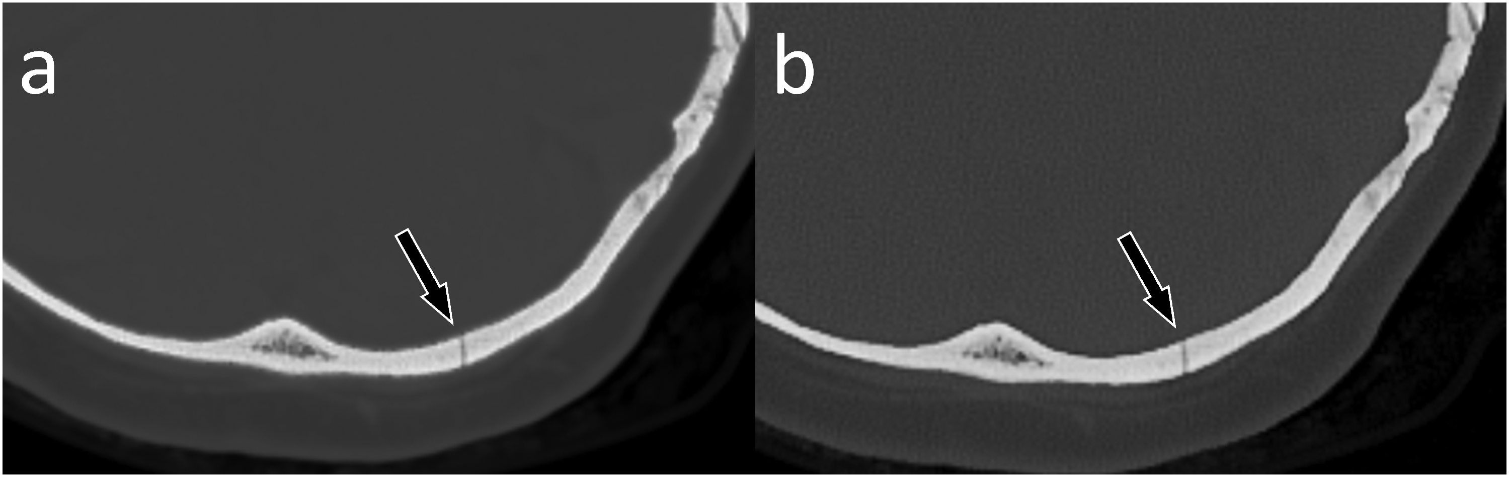
Occipital fracture (bone lesion). Head CT in a 55-year-old woman after a fall. (a, b) The fracture line is shown on (a) a 0.75 mm ZIRCON image (arrow) as well as (b) a 0.75 mm bone setting clinical image (arrow). Three and one readers identified the fracture correctly on ZIRCON and clinical images, respectively. ZIRCON, SynthesiZed Improved Resolution and Concurrent nOise reduction.
Sensitivity and positive predictive value for neuroradiologist detection of intracranial findings and fractures compared to the reference standard on a per-lesion level, according to the reference standard, for clinical images (using two different reconstruction kernels and slice thicknesses) vs. a single ZIRCON reconstruction kernel with thin slice thickness.

CT, computed tomography; GEE, generalized estimate equations; PPV, positive predictive value; ZIRCON, SynthesiZed Improved Resolution and Concurrent nOise reduction.
JAFROC FOM also considers reader confidence for individual imaging findings, taking multiple findings into account for each patient. Figure 4 shows the JAFROC FOM for all acute imaging findings, intracranial findings, and fractures across readers with their corresponding 95% CI for detection on both ZIRCON and conventional CT images. For each detection task, the JAFROC point estimates were similar and the 95% CIs widely overlapped (Fig. 4a–c).
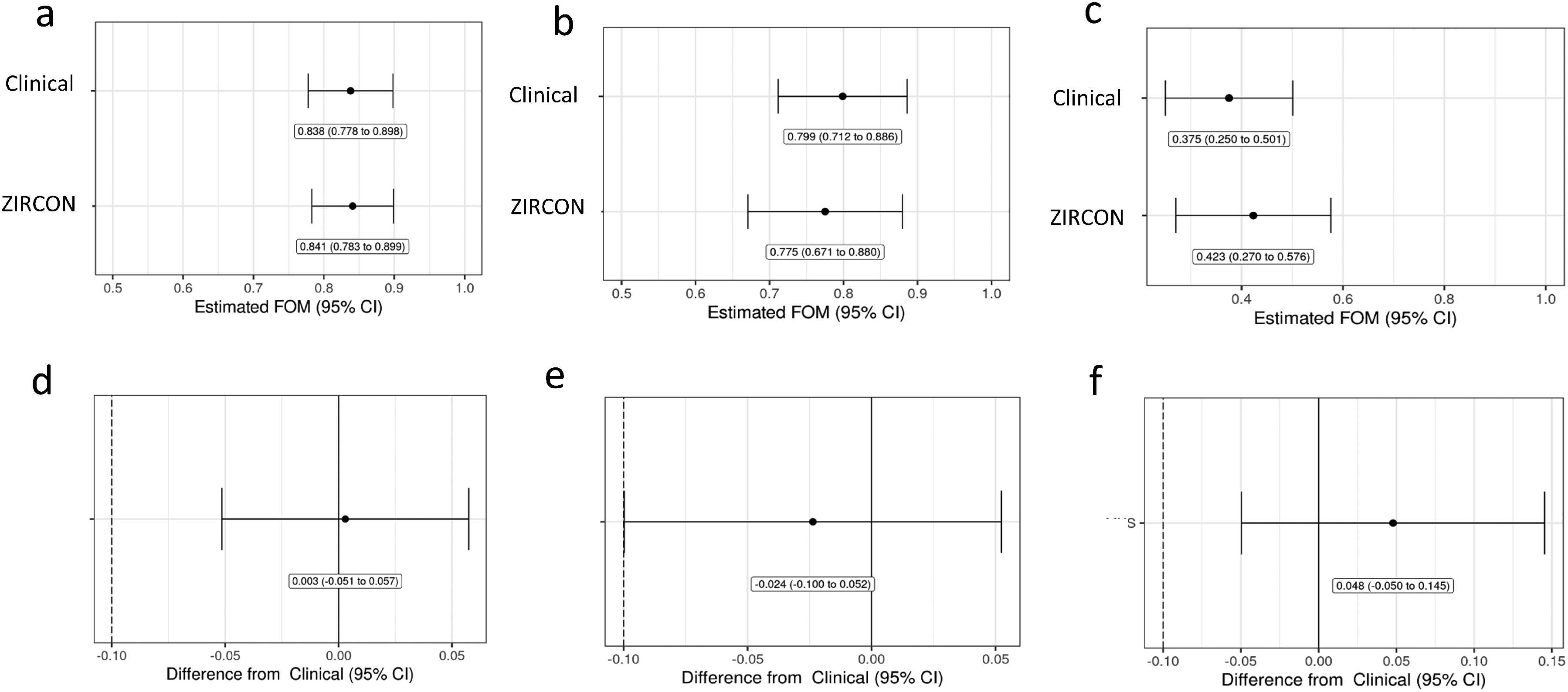
JAFROC FOM for clinical routine and ZIRCON images along with 95% confidence intervals in diagnosing (a) all acute findings, (b) intracranial findings, and (c) fractures. Forest plots demonstrate estimated differences in JAFROC FOM between ZIRCON CT images compared to routine clinical CT images in (d) all acute imaging findings, (e) intracranial findings, and (f) fractures, taking into account reader confidence, correct localization, and potential for multiple findings per patient. When the lower limit of the 95% confidence interval of the estimated difference between modalities exceeds the preset threshold of non-inferiority (−0.10, which is denoted by the dotted line in panels d, e, and f), ZIRCON CT images are non-inferior to routine clinical CT images. (d and f) Forest plots show that considering all acute imaging findings as well as fractures alone, respectively, ZIRCON CT images are non-inferior. (e) For intracranial findings, the lower limits of the 95% CI overlap with the preset threshold, so non-inferiority is not demonstrated. CT, computed tomography; FOM, figure of merit; JAFROC, jackknife free-response; ZIRCON, SynthesiZed Improved Resolution and Concurrent nOise reduction.
Forest plots were used to show the estimated difference in JAFROC FOM performance between ZIRCON and conventional CT images, with these plots also displaying the 95% CI of the estimated difference (Fig. 4d–f). When the lower limit of the 95% CI was greater than the preset limit of non-inferiority, non-inferiority was demonstrated. Overall, ZIRCON was non-inferior to clinical images for all acute imaging findings, as the lower limit of 95% CI of estimated difference in JAFROC FOM between modalities (i.e. −0.051) was greater than the non-inferiority limit (−0.10) (Fig. 4d).
For acute intracranial findings, the JAFROC FOM for intracranial findings in ZIRCON images was slightly lower than that for clinical routine images (i.e. 0.755 vs. 0.799, respectively) (Fig. 4b). The lower limit of the 95% CI of the estimated difference between the imaging modalities was −0.10, which was also the limit of non-inferiority (Fig. 4e), so ZIRCON images did not meet the preset threshold for non-inferiority for intracranial findings. False-positive markings of subarachnoid hemorrhage and subdural hematoma were more frequently seen in ZIRCON than clinical images (R1: 6 vs. 3, R2: 9 vs. 4, R3: 1 vs. 0). Subsequent evaluation of these markings by the senior non-reader neuroradiologist demonstrated that ZIRCON images revealed a high-attenuation edge artifact adjacent to the calvarium in some patient images, which mimicked subarachnoid hemorrhage or subdural hematoma as shown in Fig. 5. Careful inspection of the input clinical images suggests such artifacts are present in the smooth thin image series from the enhanced soft-tissue postprocessing but are made more conspicuous after noise removal by ZIRCON.
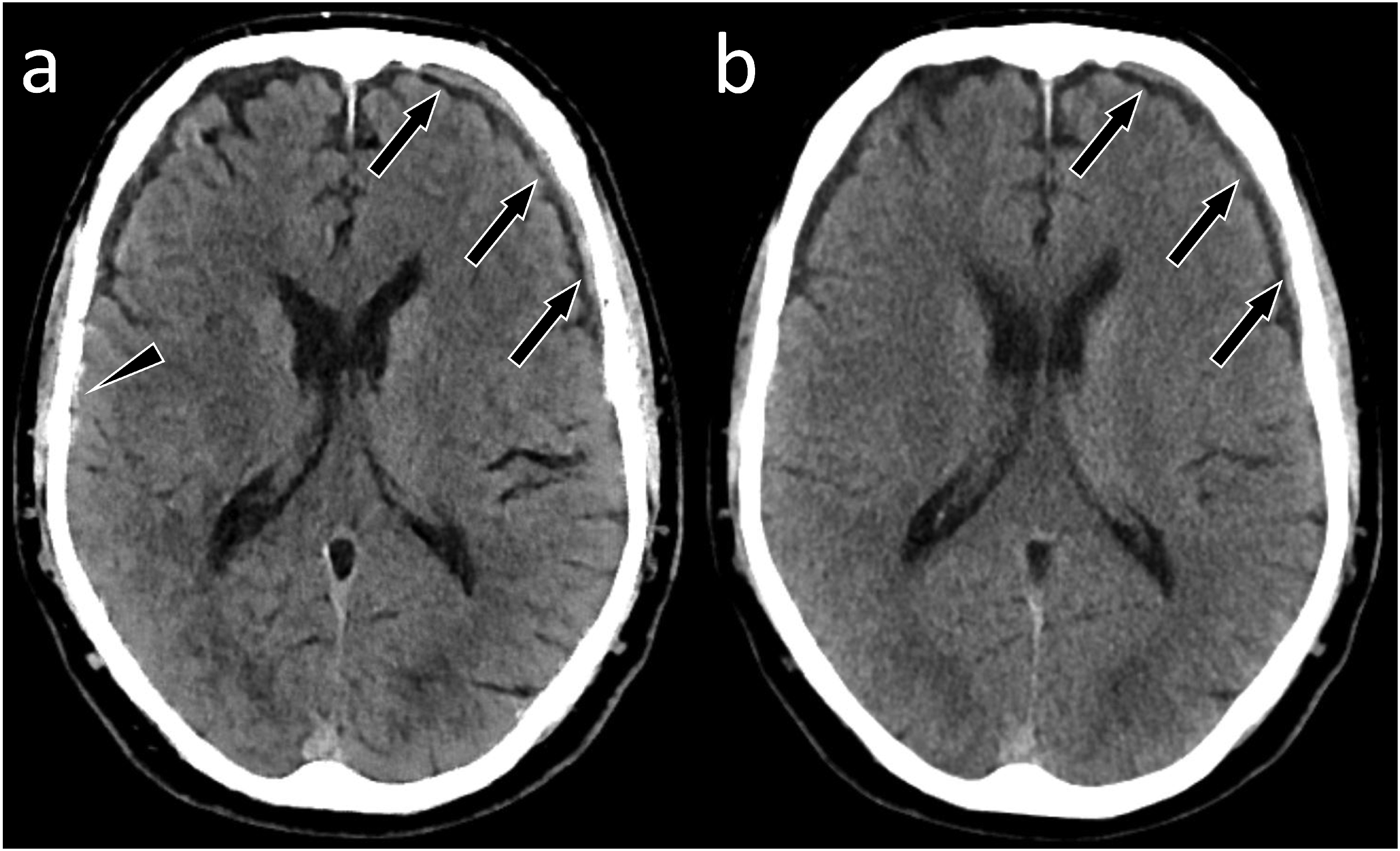
Small subdural hematoma (high attenuation lesion) and edge artifact on ZIRCON causing a false positive. ZIRCON and clinical CT images in a 74-year-old man after left-sided subdural hematoma. He had an episode of transient speech difficulty. The thin residual left frontal subdural hematoma is depicted on (a) a 0.75 mm ZIRCON image (arrows) as well as (b) a 5 mm brain setting clinical image (arrows). All three readers diagnosed this lesion correctly, with confidence score of 100 on both routine and ZIRCON images for two readers, and a third reader having confidence scores of 100 on routine and 64 on ZIRCON images. Note that a thin high-attenuation region is displayed at the edge of the right temporal lobe on ZIRCON images (panel a, arrowhead), which may mimic subdural hematoma or subarachnoid hemorrhage, whereas the clinical image shows no corresponding imaging finding. One reader misdiagnosed this apparent lesion as a subdural hematoma, with confidence level of 75 on ZIRCON. CT, computed tomography; ZIRCON, SynthesiZed Improved Resolution and Concurrent nOise reduction.
For fractures, the point estimates for JAFROC FOM for ZIRCON images was higher than conventional CT images, so the point estimate for the difference was positive in favor of ZIRCON (Fig. 4f). The lower limit of the confidence interval was −0.05, so ZIRCON images are non-inferior for fracture detection.
Because the JAFROC FOM was lower for fractures compared to intracranial findings, the senior non-reader neuroradiologist reviewed all false-negative fracture interpretations by readers using both types of CT images (18 false-negative fractures missed on conventional images by at least one neuroradiologist; 16 missed on ZIRCON images). The majority of missed fractures were very subtle, non- or minimally displaced fractures without soft tissue swelling in the setting of larger, more obvious findings (Fig. 6). Regarding false-positive markings, there were 15 patients with 34 false-positive markings for fractures by at least one of the neuroradiologists on conventional CT images; similarly, 12 patients had 39 false-positive markings on ZIRCON images.
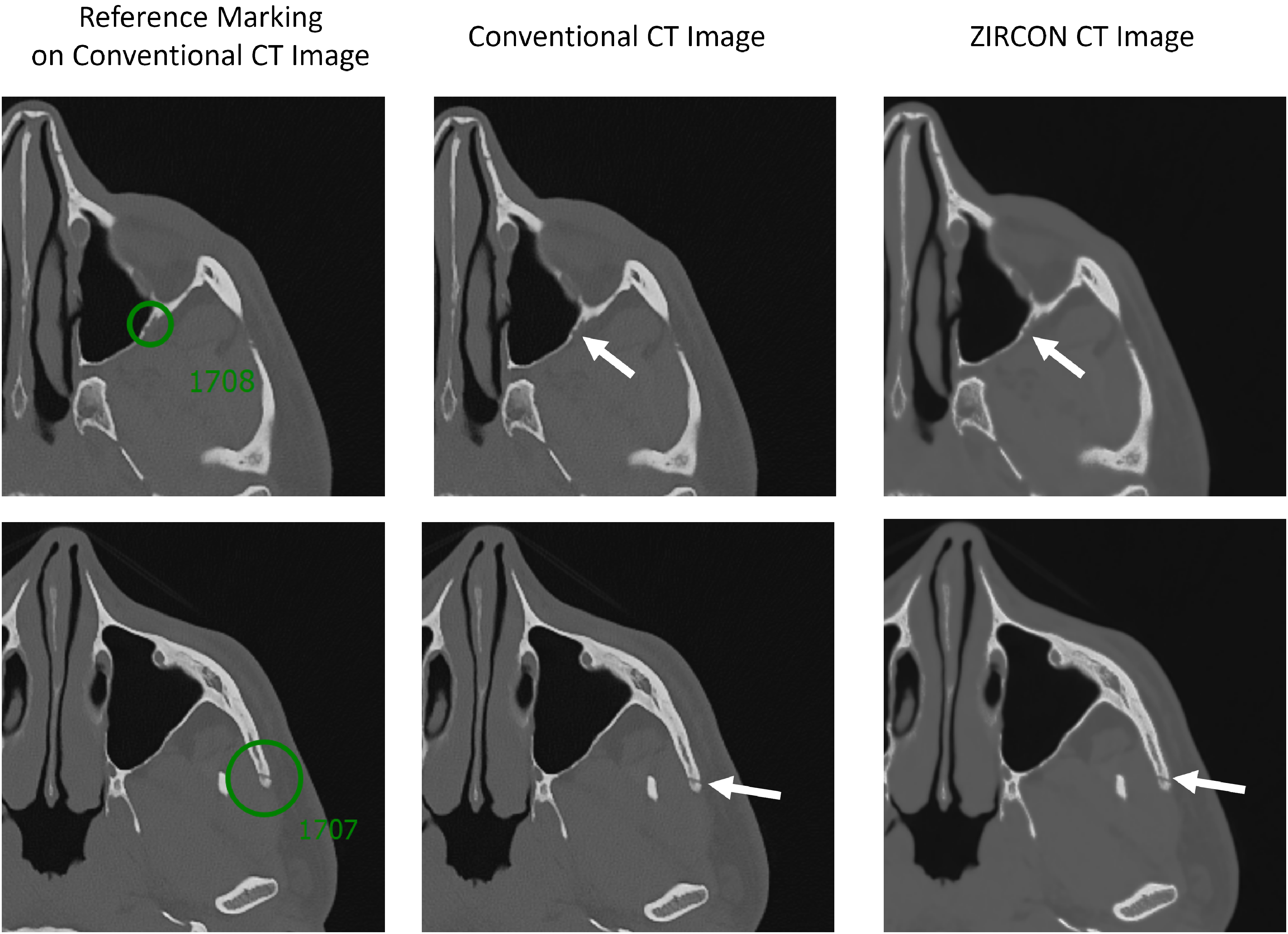
Conventional and ZIRCON images showing reference reader markings and false-negative interpretations by readers in a subject with multiple other traumatic findings including other fractures. Reference markings are denoted by the computer software in the left column using green circles and numbers. Top: a subtle non-displaced fracture along the posterior wall of the left maxillary sinus (arrows), which was missed by all three readers examining both types of CT images. Bottom: non-displaced left zygomatic arch fracture without overlying soft tissue swelling (arrows), which was missed by 2 of 3 readers using both imaging modalities. CT, computed tomography; ZIRCON, SynthesiZed Improved Resolution and Concurrent nOise reduction.
Image quality analyses
ZIRCON images were rated slightly but significantly lower than clinical images in scores of overall image quality (intracranial [3.9 vs. 4.9; P < 0.001]; bone [4.3 vs. 4.9; P < 0.001]), sharpness (intracranial [3.7 vs. 4.8; P < 0.001]; bone [4.3 vs. 4.9; P < 0.001]), and image noise (intracranial [2.0 vs. 2.9; P < 0.001]; bone [2.6 vs. 3.0; P < 0.001]).
Discussion
ZIRCON is a multi-kernel synthesis denoising method that creates a single thin-slice image series that is optimized both for display of low contrast intracranial lesions in the brain and high spatial resolution image features in the bone such as fractures. In the setting of trauma, use of a single thin-slice image series for multiple diagnostic tasks is highly desired for reader efficiency, as well as for minimizing partial volume averaging. Our multi-reader study was subsequently conducted to compare the relative performance of ZIRCON images versus conventional CT (multiple kernels and slice thicknesses) in head CT for patients in the emergency department in a highly constrained and challenging patient population. We found that ZIRCON images demonstrated comparable diagnostic performance for acute pathologies on both a per-patient and per-lesion level. Using a non-inferiority study design and the JAFROC FOM, ZIRCON demonstrated non-inferiority for the detection of all acute findings and fractures. For intracranial findings alone, however, the lower limit of the 95% CI of the estimated difference between ZIRCON and conventional images overlapped with the preset limit of non-inferiority, so non-inferiority was not demonstrated.
Combining the ability to simultaneously display low contrast structures in the brain and high spatial resolution in the bone at thin slices has substantial promise for decreasing the resource inefficiency of reconstructing, transmitting, storing, presenting, and interpreting multiple image series, as well as minimizing partial volume effect at CT head imaging, while preserving observer performance. Since image sharpness must often be sacrificed for reduced noise and vice versa (7), it is challenging to simultaneously maintain the diagnostic performance for both bone and brain pathologies, which are required in patients presenting with trauma/acute neurologic deficit (20). Previous efforts at balancing these trade-offs in a single image have used threshold-based methods to optimize display of the bone but not brain (9), or to replace bone pixels in a smooth kernel image with sharp kernel pixels (8). The CNN denoising model approach of ZIRCON is well suited to the multi-kernel imaging task by removing noise from a sharp image while providing flexibility to incorporate image quality optimizations, such as enhanced soft tissue contrast and thinner images than in previous studies (8,9).
The per fracture analysis are consistent with a previous study combining CT image kernel images (9) and showed that the single ZIRCON image series demonstrated comparable diagnostic performance to the routine clinical image series evaluated by each reader. The sensitivities of both clinical and ZIRCON images in our study were lower than expected compared to the previous study estimating the performance of CT for calvarial fracture detection (21); these findings may have occurred because we included facial fractures (9), excluded patient with obvious abnormalities, and examined only axial images. Since the aim of this study was to compare the diagnostic performance between modalities, the relative difference in diagnostic performance and non-inferiority analyses were study endpoints.
For the comparison of intracranial imaging findings, although sensitivity and PPV were comparable between clinical and ZIRCON images, the JAFROC analysis did not show non-inferiority of ZIRCON, as the difference in JAFROC FOM between the two types of images just reached the limit of inferiority (0.10). Image quality factors potentially affecting reader confidence and JAFROC include unsatisfactory image texture from applying too much denoising strength and artifacts at the brain–skull interface potentially mimicking pathology. The denoising strength used in this study was determined from non-reader (A.I.) expert feedback during hyperparameter tuning to balance noise reduction and image texture. Our analysis demonstrated an edge artifact present in the input of Hr40 smooth kernel images that was exacerbated after ZIRCON denoising and possibly decreased the reader's confidence for extra-axial blood products near the calvarium, potentially resulting in false positive findings on ZIRCON intracranial images, which mimicked subarachnoid hemorrhage and/or subdural hematoma. Further tuning of ZIRCON is needed to suppress these artifacts and avoid false positives.
The present study has some limitations. First, the number of patients and lesions was relatively small and limited to bone or intracranial findings from patients in the emergency department. We did not perform an a priori power calculation given the preliminary nature of our study. As this feasibility study was undertaken to assess a new image type combining the advantages of two reconstruction kernels, a performance estimate was not available. In addition, our study was constrained by practical resources available. We consequently chose a non-inferiority study design that takes power into account by using 95% confidence intervals. It is unclear if additional cases would have narrowed the confidence interval associated with the estimated difference in performance between conventional and ZIRCON images relating to intracranial findings. Second, the readers could recognize image type despite being blinded to this information. Because the reader neuroradiologists were familiar with the clinical routine head CT image quality and because clinical and ZIRCON image sets had three panels and two panels, respectively, it was clear which were a clinical routine or ZIRCON series. Third, only axial images were shown in the reading session to simplify the evaluation. In real clinical practice, multiple image series including coronal and sagittal planes of brain and bone setting are generally used (22). We reported reader results individually to address reader performance and perceptual differences between conventional and ZIRCON images and set the time period between reading sessions at 3 weeks; a longer period of memory extinction may have minimized recall bias to a greater extent. Our study design did not address intra-rater and inter-rater reliability of radiologists’ detection of intracranial findings and fractures using ZIRCON images.
In conclusion, ZIRCON is a CNN-based multi-kernel method to create low noise, high spatial resolution images that display intracranial and bony pathology in a single thin-slice image series, with a similar performance compared to multiple conventional CT image series. While further optimization is needed, ZIRCON may help to reduce the workload in radiology departments by reducing the amounts of images needing to be reconstructed, transferred, stored, and interpreted. Future studies in larger numbers of patients after algorithmic improvement appear warranted. In this work, ZIRCON displayed acute intracranial findings and osseous anatomy in a single series of thin images with comparable diagnostic performance to viewing three clinical image series.
Footnotes
Acknowledgments
The authors want to thank Mr Kevin Kimlinger for his assistance with manuscript preparation. This research was presented at the 2021 RSNA meeting (28 November 2021 to 2 December 2021).
Declaration of conflicting interests
The authors declared the following potential conflicts of interest with respect to the research, authorship, and/or publication of this article: CHM and JGF are the recipients of a research grant to the institution from Siemens Healthineers, unrelated to this work. The other authors have no relevant conflicts of interest to disclose.
Funding
The authors received the following financial support for the research, authorship, and/or publication of this article: This work was supported by the CT Clinical Innovation Center, the Mayo Clinic Graduate School of Biomedical Sciences, and with funding from the 2020 Mayo Discovery Translation Grant.
