



Abstract
Background
Concurrent chemoradiotherapy (CCRT) is used as the primary treatment modality for currently limited cervical cancer and lacks non-invasive quantitative parameters to assess clinical outcomes of treatment for cervical cancer treatment.
Purpose
To develop nomograms based on clinical prognostic factors and apparent diffusion coefficient (ADC) in predicting downstaging and progression-free survival (PFS) after CCRT for cervical cancer.
Material and Methods
X-tile was used to calculate the optimal threshold for ΔADCmean(%) for prognostic stratification. Kaplan–Meier curves were used to calculate the difference in PFS between high- and low-risk groups. Univariate and multivariate Cox proportional risk regression models were used to identify clinical and radiological risk factors for prognosis and construct a prognostic nomogram model.
Results
ΔADCmean(%) was significantly correlated with tumor downstaging; the area under the receiver operating characteristic curve (AUC) was 0.868. X-tile showed that the optimal threshold for ΔADCmean(%) to diagnose prognosis was 40.8. Kaplan–Meier curves showed that the low-risk population in the training group had significantly longer PFS within 3 years (P < 0.001). Multivariate Cox regression showed that ΔADC (%) is independent risk factor for PFS. The C-index of ΔADC(%) predicting 3-year PFS in the training set is 0.761 and the C-index of the nomogram model is 0.862.
Conclusion
ΔADCmean(%) is a non-invasive biomarker for predicting tumor downstaging in cervical cancer after CCRT. The nomograms based on ΔADCmean(%) predict PFS of patients with cervical cancer with moderate accuracy.
Introduction
Cervical cancer is the most common malignancy in the female reproductive system. Due to the lack of specific clinical symptoms in the early stages, some patients are already at the locally advanced or advanced stage when they are first diagnosed. The International Federation of Gynecology and Obstetrics (FIGO) 2018 Cancer Report: Guidelines for New Staging and Diagnosis and Treatment of Cervical Cancer states that concurrent chemoradiotherapy (CCRT), which includes external beam radiation therapy (EBRT), platinum-based chemotherapy (PBCT), and intracavitary brachytherapy (ICR), is the preferred treatment for locally advanced (IB3∼IIA2) and mid-advanced (IIB∼IVA) cervical cancer (1). Reducing tumor stage by CCRT helps to alleviate patients’ clinical symptoms (2), but the risk of recurrence and metastasis in locally advanced cervical cancer is still more than 20% due to initial invasive tumor growth (3), with a median survival of only 16.8 months (4,5). Therefore, monitoring the early efficacy of CCRT for cervical tumors is of vital importance in guiding clinical control of the dosage, duration of chemoradiotherapy, and timely adjustment of treatment plans to improve patient outcomes and long-term prognosis.
CCRT resistance and residual lesions are high-risk factors for recurrence and distant metastasis in patients with cervical cancer, which was markedly associated with efficacy and short-term prognosis. Currently, tumor volume change analysis is widely used to evaluate CCRT efficacy, but this method only reflects changes in tumor dimensions and cannot show changes in the internal metabolism and invasiveness to surrounding tissues of the tumor. Magnetic resonance imaging (MRI) provides high soft tissue resolution, enabling the visualization of tumor morphology and the extent of tumor infiltration into the surrounding soft tissues, making it the preferred imaging modality for cervical cancer (6,7). The apparent diffusion coefficient (ADC) was significantly related to cell density and tissue water molecule mobility. It can provide effective information for clinical assessment of tumor malignancy, invasiveness, and treatment efficacy, and is a potential imaging biomarker for most tumor treatment outcomes and prognosis (8–11). ΔADCmean(%) is based on the changes within the tumor to reflect its sensitivity to treatment. Research employing the percentage change in ADC (ΔADCmean(%)) to predict the therapeutic efficacy of CCRT in cervical cancer remains scarce and is predominantly characterized by limited sample sizes and short-term studies. Furthermore, these investigations often lack rigorous internal validation. In addition, there are no existing reports on the use of ΔADCmean(%) to predict tumor regression in cervical cancer after CCRT. Therefore, the aim of our study was to investigate the clinical value of ΔADCmean and clinical prognostic factors (CPF) to predict tumor downstaging after CCRT and 3-year postoperative progression-free survival (PFS) in locally advanced cervical cancer, and to produce a nomogram to construct a visual prognostic model.
Material and Methods
Patients
The present study was approved by the ethics committee (reference no. 2023YY095). Due to the retrospective nature of this study, the requirement for informed consent from participants was waived. Clinical and imaging data were collected from 148 patients with cervical cancer diagnosed by pathological biopsy and treated with CCRT between February 2017 and February 2022. Among them, 27 cases were excluded due to incomplete or poor-quality imaging (n = 20) and artifacts from intrauterine devices or hip replacement surgery (n = 7). In total, 121 patients who met the inclusion criteria were included in this study.
Image analysis
MRI scans were acquired before the start of CCRT and approximately 1 month after the end of treatment. All images were analyzed in consensus by two radiologists (A and B, with 10 years and 3 years of experience in MRI of the abdominopelvic region, respectively) on our PACS system to observe and record the tumor staging before and after CCRT by selecting the coronal, sagittal, and organ-axis T2-weighted (T2W) imaging non-compacted lipid sequences, respectively. They selected the largest cross-section of the tumor on the ADC map phase, measured the mean ADC value at that level avoiding the cystic necrotic area, then calculated the rate of change between the two means. The maximum tumor diameter, pelvic lymph node metastasis, and involvement of the surrounding pelvic soft tissues were also measured. Consistency analysis was performed on the measurements of the two radiologists. The formula for calculating the pre-treatment ADCmean and post-treatment ADCmean change rate (ΔADCmean(%)) was as follows: (Post-treatment ADCmean–Pre-treatment ADCmean) / Pre-treatment ADCmean × 100 (%).
MRI method
All MRI scans were performed using a 1.5-T MRI device (Avanto; Siemens, Germany) equipped with a phased-array body coil, and the scanning range extended from the superior border of the ilium to the lower border of the pubic symphysis. The scan sequences and parameters included routine T1-weighted (T1W) imaging in the body axis, fat-suppressed T2W imaging in the body axis, non-fat-suppressed T2W imaging in sagittal, coronal, and organ-axis orientations, and diffusion-weighted imaging (DWI) in the body axis. Gradient echo sequences were harnessed at b-values of 0–1000. The sequence parameters were set with a repetition time (TR) of 4000 ms and an echo time (TE) of 87.0 ms. The slice thickness was defined at 3.0 mm with an inter-slice gap of 2.0 mm, and a field of view (FOV) meticulously calibrated at 240 × 240 mm.
CCRT protocol
All patients received extracorporeal radiation therapy along with chemotherapy. External irradiation was prescribed at a total dose of 45 Gy, 1.3–2 Gy per session, five times per week. Concurrent platinum-containing chemotherapy was administered during radiotherapy at 30–40 mg/m2 per session, once a week, for a total of 4–6 sessions.
Clinical and histopathologic factors
Approximately 1 month after completing CCRT (detailed treatment protocols can be found in the supplementary material), all patients underwent pelvic MRI scans for clinical restaging and treatment response assessment. Follow-up pelvic MRI and abdominal CT examinations were performed every 3–4 months during the first 2 years, and every 6 months for the next 3 years, with physical examination and tumor marker serology performed concurrently at each follow-up visit to monitor for tumor recurrence. Suspicious lesions were subjected to pathological examination to confirm recurrence and record the time of recurrence.
Clinical outcomes
Successful downstaging was defined as a reduction in viable tumor burden after CCRT to within RECIST. PFS was defined as the time from the start of treatment until the first suspicion of recurrence or metastasis on imaging confirmed by pathological examination.
Statistical analysis
Prism was used to analyze the relationship between downstaging and prognosis. Receiver operating characteristic (ROC) curves were used to calculated the area under the ROC curve (AUC), sensitivity, and specificity of baseline ADC parameters for predicting tumor downstaging, and X-tile was used to calculate the optimal threshold for ΔADCmean(%) to predict tumor downstaging.
The population eligible for follow-up time was selected and randomized into training and validation groups in a 2:1 ratio. Imaging parameters were analyzed using LASSO Cox regression to calculate the corresponding risk index. The optimal threshold for the risk index was assessed using X-tile to derive the corresponding ΔADC% value. The Kaplan–Meier analysis was used to assess the difference in 1–3-year PFS within each group under the threshold. Univariate Cox regression analysis was performed to screen for high-risk factors for imaging parameters and clinical information associated with PFS (P < 0.01) and multivariate analysis was performed to screen for independent risk factors associated with PFS (P < 0.05) to produce a nomogram for prognosis. ROC curves and decision curves were plotted for the training group and the AUC and consistency index (C-index) were calculated. Validation groups are used for validation.
Results
Baseline characteristics
ΔADCmean(%) value predicts tumor downstaging value
Among 121 patients, 93 showed tumor downstaging 1 month after completing CCRT. An example of downstaging is shown in Fig. 1. The patient’s imaging parameters are shown in Table 1. Kaplan–Meier curves showed that PFS was significantly higher in patients with declining tumor re-staging than in those without (P < 0.004) (Fig. 2). ROC curves calculated that ΔADC, ΔADCmean(%), pre-treatment ADC value, and post-treatment ADC value could be used to predict tumor downstaging, and ΔADCmean(%) had the highest predictive value, with an AUC value of 0.868 (95% confidence interval [CI] = 80.0–91.9), sensitivity of 83.33%, and specificity of 81.58%. The optimal threshold of ΔADCmean(%) for predicting tumor downstaging was 26.8%.
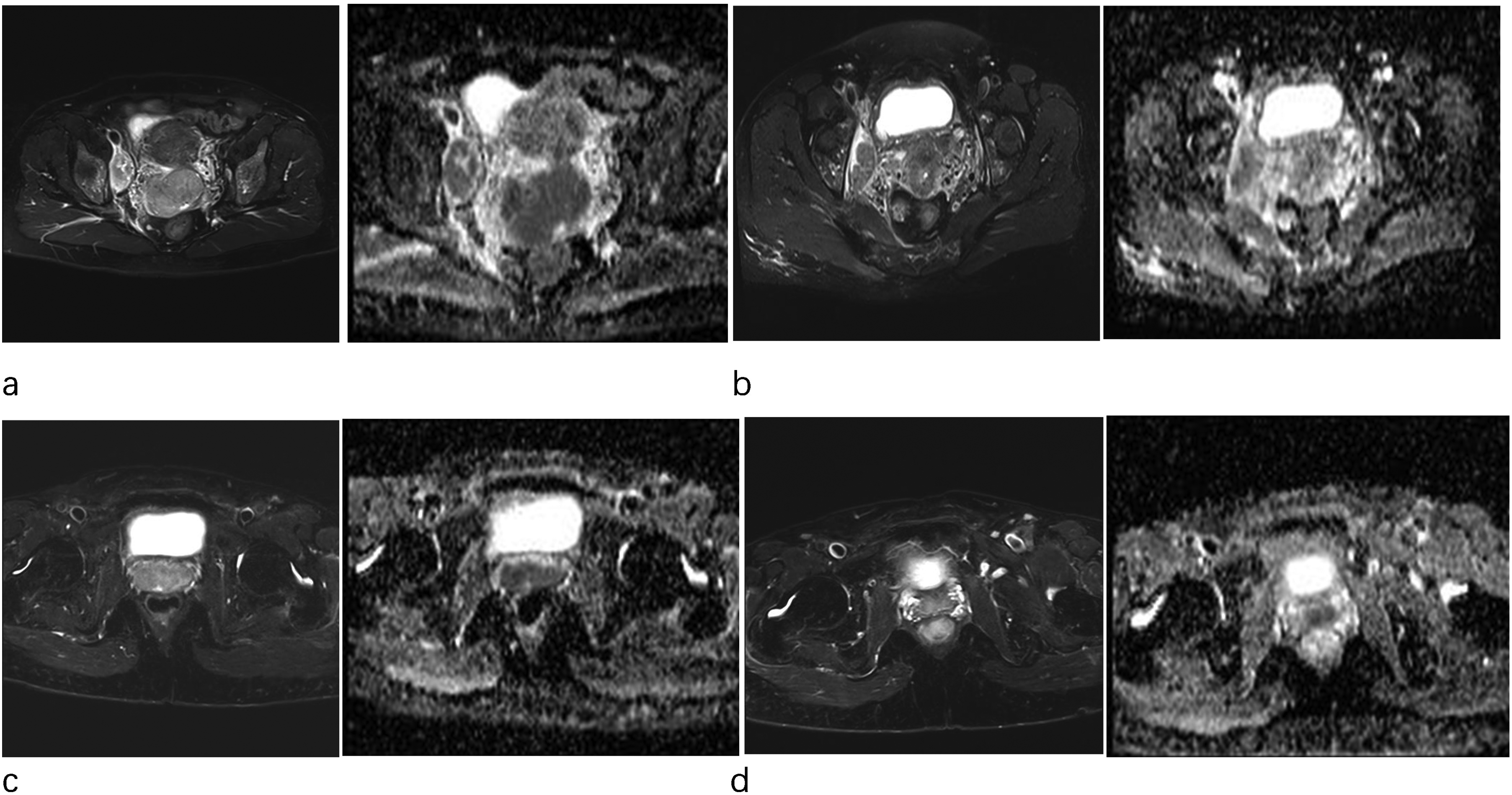
Patient A, stage IIIC1. (a) Before CCRT. (b) After CCRT, the tumor volume was reduced, but still had lymph node metastasis, re-staged as IIIC1, we defined as non-downstaging. PFS = 819 days, ΔADCmean(%) = 25.8. Patient B, stage IIIC1. (c) Before CCRT. (d) After CCRT, the tumor volume was reduced and with no lymph node metastasis, re-staged as IIA1, we defined as downstaging. PFS = 1208 days, ΔADCmean(%) = 28.2. CCRT, concurrent chemoradiotherapy; PFS, progression-free survival.

(a) Comparison of PFS between tumors downstaging and non-downstaging (b) ROC curves using ΔADC, ΔADC% mean, pre-treatment ADC values, and post-treatment ADC values to predict cervical cancer downstaging. ADC, apparent diffusion coefficient; PFS, progression-free survival; ROC, receiver operating characteristic.
Relationship between ADC value parameters and tumor downstaging.

Values are given as mean ± SD unless otherwise indicated.
ADC, apparent diffusion coefficient.
ΔADCmean(%) value predicts PFS and prognosis-related risk factors after CCRT treatment
A total of 121 patients were included in this study. The median follow-up time was 41.2 months (range = 18.2–78.5 months) and the median PFS time was 35.3 months (range = 5.4–78.5 months). During the follow-up period, 36 (29.8%) patients experienced tumor recurrence: locoregional recurrence in nine patients and distant metastasis in 26 patients, or both (Table 1). Using X-tile, it was discovered that when the corresponding ΔADCmean(%) value was 40.8, the prognosis of the patients was significantly stratified (P < 0.001). The 121 patients were randomly divided into a training group (n = 81) and a validation group (n = 40) in a 2:1 ratio. Clinical and imaging parameters of the patients showed no statistically significant differences (P > 0.05) (Table 2). Kaplan–Meier curves indicated that patients can be significantly differentiated into low-risk and high-risk groups in the training group with ΔADCmean(%) cutoff value determined by 40.8; the low-risk group had a lower recurrence rate and significantly longer PFS within 3 years (P < 0.001), which was validated in the validation group (P < 0.012) (Fig. 3). Univariate Cox regression analysis in the training group revealed that ΔADCmean(%) (P < 0.01), pathological grade (P = 0.024), FIGO stage (P < 0.01), tumor diameter change rate (P < 0.01), pelvic lymph node metastasis (P < 0.01), and para-aortic lymph node metastasis (P < 0.01) were all high-risk factors for recurrence after CCRT treatment for cervical cancer. Multivariate analysis demonstrated that ΔADCmean(%) (P < 0.01), pathological grade (P = 0.01), pelvic lymph node metastasis (P = 0.04), and para-aortic lymph node metastasis (P < 0.01) were all independent risk factors for predicting recurrence after CCRT treatment for cervical cancer (Tables 3 and 4). Independent risk factors were used to produce the prognostic nomogram model in Fig. 4. The ROC curves showed that in the training group, the AUC of ΔADCmean(%) was 0.761 and the AUC of the nomogram model was 0.862; in the validation group, the AUC of ΔADCmean(%) was 0.734 and the AUC of the nomogram model was 0.787 (Table 5). The results showed that the nomogram model constructed using ΔADCmean(%) in combination with the clinical information was more effective in predicting 3-year PFS in patients with locally advanced cervical cancer, as depicted in Fig. 5.

Kaplan–Meier showed a significant difference in progression-free survival at 3 years between high- and low-risk groups in the training and validation groups when the ΔADC (%) value was 0.408.

ΔADC nomogram for predicting postoperative progression at 1–3 years after CCRT for cervical cancer. No pelvic or para-aortic LNMs were seen in this patient, the pathology graded was moderately differentiated, the ΔADCmean(%) was 1.25, and the cumulative score corresponding to each component was 285, and the 3 years of PFS in this patient was 0.968. The gray area shows the distribution area. ADC, apparent diffusion coefficient; CCRT, concurrent chemoradiotherapy; LNM, lymph node metastasis.

Comparison of the decision curves of the ΔADCmean(%) and ΔADCmean(%)-nomogram in the training and validation groups.
Clinical prognostic factors of patients in training and validation groups.

Values are given as n or mean ± SD. (+) means have this characteristic.
ADC, apparent diffusion coefficient; FIGO, International Federation of Gynecology and Obstetrics; LNM, lymph node metastasis; MLR, monocyte-lymphocyte ratio; NLR, neutrophil-lymphocyte ratio; SCC, squamous cell carcinoma.
Cox regression analysis of clinical and MRI variables for PFS.

ADC, apparent diffusion coefficient; FIGO, International Federation of Gynecology and Obstetrics; LNM, lymph node metastasis; MRI, magnetic resonance imaging; PFS, progression-free survival.
The ADC change values of the progressed and non-progressed in the training and validation groups.

Values are given as n or mean ± SD.
ADC, apparent diffusion coefficient.
Comparison of different models predicting 3-year PFS in the training and validation groups.

Values in parentheses are 95% confidence intervals.
ADC, apparent diffusion coefficient; AUC, area under the receiver operating characteristic curve; PFS, progression-free survival.
Inter-reader reliability and variability
The inter-reader reliability and variability of tumor downstaging was 0.853 (95% CI = 0.800–0.893), and the reliability and variability of ΔADCmean(%) was 0.954 (95% CI = 0.940–0.966).
Discussion
Our research results show that patients who were downstaged in tumor after CCRT had a significantly higher disease-free survival (DFS) compared to those who did not (P = 0.0039). When the ΔADCmean(%) threshold was set at 26.8, the AUC value for the tumor restaging decrease was 0.868 (95% CI = 80.0–91.9%). In this study, the PFS of patients with cervical cancer was significantly stratified when the ΔADCmean(%) threshold was 40.8, and the PFS of patients with lower ΔADCmean(%) was significantly shorter than that of patients with a higher mean. ΔADCmean(%), pathological grade, pelvic lymph node metastasis (P = 0.04), and para-aortic lymph node metastasis are independent risk factors affecting the PFS of patients with cervical cancer. A nomogram model using ΔADC (%) combined with clinical information yielded the optimal prediction for 3-year PFS with locally advanced cervical cancer (AUC = 0.862), which is higher than the prediction using ΔADC (%) alone (AUC = 0.761), and the same results were achieved in both the training group and the verification group.
CCRT can restore the immune equilibrium and improve the tumor immune microenvironment (12), making it a primary clinical treatment for locally advanced and advanced cervical cancer. Numerous studies (13–15) have shown that the tumor downstaging after neoadjuvant chemotherapy or CCRT therapy is considered to be a factor in the favorable prognosis of many tumors. Wang et al. (14) conducted a retrospective analysis of 226 patients with late-stage nasopharyngeal cancer who underwent CCRT treatment and found that patients with downstaging after CCRT had a significantly lower rate of distant metastasis (10.8%) compared to the non-downstaging group (22.9%) and exhibited significantly improved survival time, which was similar to our results. Han et al. (15) showed that preoperative CCRT treatment for rectal cancer can reduce T and N stages and achieve complete pathological remission. By predicting tumor response after CCRT and adjusting the treatment modality, the overall survival time can be prolonged. Although previous research has evaluated the relationship between tumor downstaging and prognosis, it did not comprehensively consider imaging parameters and failed to reflect changes in the molecular biology within the tumor. MRI has the advantages of non-invasive and high soft tissue resolution and is the first imaging method to evaluate the curative effect. ADC can quantify the movement of water molecules within cells, which is related to tumor density. Radiation and chemotherapy kill tumor cells and reduce their activity, altering water molecule diffusion. Prior research (6,16) has confirmed that ΔADC can reflect internal tumor changes before morphological alterations occur, which can be used to predict tumor regression rates and patient survival time after CCRT, assess treatment efficacy, monitor changes in disease status. However, no research has connected ΔADCmean(%) with tumor downstaging after CCRT. Our study demonstrates a significant correlation between ΔADCmean(%) and tumor downstaging.
Previous studies (17–19) have suggested that tumor size, clinical stage, lymph node metastasis, and SCC are prognostic factors related to cervical cancer outcomes, but they have not been combined with imaging parameters. Several studies (20–22) have now shown that ADC values can be used to predict the prognosis of patients with cervical cancer. Gu et al. (20). studied 124 patients and found that the ADC value before treatment had no clear relationship with tumor progression and that ΔADCmean(%) was an independent risk factor for prognosis, and ΔADC (%), pelvic lymph node metastasis, and type of pathology were imaging-independent risk factors for prognosis, which is consistent with our study. However, this study concluded that neither squamous carcinoma antigen nor histopathological type was an independent risk factor for tumor progression, which is inconsistent with the findings of Gu et al. One consideration regarding our study is that only 9 (7.4%) patients with non-squamous carcinoma were included, which was significantly fewer than the 21 (16.9%) patients in that study. Previous studies (23,24) with large sample sizes have demonstrated that cervical cancer survival is closely related to the status of lymph node metastasis, and patients with concomitant lymph node metastasis have a lower overall survival rate. Our study confirmed that para-aortic and pelvic lymph node metastasis is an independent risk factor for prognosis, which is consistent with the studies of Gouy et al. and Chen et al. (25,26). Himoto et al. (19) showed that ΔADCmean(%) and tumor volume change rate are high-risk factors for predicting the recurrence of cervical cancer, but tumor volume change was not an independent risk factor for prognosis, which is consistent with the results of our study. A previous study (27) also found that different tumor volumes affect the prognosis of patients with cervical cancer, but multivariate regression analysis in our study showed that pretreatment tumor size was not an independent risk factor for prognosis, and Zhang et al.'s (28) study reached with the same conclusion. It is considered that although the tumor size can directly express the extent of tumor involvement, it cannot reflect the degree of internal invasion of the tumor. The relationship between pre-treatment ADC value and tumor prognosis in CCRT for cervical cancer is highly controversial, and several previous studies (29–31) have shown that the pre-treatment ADC value is also a sensitive factor for assessing the risk of progression of cervical cancer. Due to various factors affecting the ADC value before treatment, such as changes in the tumor microenvironment, it may not be an effective marker for evaluating prognosis, our study is consistent with the findings of recent years.
Due to the heterogeneity of tumor cells and individual differences, there is clinical variation in the efficacy of CCRT. Lee et al. (32) found that increasing the treatment dose for specific tumor populations while keeping the total treatment duration can effectively improve patient prognosis. Therefore, we need to evaluate its efficacy at an early stage and adjust the course and dose of radiotherapy appropriately, to reduce the postoperative recurrence rate of patients and prolong the length of DFS. Our study helps to screen groups with therapeutic advantages, develop individualized protocols, and improve prognosis.
The present study has some limitations. First, our study is a single-center retrospective analysis, which is prone to selection bias, but it is internally validated and the conclusions are consistent between groups. Second, different pathological types of cervical cancer have different ADC values. This factor was not examined because the number of patients included with non-squamous cancers is small. Third, our study constructs a generalized model and does not target specific subgroups. It is hoped that the sample will be expanded for targeted analysis in the future. Fourth, the study time nodes were 1 month after CCRT; however, a study (33) reported that the optimal window to reflect the efficacy of CCRT is 2 weeks after the start of CCRT. Further studies are needed to determine the optimal time window for MRI-ADC examination for early prediction of clinical outcome.
In conclusion, our results demonstrated that ΔADCmean(%) is an effective predictor of tumor downstaging. Nomograms incorporating clinical factors and ΔADCmean(%) hold significant clinical value in predicting downstaging and PFS in patients with cervical cancer after CCRT, offering a valuable reference for prognostic assessment and personalized treatment strategies.
Supplemental Material
sj-doc-1-acr-10.1177_02841851241283042 - Supplemental material for Nomograms combining clinical factors and apparent diffusion coefficient to predict downstaging and progression-free survival after concurrent chemoradiotherapy in patients with cervical cancer
Supplemental material, sj-doc-1-acr-10.1177_02841851241283042 for Nomograms combining clinical factors and apparent diffusion coefficient to predict downstaging and progression-free survival after concurrent chemoradiotherapy in patients with cervical cancer by Jiawei Fan, Wenfei Li, Mengyu Cheng, Zhehan Wang, Zhanqiu Wang, Tao Chen and Tao Gu in Acta Radiologica
Footnotes
Acknowledgments
Thanks to all who participated in the publication of this study.
Declaration of conflicting interests
The authors declared no potential conflicts of interest with respect to the research, authorship, and/or publication of this article.
Funding
This study is supported by the Science and Technology Program of Qinhuangdao (Project Number: 202401A114 by Wenfei Li).
Data availability
The datasets used and/or analyzed during the current study are available from the corresponding author on the reasonable request.

Supplementary material
Supplementary material for this article is available online.
References
Supplementary Material
Please find the following supplemental material available below.
For Open Access articles published under a Creative Commons License, all supplemental material carries the same license as the article it is associated with.
For non-Open Access articles published, all supplemental material carries a non-exclusive license, and permission requests for re-use of supplemental material or any part of supplemental material shall be sent directly to the copyright owner as specified in the copyright notice associated with the article.
