



Abstract
Background
Intrahepatic cholangiocarcinoma (ICC) is an aggressive liver malignancy, and Ki67 is associated with prognosis in patients with ICC and is an attractive therapeutic target.
Purpose
To predict Ki67 expression based on multiparametric magnetic resonance imaging (MRI) radiomics multiscale tumor region in patients with ICC.
Material and Methods
A total of 191 patients (training cohort, n = 133; validation cohort, n = 58) with pathologically confirmed ICC were enrolled in this retrospective study. All patients underwent baseline abdominal MR scans in our institution. Univariate logistic analysis was conducted of the correlation between clinical and MRI characteristics and Ki67 expression. Radiomics features were extracted from the image of six MRI sequences (T1-weighted imaging, fat-suppression T2-weighted imaging, diffusion-weighted imaging, and 3-phases contrast-enhanced T1-weighted imaging sequences). Using the least absolute shrinkage and selection operator (LASSO) to select Ki67-related radiomics features in four different tumor volumes (VOItumor, VOI+8mm, VOI+10mm, VOI+12mm). The Rad-score was calculated with logistic regression, and models for prediction of Ki67 expression were constructed. The receiver operating curve was used to analyze the predictive performance of each model.
Results
Clinical and regular MRI characteristics were independent of Ki67 expression. Four Rad-scores all showed favorable prediction efficiency in both the training and validation cohorts (AUC = 0.849–0.912 vs. 0.789–0.838). DeLong’s test showed that there was no significant difference between the AUC of the radiomics scores, while the Rad-score (VOI+10mm) performed the most stable predictive efficiency with △AUC 0.033.
Conclusion
Multiparametric MRI radiomics based on multiscale tumor regions can help predict the expression status of Ki67 in ICC patients.
Introduction
Intrahepatic cholangiocarcinoma (ICC) is the second most common primary malignant liver cancer after hepatocellular carcinoma (HCC) (1). Compared to HCC, ICC is a more aggressive malignancy and has a worse prognosis, with an overall 5-year survival rate of less than 10% (2). Early radical surgical resection is currently the cornerstone of treatment for ICC, but due to atypical clinical symptoms and high misdiagnosis rates, only a few patients have the opportunity for surgery, the recurrence and metastasis rate after surgery is still high, resulting in a low 5-year survival rate in the range of 20%–35% (3). While the 5-year survival rate for patients who did not undergo surgery was only 0%–5% (4).
Ki67 is a nuclear antigen associated with tumor cell proliferation, and a sign of cell division and proliferation activity. It is abnormally expressed in breast cancer, lung cancer, bladder cancer, and other malignant tumors, and participates in tumor proliferation and metastasis (5–7). A high level of Ki67 expression is generally associated with high proliferative activity of tumor cells and worse prognosis. The present study shows that a high level of Ki67 expression (Ki67 index ≥ 25%) was an independent prognostic factor for ICC patients, which was bound up with tumor number, lymph node metastasis (LNM), and vascular invasion (MVI) in ICC patients (8,9). Besides, the Ki67 index found a positive correlation between hENT-1, eIF3C, and CXCL7 in ICC (10–12), serving as a potential target for targeted and immunological therapy.
Therefore, the preoperative diagnosis of Ki67 expression in ICC patients can more accurately guide clinical individualized medical treatments. Currently, immunohistochemical testing using biopsy tumor tissue or surgical resection specimens is the gold standard for evaluating Ki67 expression in patients with ICC, but both are invasive and limited by whether surgical treatment is performed, as well as the accuracy of puncture sampling. Magnetic resonance imaging (MRI), as a non-invasive examination method with high soft-tissue resolution, is an important tool for preoperative diagnosis, staging, and efficacy assessment of ICC patients (13). In addition to macroscopic information, MRI radiomics can also extract deep quantitative features from medical images to more accurately and comprehensively assess tumor heterogeneity and biological behavior. Previous studies (14–16) have shown that MRI radiomics has achieved better results in ICC diagnosis, microvascular invasion prediction, efficacy evaluation, and prognostic risk assessment, but relatively few studies have been conducted on ICC Ki67 expression and most of those studies only extracted the imaging features of entire volumetric tumor region and neglected the peritumoral information.
The aim of the present study was to develop a radiomics signature for predicting Ki67 expression based on MRI radiomics multiscale tumor region in ICC patients.
Material and Methods
This retrospective diagnostic study received ethical approval from the Xuzhou Central Hospital (ethics approval no. XZXY-LK-20221230-116) and Zhongshan Hospital, Fudan University (ethics approval no. B2021-325R). The requirement for written informed consent was waived. All authors provided consent to publication.
Patients
Between January 2016 and June 2022, through a review of the hospital pathology and radiology database, 376 patients with pathologically confirmed ICC after hepatectomy and who underwent preoperative liver MR examination were retrospectively identified. Patients were enrolled if they met the following inclusion criteria (Fig. 1): (i) single mass; (ii) no prior history of liver surgery; (iii) MRI scan within 30 days before surgery; (iv) longest tumor diameter ≥1.0 cm; (v) lesion with clear edges; (vi) MRI scans meeting diagnostic criteria; and (vii) pathological report involving Ki67 expression. Finally, a total of 191 patients (52 in the low Ki67 status group and 139 in the high Ki67 status group) were enrolled and randomly allocated to a training cohort (n = 133, 97 in the high Ki67 status group and 36 in the low Ki67 status group) and a validation cohort (n = 58, 42 in the high Ki67 status group and 16 in the low Ki67 status group) with a proportion of 7:3.
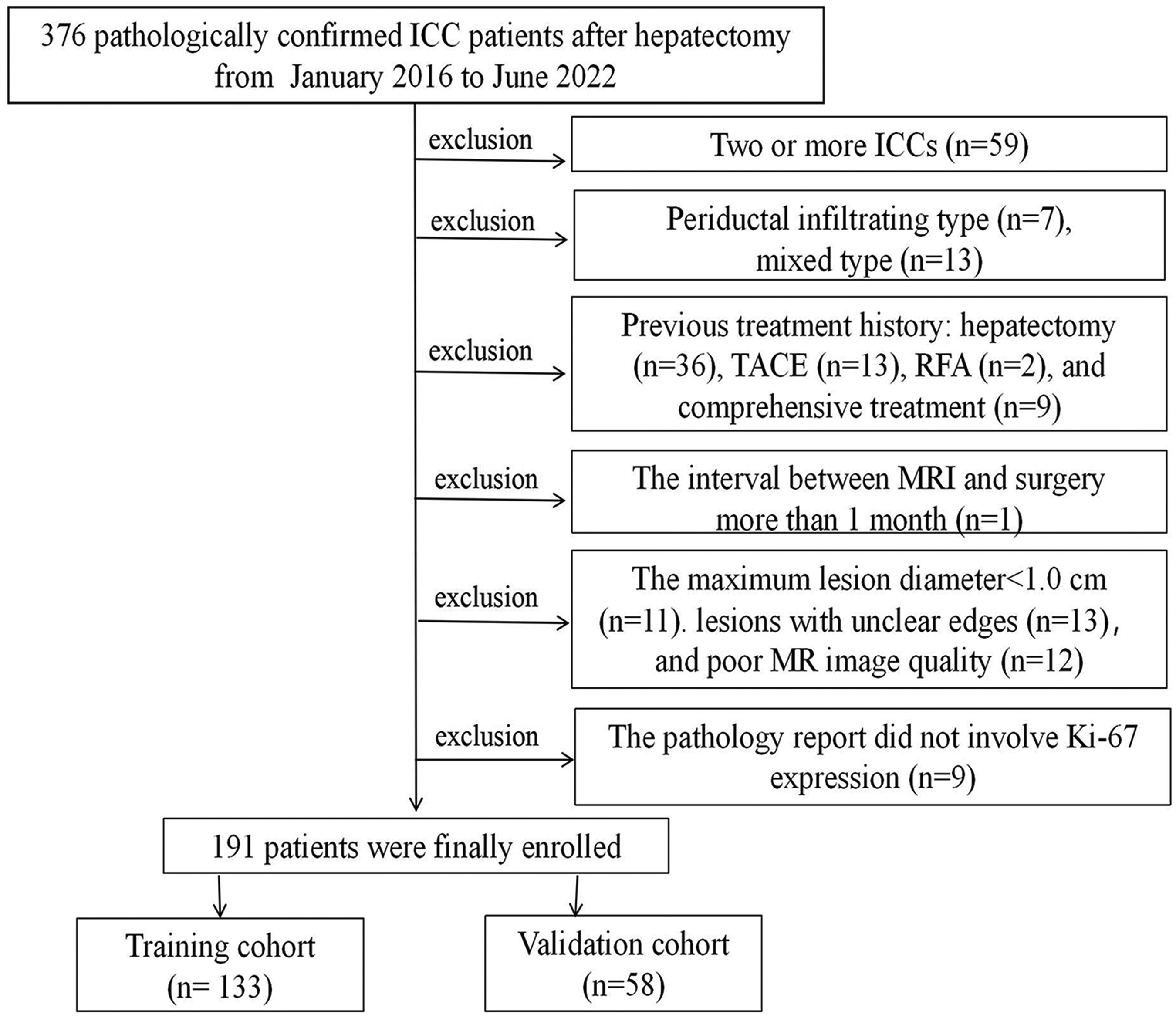
Study flowchart of the enrolled patients.
Clinical and histopathological features
The following clinical features, including sex, age, hepatitis B virus (HBV), alpha fetoprotein (AFP), carcinoembryonic antigen (CEA), and carbohydrate antigen 19-9 (CA19-9), were obtained from the medical record system. Two experienced pathologists evaluated Ki67 status, while immunohistochemical staining was employed to verify Ki67 expression. The labeling index for Ki67 expression was determined through assessing the percentage of positive cells. ICC lesions were classified into two groups based on Ki67 labeling index, namely the low-expression group (Ki67 labeling index < 25%) and high-expression group (Ki67 labeling index ≥ 25%) (8,9).
Imaging acquisition
All patients were examined with enhanced MRI scanning. Taking the 3.0-T Discovery 750 MRI Systems (GE Healthcare, Milwaukee, WI, USA) as an example, the scanning sequences and parameters were as follows: (i) fat-suppression T2-weighted (T2WI-FS) imaging: TR = 4500 ms, TE = 88 ms, section thickness = 6–6.5 mm, gap = 1–2 mm, matrix size = 320 × 224–448 × 448; (ii) diffusion-weighted imaging (DWI): b = 0 and 800 s/mm2: TR = 5454 ms, TE = 49 ms, section thickness = 6–8 mm, gap = 1–2 mm, matrix size = 130 × 96; (iii) T1-weighted (T1W) imaging and three-phase contrast-enhanced T1W imaging: TR = 3.54 ms, TE = 1.67 ms, section thickness = 4.8–5 mm, gap = 0 mm. For contrast-enhanced MRI, a dose of 20 μmol per kg of body weight of Gd-DTPA was injected as a rapid bolus and immediately followed by 20 mL saline at a rate of 2 mL/s. Images in the arterial phase (AP), portal venous phase (PVP), and delayed phase (DP) were obtained at 20–30 s, 60–70 s, and 180 s after contrast medium administration, respectively.
Imaging analysis
Routine MRI features
Two experienced abdominal radiologists who were blinded to the clinical results independently evaluated all MRI scans. Any discrepancy was resolved after a discussion between the two radiologists. All evaluated MRI features included: (i) tumor size; (ii) tumor morphology; (iii) signal intensity on T1W and T2W-FS imaging (relative to the surrounding liver); (iv) target sign on T2W-FS and DW imaging (17); (v) arterial edge enhancement ratio (18); (vi) arterial phase enhancement pattern (18,19): overall enhancement, edge enhancement, no/mild enhancement, and partial enhancement; (vii) enhancement pattern (gradual and filling, arterial and persistent, wash-in and wash-out); (viii) the Liver Imaging Reporting and Data System (LI-RADS) (version 2018) (20); (ix) intrahepatic duct dilatation; (x) hepatic capsular retraction (retraction of the hepatic capsular adjacent to the lesion); (xi) visible vessel (hepatic artery, portal vein, or hepatic vein) penetration inside the lesion (18); and (xii) peritumoral enhancement (at any phase).
Volume of interest segmentation
In this study, the volume of interest (VOI) was delineated according to the overall segmentation of intratumoral and peritumoral regions (21,22). For the MRI scans of each included patient, two radiologists with more than 5 years of experience in abdominal imaging were outlined in the double-blind method using the open source software 3D Slicer version 4.13.0 (https://www.slicer.org/), knowing that the patient had ICC, but did not understand the pathological results and clinical manifestations. The two physicians manually delineated the lesions along the tumor boundary on six sequence images. The resulting VOI was labeled VOItumor, and then used the “margin” module to expand from the tumor boundary by 8, 10, and 12 mm. After manual removal of the extraparenchyma of the liver, the area containing the intratumor and peritumoral area was labeled VOI+8mm, VOI+10mm, and VOI+12mm, respectively. If the two radiologists disagreed regarding segmentation, agreement was reached through discussion.
Radiomic feature extraction
Radiomic features were extracted from the VOI of six sequences using the Radiomics module of 3D Slicer version 4.13 software (https://www.slicer.org/). Before feature screening, radiomics features were standardized using the Z-score to improve data comparability. To evaluate the reproducibility of VOI delineation, intraclass and interclass correlation tests were conducted. A total of 30 cases were randomly selected from the collected data. Readers 1 and 2 independently delineated the images, and the resulting feature values were analyzed to assess inter-observer consistency. In addition, reader 1 re-delineated the same 30 cases 1 month later. The extracted feature values from this re-delineation were compared with those from 1 month before calculating another set of intraclass correlation coefficient values, assessing intra-observer consistency. The consistency and reliability of the segmentation algorithm depend on the intra- and interclass correlation coefficients, which are in the range of −1 to 1, where 1 indicates perfect consistency and −1 indicates perfect inconsistency. Then, SelectKBest was used to analyze the relationship between the features and classification results. Radiomics features with P < 0.05 were selected. Subsequently, through the least absolute shrinkage and selection operator (LASSO) algorithm, the radiomics features with a non-zero coefficient were chosen, and the selected features were weighted according to the feature coefficient to obtain the corresponding Rad-score of each VOI.
Model construction and evaluation
Models were established and verified by the validation cohorts using logical regression (LR). The model predictive efficacy was validated by receiver operating characteristic curve (ROC) analysis. The area under the ROC curve (AUC), sensitivity, specificity, and accuracy were calculated to evaluate the performance of the models. The Hosmer–Lemeshow test was used to assess the model goodness of fit and plot the calibration curve. The decision curve analysis (DCA) assessed the clinical utility of the model. The workflow of the above radiomics analysis is shown in Fig. 2.

Workflow of the development of the radiomics analysis.
Statistical analysis
The statistical analyses were performed using R (version 4.1.2; R Foundation, Vienna, Austria) and Python (version 3.6; Python Software Foundation, Wilmington, DE, USA). Continuous variables were compared using the Student's t-test or Mann–Whitney U-test. The chi-square test and Fisher's exact test were applied to categorical variables. A two-sided P value < 0.05 was considered statistically significant. Delong’s test was used to compare predictive performance between multiple models.
Results
Clinical imaging features
Table 1 summarizes the clinical imaging characteristics of patients in the training and validation cohorts. Among the 191 individuals, 137 men (mean age = 60.10 ± 11.02 years; age range = 29–86 years) and 54 women (mean age = 62 ± 11.26 years; age range = 36–84 years) were analyzed. There was no significant difference in Ki67 expression between patients in the training and validation groups (P > 0.05). In the univariate logistic analyses, clinical and regular MRI characteristics were independent of Ki67 expression (Table 2).
Baseline clinicoradiological features of ICC patients in training and validation cohorts.

Values are given as n (%), mean ± SD, or median (IQR).
AFP, alpha fetoprotein; CEA, carcinoembryonic antigen; DWI, diffusion-weighted imaging; FS, fat suppression; MRI, magnetic resonance imaging; SI, signal intensity; T1W, T1-weighted; T2W, T2-weighted.
Univariate logistic analyses of clinicoradiological features related to Ki67 status in ICC.

AFP, alpha fetoprotein; CEA, carcinoembryonic antigen; CI, confidence interval; DWI, diffusion-weighted imaging; FS, fat suppression; HBV, hepatitis B virus; ICC, intrahepatic cholangiocarcinoma; OR, odds ratio; SI, signal intensity; T1W, T1-weighted; T2W, T2-weighted.
Radiomics feature selection
For each patient, 851 radiomics features were extracted from each sequence in each VOI range. The main types of these radiomics features include shape-based, first order, gray-level co-occurrence matrix (GLCM), gray-level size zone matrix (GLSZM), gray-level run length matrix (GLRLM), neighboring gray tone difference matrix (NGTDM), and gray-level dependence matrix (GLDM) features. In total, 5106 features were extracted for each VOI range across the six sequences by feature fusion. The reproducibility of VOI delineation is satisfactory, with both intra- and inter-class correlation coefficient values greater than 0.8. After SelectKBest analysis, 215, 155, 164 and 168 features were selected for VOItumor, VOI+8mm, VOI+10mm, and VOI+12mm, respectively. Finally, after LASSO regression, 29, 26, 23 and 23 radiomics features related to Ki67 expression of ICC were selected in VOItumor, VOI+8mm, VOI+10mm, and VOI+12mm, respectively (Table 3). The selection procedure of features and detail feature coefficient are illustrated in Fig. S1 and Table S1.
The feature numbers and of each VOI during the procedure of feature selection.

VOI, volume of interest.
Model construction and evaluation
Table 4 summarizes the AUCs of four VOI models, and matched ROCs are provided in Fig. 3. The AUC of each VOI model is greater than 0.75 in both the training cohort and validation cohort. This indicates four multiparametric MRI-based radiomics models all had favorable prediction efficiency. The VOItumor, VOI+8mm, and VOI+12mm models performed well in the training cohort (AUC = 0.849∼0.912), but could not achieve stable performance in the validation cohort (AUC = 0.789∼0.829). The VOI+10mm model yielded the most stable predictive efficiency between the training and validation cohorts (△AUC = 0.033). In the validation cohort, the AUC values for the VOI+10mm model were higher compared to the VOItumor, VOI+8mm, and VOI+12mm models. Although the differences were not statistically significant (P = 0.8822, 0.8089, 0.2537), the VOI+10mm model showed stable performance in predicting the Ki67 task, underscoring its potential and feasibility for practical applications.
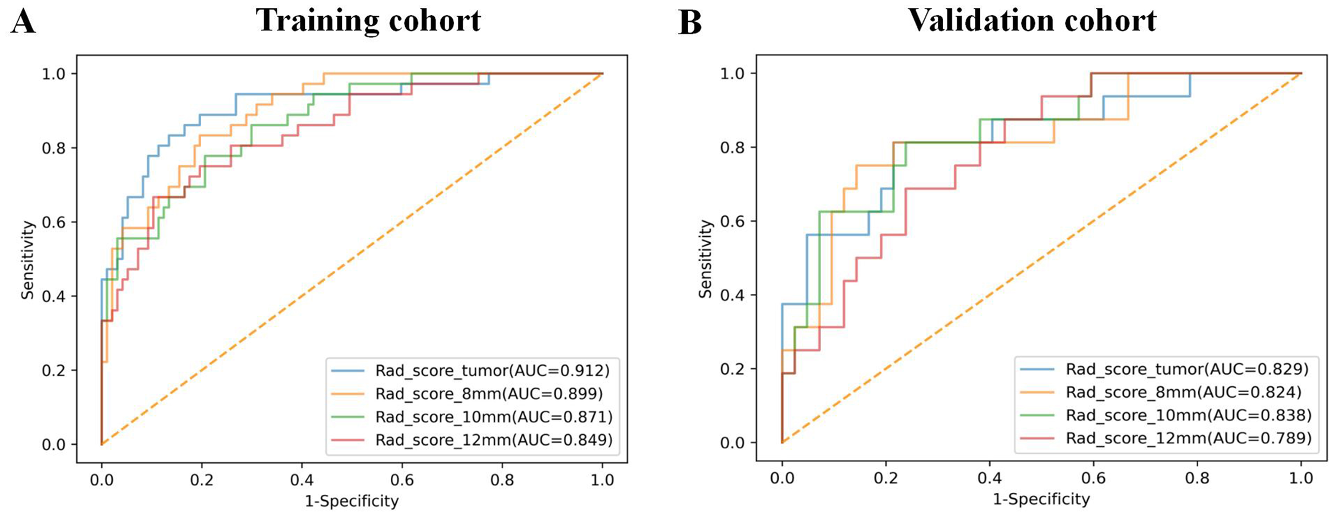
Comparison of ROC curves for Ki67 status prediction in (a) training and (b) validation cohorts by logistic regression. ROC, receiver operating characteristic.
The performance of four-group VOI models for predicting Ki67 status in ICC patients.

Acc, accuracy; AUC, area under the receiver operating characteristic curve; CI, confidence interval; ICC, intrahepatic cholangiocarcinoma; Sen, sensitivity; Spe, specificity; VOI, volume of interest. The bold value represents the model with stable and desirable predictive performance.
The calibration curves of four models exhibit satisfactory predictive performances in the training and validation groups (Fig. 4a, b). After the Hosmer–Lemeshow test, all models have good calibration capability (P > 0.05); the predicted risk probability coincides with the actual risk. The decision curve (Fig. 4c, d) showed that the four groups were significantly higher than the clinical net benefit at total biopsy, indicating that four models were valuable. The DCA curves plotted for the four-group model in both training and validation cohorts are significantly higher overall than the net clinical benefit at full biopsy, suggesting that all four-group models are valuable. In the training set, the four-group model thresholds were in the range of 0.05–0.9, with a maximum net gain of 0.28. When the threshold was 0.28, the benefits were tumor, 8 mm, 10 mm, and 12 mm in order from high to low, which is consistent with AUC results. In the validation cohort, the range of thresholds for the four-group model was 0.05–0.53, with the highest net gain of 0.28. when the threshold was 0.4. The benefits were 10 mm, tumor, 8 mm, and 12 mm in descending order, which is consistent with the AUC results.
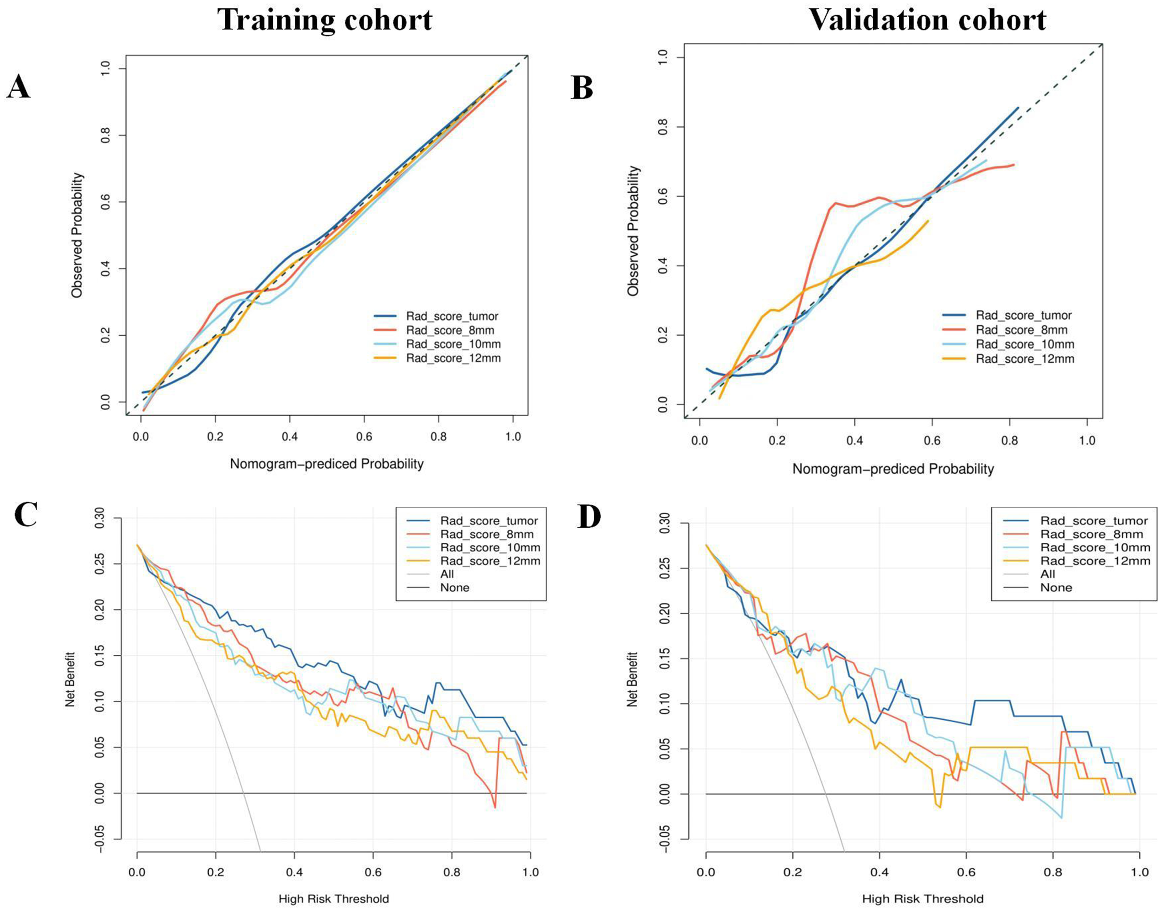
(a, b) Calibration and (c, d) decision curves in the training and validation cohorts.
Discussion
In the present study, we constructed and validated a radiomics signature based on multiparametric MRI multiscale tumor regions for preoperative prediction of Ki67 expression status in patients with ICC. The results showed that the models built based on the tumors and peritumor-containing VOI (VOItumor, VOI+8mm, VOI+10mm, VOI+12mm), respectively, demonstrated good predictive ability for predicting Ki67 expression in ICC patients in both the training and validation cohorts. Among them, the model based on the VOI+10mm showed the most stable performance, with AUCs of 0.871 and 0.838 in the training and validation cohorts, respectively. Thus, multiparametric MRI-based radiomics might assist in preoperatively predicting the Ki67 expression in ICC, which is expected to provide significant value in guiding personalized treatment for patients.
A previous study (23) showed that Ki67 expression in ICC patients correlates with HBV, arterial rim enhancement, and enhancement pattern. However, no independent characteristics of Ki67 were found for any of the clinical and regular MRI characteristics included in our study. Several studies (24,25) have confirmed that HBV infection is an important risk factor for the development of ICC, and the prognosis of HBV-associated ICC is inconsistent across studies. Jeong et al. (26) found that HBV-associated ICC patients had a relatively high proportion of microvascular infiltration and poor differentiation, implying a relatively high likelihood of Ki67 high expression. However, Zhang et al. and Wu et al. (27,28) concluded that the activation of the immune response by HBV infection may have a protective effect on patients with ICC, and that patients with HBV-associated ICC have a better prognosis than negative patients. Therefore, further studies are needed on the prognosis of HBV infection and ICC and its correlation with Ki67. ICC patients with arterial rim enhancement have a greater chance of vascular invasion and necrosis and a relatively worse prognosis (19). In the present study, although there was not statistically significant between the high and low Ki67 expression groups, the incidence of arterial rim enhancement was still higher in the Ki67 high-expression group than in the low-expression group. A previous study (23) suggested that the Ki67 index is relatively high in ICC patients with the enhancement pattern of “wash-in and wash-out,” and similar results were not obtained in the present study by univariate logistic analysis. It is hypothesized that due to the relatively small percentage of ICC case phases (14.3% for the training cohorts, 10.3% for the validation cohorts) that demonstrated the “wash-in and wash-out” enhancement pattern in this study, the sample size still needs to be increased for further study at a later stage.
Radiomics can non-invasively extract features within the VOI that are associated with tumor heterogeneity to build models, thus assessing the biological behavior of tumors more accurately and objectively. However, tumor heterogeneity is not only limited to cancer cells, and highly aggressive malignant tumors often invade the peritumoral region, resulting in peritumoral vasculo-lymphangiogenesis, mesenchymal reaction, and so on (29,30). Most of the current radiomics studies are focused on the analysis of the lesion itself, while relatively few studies have examined peritumoral features, and in liver tumors have mostly focused on the prediction of MVI. In HCC, Zhang et al. (31) simultaneously extracted the radiomics features of intra- and peritumoral dual regions based on Gd-DTPA-enhanced MRI scans, and the results showed that the efficacy of predicting MVI based on the dual-region fusion model was better, with an AUC of 0.820 in the validation cohorts. With the combined clinical and imaging features, the prediction efficacy was further improved, and the AUC was 0.858 in the validation cohorts. In ICC, we extracted the radiomics features of six MRI sequences images from four different VOIs, respectively, and the comprehensive analysis showed that the efficacy of multisequence MRI (T1W + T1W-D + DWI), intra- and peritumor 10 mm (VOI+10mm) for predicting MVI was superior to that of the intratumor model, with AUCs of 0.987 and 0.858 in the training and validation cohorts, respectively (32). In this study, we extracted the radiomics features of the four VOI regions, established Rad-scores, and incorporated the logistic regression model. The results showed that the four groups of models predicted the efficacy of Ki67 expression better. In the validation cohorts, the AUC of the VOI+10mm was the highest, which was 0.838, but the differences were not statistically significant. The results showed that multiparametric-based MRI radiomics has the potential to predict ICC Ki67 expression, but the peritumor features did not provide added value to the diagnostic efficacy.
The reason for this may be that the peritumor region is considered to be the primary site of MVI, and both the intra- and peritumoral regions contain more information related to MVI (33). Therefore, the intratumor combined with the peritumor model may provide better predictive efficacy in predicting MVI, and it is hypothesized that the information represented by the peritumor region of the ICC is not complementary to that of intra-tumor features in identifying Ki67. It is speculated that the information represented by ICC peritumor region is not complementary to the intratumoral features in in terms of identifying Ki67. In addition, the peritumor region in this study was automatically expanded based on the intratumor region, and some of the tumors were larger in size and involved the hepatic margin region, at which time the automatically extracted peritumor region might be mixed with some extra-hepatic features, increasing the heterogeneity of the peritumor region.
The present study has some limitations. First, it is a retrospective study, the sample size was relatively small, and the proportion of the Ki67 low-expression group was relatively low, so there may be some selection bias. At a later stage, we will increase the sample size of the study. Second, this study was a single-center study and lacked multicenter data validation. Third, the VOIs were sketched by manual segmentation; it is difficult to avoid the influence of subjective factors of the outlining person.
In conclusion, multiparametric MRI radiomics based on multiscale tumor regions can be used to predict Ki67 expression in patients with ICC, thereby facilitating individualized clinical diagnosis and treatment.
Supplemental Material
sj-docx-1-acr-10.1177_02841851241310394 - Supplemental material for Multiparametric magnetic resonance imaging-derived radiomics for the prediction of Ki67 expression in intrahepatic cholangiocarcinoma
Supplemental material, sj-docx-1-acr-10.1177_02841851241310394 for Multiparametric magnetic resonance imaging-derived radiomics for the prediction of Ki67 expression in intrahepatic cholangiocarcinoma by QingWang, ChenWang, Xianling Qian, Baoxin Qian, Xijuan Ma, Chun Yang and Yibing Shi in Acta Radiologica
Footnotes
Acknowledgements
We sincerely thank Jia Zhang and Lulu Lv for their assistance with radiomics analysis.
Data availability
Datasets and material analyzed are available on reasonable request from the corresponding author.
Declaration of conflicting interests
The authors declared no potential conflicts of interest with respect to the research, authorship, and/or publication of this article.
Funding
The authors disclosed receipt of the following financial support for the research, authorship, and/or publication of this article: This work was supported by the Medical Scientific Research Program of Jiangsu Commission of Health, Xuzhou Leading Talent Training Project, Clinical Research Special Fund Project of Wu Jieping Medical Foundation, and Clinical Research Project of Zhongshan Hospital, Fudan University (grant nos. M2021014, XWRCHT20210030, 320.6750.2023-11-24, and 2020ZSLC61).

Supplementary material
Supplementary material for this article is available online.
References
Supplementary Material
Please find the following supplemental material available below.
For Open Access articles published under a Creative Commons License, all supplemental material carries the same license as the article it is associated with.
For non-Open Access articles published, all supplemental material carries a non-exclusive license, and permission requests for re-use of supplemental material or any part of supplemental material shall be sent directly to the copyright owner as specified in the copyright notice associated with the article.
