



Abstract
Background
Primary insomnia (PI) is one of the most common sleep disorders. Diagnosis of insomnia is mainly based on subjective sleep difficulties, and it is still necessary to find objective neurobiological markers.
Purpose
To investigate the functional connectivity (FC) of frontal hub regions important for PI.
Material and Methods
We enrolled 20 patients (5 men, 15 women) with PI and 20 controls (5 men, 15 women), matching age, sex. We used resting-state functional magnetic resonance imaging (fMRI) and voxel-mirrored homotopic connectivity (VMHC) to analyze the abnormal changes of FC in the frontal lobe of PI patients.
Results
Compared to controls, abnormal FC regions were mainly concentrated in the superior frontal gyrus (L/R), middle frontal gyrus (L/R), and inferior frontal gyrus (L) of the orbital region and the inferior frontal gyrus of the opercular region (L) (P < 0.05). The VMHC results showed abnormal FC in the middle frontal gyrus of the orbital region (GFR correction, voxel P < 0.01, cluster P < 0.025) in PI patients. The FC between the orbitofrontal gyrus and the inferior frontal gyrus of the opercular region with the frontal gyrus of the medial orbital region demonstrated a significant correlation with the clinical scale (p < 0.05).
Conclusion
Our study identified abnormal FC, which was mainly located in the orbitofrontal gyrus and the inferior frontal gyrus of the opercular region, in the frontal lobe of patients with insomnia using resting-state fMRI. This is helpful to understand the abnormal neural activity mechanism of insomnia in the frontal lobe and provide a relatively accurate brain region basis for future prevention, diagnosis, and treatment.
Introduction
Primary insomnia (PI) is one of the most common sleep disorders. Insomnia is defined as dissatisfaction with the duration or quality of sleep, which can be manifested as difficulty falling asleep, easily waking up at night, and/or waking up early (1). It is usually accompanied with daytime cognitive impairment and memory consolidation impairment during sleep (2). The prevalence of insomnia is in the range of 6%–10%, and the incidence rate is 4% per year (3). It is also significantly higher in women than in men, and the prevalence increases with age (4). Insomnia seriously affects the mood and quality of life of patients, easily leading to negative emotions, such as depression and anxiety. At present, the diagnosis of insomnia is mainly based on subjective sleep difficulties, and it is still necessary to find objective neurobiological markers. At the same time, the neuropathological mechanism of insomnia also needs to be studied further, so that we can determine the more superior treatment methods.
Functional magnetic resonance imaging (fMRI) measurement is an indirect and non-invasive measurement of brain activity via blood oxygen level dependent (BOLD) contrast, which has the characteristics of high spatial resolution. fMRI technology provides a new imaging method for the study of neuropsychiatric diseases including insomnia (5). Previous fMRI studies have linked insomnia disorder to cortical dysfunction. Studies have found abnormal brain activity or functional connectivity (FC) in the prefrontal cortex, insular cortex, amygdala, precuneus, and caudate nucleus in PI, as well as abnormal FC in the default mode network, including the anterior and posterior cingulate cortex, inferior parietal lobule, ventromedial prefrontal gyrus, posterior splenium cortex, precuneus, and hippocampus (6). A recent animal experiment also showed that prefrontal dysfunction may be related to neural fatigue of locus coeruleus neurons projecting to the prefrontal cortex under chronic sleep deprivation (7). The prefrontal cortex and its relationship with other cortexes play a key role in the sleep-wake pattern. Many fMRI studies about insomnia have involved abnormalities of the frontal lobe, but the studies on the divisions of the frontal lobe are not consistent, and the interaction between the brain activities of the internal regions of the frontal lobe is also unknown. FC methods in fMRI can identify the spatiotemporal association patterns of the brain at rest or while performing tasks. These association patterns are a measure of co-activation in the time series of FC between anatomically distinct brain regions (8). VMHC is a resting state (rs)-fMRI method that analyses synchronized activity between the two cerebral hemispheres. That is the time series correlation between each voxel in one hemisphere and its allelic voxel in the other hemisphere (9). The aim of the present study was to use FC and VMHC methods to examine the abnormal brain activity changes in the frontal lobe to study the mechanism of insomnia or find more accurate neurobiological markers related to the frontal lobe.
Material and Methods
A total of 40 participants (20 patients with PI, 20 controls) were enrolled. They were matched for age, dominant hand, and sex.
Patients with PI were included in this study according to the following criteria: (i) patients met the criteria of the Diagnostic and Statistical Manual of Mental Disorders, 4th Edition; (ii) a history of difficultly falling asleep or maintaining sleep or waking up early for at least 1 month; (iii) Pittsburgh Sleep Quality Index (PSQI) ≥ 7; (iv) right-handed patients as assessed by the Edinburgh Handedness Inventory (EHI); and (v) patients aged <65 years.
The exclusion criteria are as follows: (i) patients with abnormal signals on routine T1 or T2 fluid-attenuated inversion recovery MRI; (2) insomnia patients with psychiatric disorders, addiction, or other sleep disorders (including hypersomnia, parasomnia, sleep-disordered breathing, sleep-related dyskinesia, or circadian rhythm sleep disorders); (iii) metallic implants in the body; (iv) individuals who were underweight or obese (body mass index < 18.5 or > 28 kg/m2).
All healthy controls met the following criteria: (i) good sleep status, PSQI score <5; and (ii) no stimulant or drug use for ≥3 months before enrollment. All controls met the above exclusion criteria. The purpose and benefits of the study were explained to each individual. All participants provided written informed consent.
Assessment of sleep complaints
Sleep quality, sleep latency, sleep duration, sleep efficiency, sleep disturbance, use of hypnotics, and daytime dysfunction in the past month were measured using the PSQI. A total score ≥7 indicates poor sleep quality. The Self-rating Anxiety Scale (SAS) can evaluate the severity of anxiety symptoms. It contains 20 items that reflect the subjective feeling of anxiety. Each item is divided into four grades according to the frequency of symptoms. The Self-rating Depression Scale (SDS) can reflect depressive mood, physical symptoms, psychomotor behavior, and psychological symptom experience. It includes 20 items and is divided into four grades.
MRI and data acquisition
A Magnetom Skyra 3.0 T scanner (Siemens AG, UK) was used with the participant in the supine position; head movement was controlled using foam pads. During the scan, individuals were instructed to close their eyes and remain still. Resting-state fMRI scans were performed parallel to the anterior skull base and anteroposterior symphysis, using an echo-planar imaging (EPI) sequence. The scan parameters were as follows: TR = 3000 ms; TE = 30 ms, field of view (FOV) = 220 × 220 mm; flip angle (FA)= 90°; slice thickness = 3.0 mm; number of slices = 40; voxel size = 2.3 × 2.3 × 3.0 mm; and the scan was interleaved. The scan parameters for T1-weighted 3D magnetization preparation gradient echo sequence were as follows: TR = 2300 ms; TE = 2.32 ms; matrix = 256 × 256; FOV = 240 × 240 mm; FA = 8°. Conventional T2-weighted images were acquired to exclude other lesions. The BOLD sequence and 3D T1-weighted sequence were each acquired once.
Analysis of functional magnetic resonance data
Using SPM8 toolkit (http://www.fil.ion.ucl.ac.uk/spm/) for image preprocessing. For each participant, the first 10 time points were discarded due to signal balancing and participant adaptation to scanning noise. To minimize the effects of head motion, we first excluded individuals with maximum displacement >2 mm and 2° angular motion in any dimension throughout the fMRI session. Slice timing, head motion correction, and spatial normalization were performed in sequence using statistical parametric mapping (SPM8) and resampled to 3 × 3 × 3 mm. To eliminate the influence of extremely low frequency drift and physiological high frequency respiratory and cardiac noise, all data were filtered by linear trend of time course and time bandpass (bandpass frequency = 0.01–0.08 Hz) with REST software. Brain regions of frontal lobe was segmented into 18 regions using Anatomical Automatic Labeling (AAL) template and set as 18 nodes. The time series of the corresponding nodes were extracted for each participant. Then the Pearson correlation coefficients between the time series of all nodes were obtained to form the FC matrix of nodes. Fisher’s transformation was used to convert the FC matrix into Z scores. Then, a two-sample t-test (P < 0.05) was performed by DPABI Net software. The Pearson correlation coefficient of BOLD signals between each pair of left and right symmetrical voxels in the brain was calculated using the VMHC module of the REST software, and the correlation coefficient was converted to Z score by using Fisher’s Z transformation.
Statistical analysis
All statistical tests were analyzed using SPSS 20.0 statistical software. The chi-square test was used to compare differences between sexes. The Kolmogorov–Smirnov test was performed to test the normal distribution of the measurement data. Normally distributed data was expressed as
Results
Demographic and clinical outcomes
There were no significant differences in age or sex, but there were significant differences in PSQI, SAS, and SDS between the insomnia group and the control group (Table 1).
Demographic and clinical outcomes.

Values are given as n, mean ± SD, or median (range).
PSQI, Pittsburgh Sleep Quality Index; SAS, Self-rating Anxiety Scale; SDS, Self-rating Depression Scale.
Functional connectivity results
The frontal lobe regions in the AAL template were selected as the template, with a total of 18 brain regions: Frontal_Sup_L/R, Frontal_Sup_Orb_L/R, Frontal_Mid_L/R, Frontal_Mid_Orb_L/R, Frontal_Inf_Oper_L/R, Frontal_Inf_Tri_L/R, Frontal_sup_orb_L /R, Frontal_mid_orb_L /R, Frontal_inf_tri_L /R, Frontal_Inf_Orb_L/R, Frontal_Sup_Medial_L/R, Frontal_Med_Orb_L/R (as shown in Fig. 1). In the patients with PI, we found that the abnormal FC areas were mainly concentrated in the superior frontal gyrus (L/R), middle frontal gyrus (L/R), and inferior frontal gyrus of the orbital region (L/R) as well as the inferior frontal gyrus of the opercular region (L) (P < 0.05). The abnormal FC matrix results are shown in Fig. 2. In the VMHC results, it was found that there was abnormal FC in the middle frontal gyrus of the orbital region (GFR correction, voxel P < 0.01, cluster P < 0.025), as shown in Fig. 3.
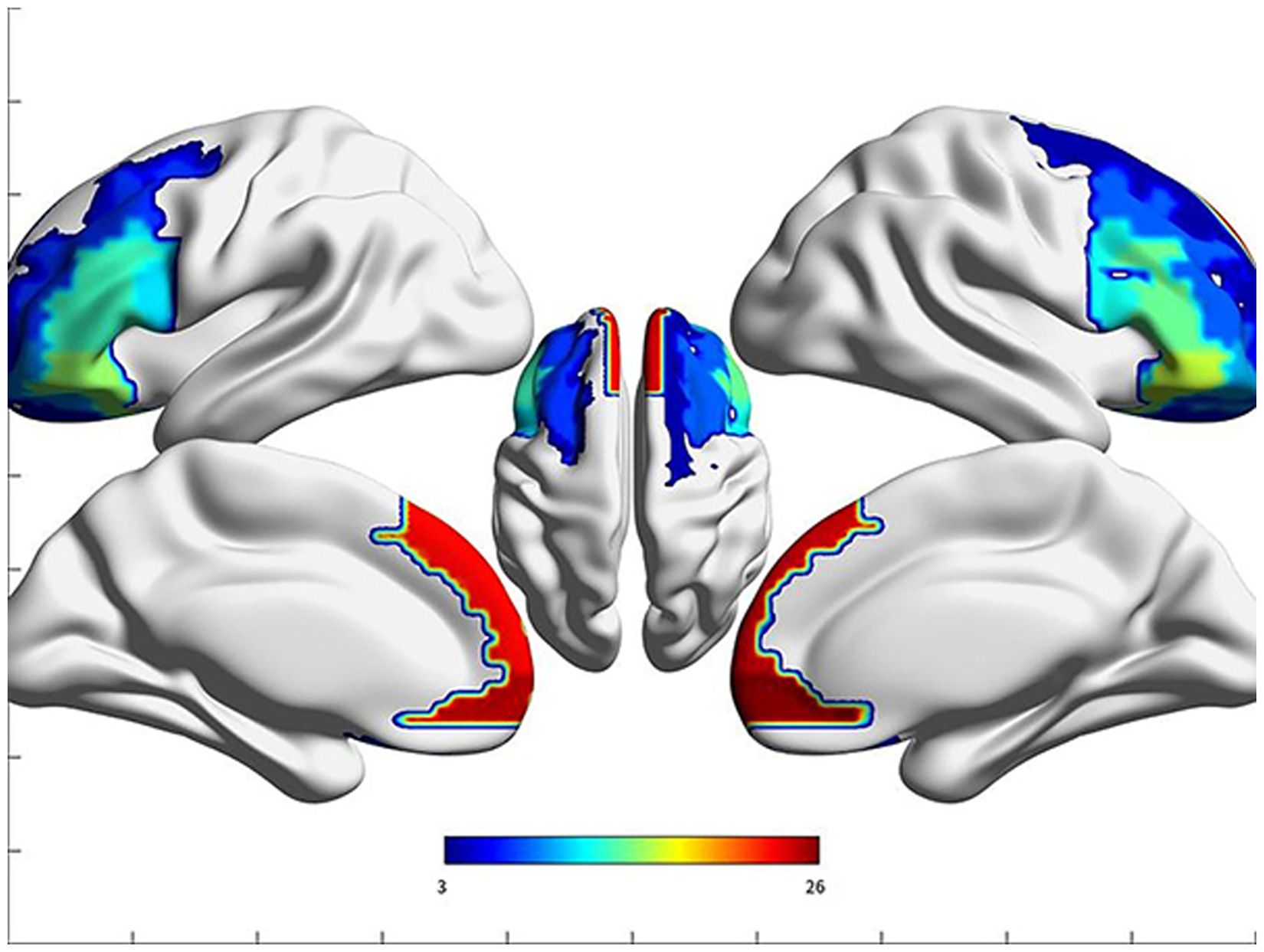
A total of 18 frontal brain regions in the AAL template.

Abnormal difference map of frontal FC maps between the insomniac and healthy individuals. In the patients with PI, we found that the abnormal FC areas were mainly concentrated in the superior frontal gyrus of orbital region (L/R), middle frontal gyrus of the orbital region (L/R), inferior frontal gyrus of the orbital region (L/R), inferior frontal gyrus of the opercular region (L) (P < 0.05). FC, functional connectivity; PI, primary insomnia.

Map of mirrored homotopy abnormalities between the insomniac and healthy individuals. The VMHC results showed that there was abnormal FC in the middle frontal gyrus of the orbital region. FC, functional connectivity.
The FC between the left superior frontal gyrus of the orbital region and left frontal gyrus of the medial orbital region exhibited a significant correlation with the course of disease (P = 0.037, r = 0.482), as well as with PSQI (P = 0.034, r = 0.489). A significant correlation existed between the FC of the left middle frontal gyrus of the orbital region and the left frontal gyrus of the medial orbital region with the course of disease (P = 0.023, r = 0.519). An association was found between the FC of the right middle frontal gyrus of the orbital region and the left frontal gyrus of the medial orbital region with the course of disease (P = 0.013, r = 0.561). There was a significant correlation observed between the FC of the left inferior frontal gyrus of the opercular region and the left frontal gyrus of the medial orbital region with SDS (P = 0.020, r = 0.541) (Fig. 4).
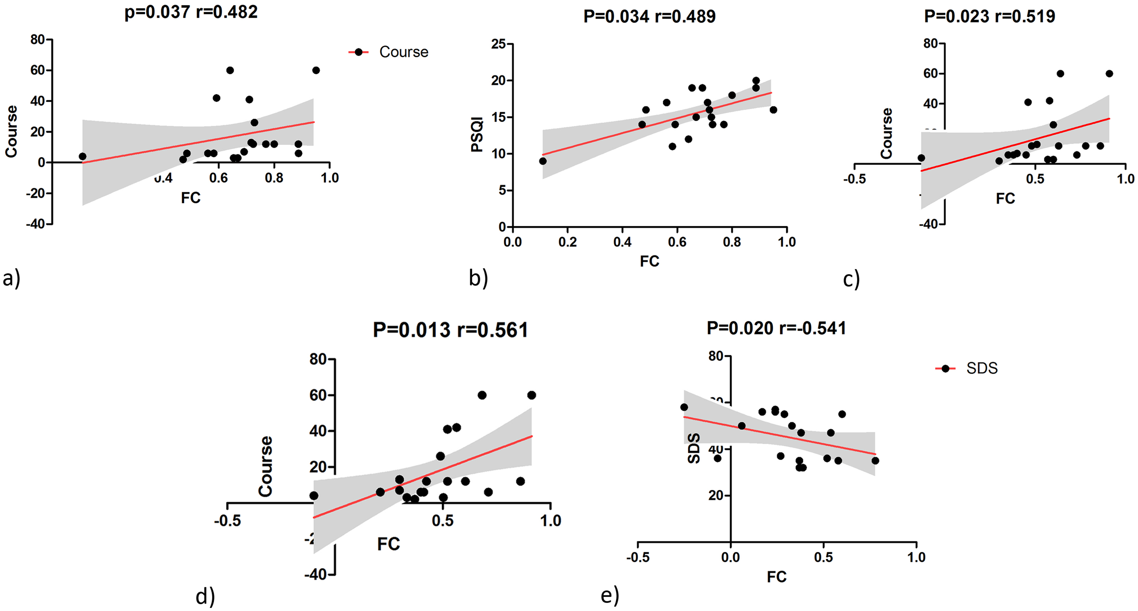
The FC between the orbitofrontal gyrus and the inferior frontal gyrus of the opercular region with frontal gyrus of medial orbital part demonstrated a significant correlation with the clinical scale (P < 0.05). (a, b) FC (Frontal_Sup_Orb_L/Frontal_Med_Orb_L), (c) FC (Frontal_Mid_Orb_L/Frontal_Med_Orb_L), (d) FC (Frontal_Mid_Orb_R/Frontal_Med_Orb_L), (e) (Frontal_Inf_Oper_L/Frontal_Med_Orb_L). FC, functional connectivity.
Discussion
In this study, the abnormal FC within the frontal lobe was found to be concentrated in the frontal gyrus of the orbital region and the inferior frontal gyrus of the opercular region. The orbitofrontal gyrus has a unique role in acquiring primary and advanced sensory information and learning complex stimulus-outcome relationships and transmitting anticipatory signals of outcome (10,11). It is also a key component in participating in value-based decision-making (12).
The prefrontal cortex is a large complex region that includes the dorsolateral prefrontal cortex, the ventrolateral prefrontal cortex, the orbitofrontal cortex, and the polar frontal region. Functional images also attribute the anterior cingulate cortex to the prefrontal cortex, but the two are not identical in terms of cellular structure. The prefrontal brain region is involved in the ventral, dorsal attention network, prominence network, frontal parietal executive network, and most importantly, the default network (13). The prominent clinical manifestation of prefrontal lesions is mental symptoms (14). This region has many connections with the thalamus, striatum, and autonomic nervous center of hypothalamus, and its projection fibers pass through the pontine nucleus to the cerebellum and may even end up in the cranial motor nucleus. The prefrontal cortex, known as the visceral motor cortex, causes synchronous changes in respiratory, metabolic, and cardiovascular functions (15), stimulates the orbital gyrus of monkeys, reduces respiration, blood pressure, and stomach peristalsis, and inhibits movement caused by motor cortex and reflexia. Insomnia patients are often accompanied by abnormal heart rate or digestive dysfunction.
Meanwhile, the orbital prefrontal cortex is involved in value judgments formed in complex conditions. which usually encodes information about the characteristics or identity of stimuli, such as food, odors, and trinkets. Studies have shown that insomnia does differ between healthy individuals on questions related to “wants” and “likes,” which are two main distinguishable dimensions of reward and hedonism (16). These findings suggest deficits in hedonism and reward processing in insomnia. Nevertheless, the orbitofrontal cortex is closely associated with hedonic assessment. The abnormal orbitofrontal FC in patients with insomnia indicates that participants have abnormal value judgment of things, which then affects the hedonic experience and abnormal reward processing (17).
Insomnia is associated with emotional regulation, and the risk of insomnia is associated with adverse childhood experiences, for instance, recent trauma or major life events, such as the death or serious illness of relative or friend, and incidents of physical, sexual, or emotional violence. Experiencing poor sleep in the face of stressful situations is known as “sleep reactivity.” People with high sleep reactivity are also more likely to develop depression and anxiety disorders (18). The orbitofrontal cortex has been implicated in the downregulation and reappraisal of emotional distress. The orbitofrontal cortex is also a major part of limbic network, which plays a central role in cognitive and emotional processing, including conflict monitoring, emotional arousal, and attentional control in motivation and emotion regulation. Previous studies have shown that patients with insomnia have reduced functional network connectivity in the prefrontal lobe, and the complexity of brain network is significantly increased after sedative and hypnotic drug treatment (19). Patients with insomnia showed stronger activation of the precentral gyrus and prefrontal cortex in response to sleep-related pictures, while this enhanced response was attenuated after cognitive behavioral therapy (20). Studies based on fMRI showed spontaneous neural activity or disrupted FC in the insula, prefrontal, and precuneus (21). People with lower gray matter density in parts of the orbitofrontal cortex are prone to early morning arousal, fragmented sleep, and poor sleep quality. People with high gray matter density in the orbitofrontal cortex sleep longer (22), and some studies have found that sleep quality and duration correlate with fractional anisotropy and mean diffusivity of white matter in the anterior cingulate, orbitofrontal, and insular regions as well as the caudate. The study of FC also includes impaired connectivity of the orbitofrontal-anterior insula and anterior cingulate cortex in insomnia patients (23).
The middle frontal gyrus is involved in the fronto-parietal attention network and is also related to associative memory function (24). Middle frontal gyrus discovered significant β and θ oscillations during REM sleep and suggested that this region may play a role in the regulation of memory consolidation. In addition, the middle frontal gyrus belongs to the dorsal-lateral prefrontal cortex, which is considered to be related to alertness, attention, and higher-order cognitive processes; all these functions are disrupted in insomnia patients (25). Therefore, the dysfunction of the middle frontal gyrus in patients with PI may also be related to abnormal memory consolidation and impaired cognitive function. Previous studies have found that patients with insomnia have decreased gray matter volume in the middle frontal gyrus. Broca's gyrus includes the inferior frontal gyrus of opercular region. Since the production and development of language are closely related to the dominance of the hand, for the right-handed participants in this study, the abnormal FC region occurred on the left side. Meanwhile, the correlation analysis revealed a significant association between the frontal gyrus of the orbital region and the inferior frontal gyrus of the opercular region on a clinical scale.
Patients with insomnia have previously been found to have intact overall cognitive function. However, no significant performance deficits were found in cognitive function, perceptual and psychomotor processes, procedural learning, language function, attention, verbal fluency, and cognitive flexibility. Small to moderate deficits were found only in episodic memory, problem solving, and working memory. Frontal lobe dysfunction in insomnia is generally considered to be associated with cognitive impairment, but further task-specific studies are needed. Moreover, insomnia itself is greatly affected by the body’s state and the surrounding environment, and there are many interference factors. We hope to increase the sample size in the future, and the inclusion and exclusion criteria should be stricter. Moreover, the FC method only studies the abnormalities of the internal brain areas of the frontal lobe and ignores the effects of other brain areas on the frontal lobe. At the same time, it is hoped that in the future, specific task-state studies are needed for the methods of frontal lobe cognitive impairment.
In conclusion, the frontal gyrus of the orbital region and inferior frontal gyrus of the opercular region are related to the impairment of decision making, value judging, reward and hedonism processing, emotional processing, and language function in patients with insomnia. This study is more helpful to reveal the neuropathological mechanism of patients with insomnia and to select more advantageous treatment methods for patients.
Footnotes
Declaration of conflicting interests
The authors declared no potential conflicts of interest with respect to the research, authorship, and/or publication of this article.
Funding
The authors disclosed receipt of the following financial support for the research, authorship, and/or publication of this article: This work was supported by the Beijing Municipal Administration of Hospitals Incubating Program (grant no. PZ2020014).
