



Abstract
Background
Automatic exposure control (AEC) devices are necessary to reduce the radiation dose and enhance image quality in radiography. However, AEC devices are not being used in portable X-ray systems due to technical issues.
Purpose
To evaluate the radiation dose and image quality of a new AEC for a wireless portable X-ray system compared to the standard fixed radiation dose of manual controls in chest radiography.
Material and Methods
This retrospective analysis evaluated the performance of a portable X-ray system equipped with software-based AEC on anteroposterior chest radiographs. A quantitative evaluation comparing the manual and AEC groups was conducted to ascertain the exposure index (EI) and dose-area product (DAP). We performed observer-based analysis of image quality for lowest exposure and average exposure images in the manual and AEC groups.
Results
Overall, 2093 examinations were conducted in 467 patients. AEC yielded a statistically significant reduction in the DAP (AEC: 1.64 dGycm2; manual: 1.99 dGycm2) and EI (AEC: 266; manual: 393) compared to manual control (P <0.001). Evaluation of the average and low exposure images revealed that the DAP and EI were consistently lower in the AEC group (P <0.01). Observer-based analysis of the lowest exposure images revealed that AEC yielded a statistically significant higher score compared to manual control (12.5 manual, 13.4 AEC; P <0.001).
Conclusion
AEC within a wireless portable X-ray system improves subjective image quality while reducing radiation exposure, as substantiated by quantitative and qualitative metrics.
Introduction
Automatic exposure control (AEC) devices are necessary to reduce the radiation dose and enhance image quality in radiography. AEC devices are mounted in front of the digital detector and enable image acquisition for patients of varying thickness, various regions of the body, and with different tube potentials using exposures that approximate the optimum (1). The radiation dose passing through the patient's body is measured in real time in the ion chamber or solid state sensor of the AEC, and radiation exposure automatically terminates at a preset radiation dose (1).
Thus, the radiation exposure to the patient can be reduced by delivering only the necessary dose according to body size, maintaining uniform and high quality of images obtained at a constant dose. It also helps more efficient radiographic acquisition by preventing retakes because of insufficient radiation doses (1,2).
AEC devices are used in fixed digital X-ray systems with wired communications. The ion chambers or solid-state sensors of the AEC and the position of the patient must be precisely aligned; otherwise, an error occurs in the calculation of the radiation dose, resulting in a higher radiation dose (1). Currently, difficulties in using the AEC system in a portable X-ray system is that latency may occur when taking X-rays in a wireless environment, and since the X-ray tube becomes thicker and heavier, it is difficult for the radiographers to properly align the X-ray detector and the patients, which increases the radiation dose.
Recently, AECs have been developed for wireless portable X-ray detectors. This AEC functions on the basis of software without the need to mount physical ion chambers in the detector and can be used in wireless environments by taking a scout shot before the main shot. Patient positioning is executed with automatic region-of-interest recognition via the new algorithm.
Therefore, the aim of the present retrospective study was to examine the efficacy of a new wireless AEC embedded in a portable X-ray system by comparing it with the fixed radiation dose of manual controls in anteroposterior (AP) chest X-ray (CXR) for radiation dose and observer-based image quality.
Material and Methods
This study was approved by the institutional review board (IRB N0. 2023-10-146). The requirement for patient consent was waived due to the retrospective nature of the study. Medical data were de-identified for anonymization.
Patient population
Consecutive hospitalized individuals who underwent AP CXR with portable X-ray system with novel AEC software attached were recruited between 17 November and 10 December 2023 at a single tertiary referral center. The exclusion criteria were as follows: (i) CXR with severe artifacts (patient movement and improper positioning) and (ii) patients aged under 18 years. Data on age, sex, body weight (kg), height (cm), and body mass index (BMI) were collected based on electrical medical records (Fig. 1).
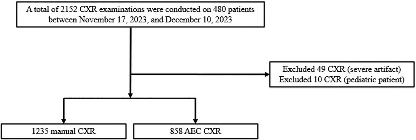
Flow chart of the study population.
AEC for wireless portable X-ray detector
The Korea Food and Drug Administration-approved S-AEC (X-ray System: GM85, Function: S-AEC, X-ray Detector: F4335-AW; Samsung Electronics Co., Ltd., Seoul, Korea) was used in the present study. S-AEC supports AP, posteroanterior, apicogram, and decubitus views, but not lateral projections.
The portable X-ray system communicates via Wi-Fi technology, but in hospitals, various wireless devices such as medical equipment and mobile phones can cause latency ranging from a few to tens of milliseconds. The exposure time for AP CXR is approximately 10 ms, and when latency occurs, the existing AEC devices that measure radiation dose in real-time and block the X-ray can result in increased radiation exposure, leading to potential overexposure for patients. To ensure stable application in a wireless environment, the S-AEC incorporates a scout shot. In the scout data, the lung area is segmented using S-AEC algorithm and the main exposure time is calculated from the pixel value of the lung area to meet the target dose and the optimized radiation exposure condition is calculated in advance according to the patient's body size, which is followed by the main shot (Fig. 2). When the radiographer presses the X-ray switch, the scout shot and the main shot are examined in succession. The interval between the two shots is very short, so the operating feeling felt by the radiographer is similar to that of a conventional single shot. Because CXR is mainly aimed at diagnosing the lung area, it should receive the appropriate radiation dose.

Overall operation principle of the S-AEC. In the scout data, the lung area is segmented using the S-AEC algorithm and the main exposure time is calculated from the pixel value of the lung area. Then, the main shot is automatically irradiated under the calculated condition. S-AEC, automatic exposure control.
The exposure index (EI) is used to verify the incident dose that penetrates the patient's body. The EI is a dose index that is extracted from the image and has been standardized to manage image quality and radiation dose in digital radiography (3). As the EI increases, image quality improves, but the radiation dose also increases. Therefore, determining the range of EI required for imaging diagnosis and managing its deviation are important for calibrating the patient's radiation dose (4). The radiation dose of the CXR was measured by adding the doses of the scout shot and main shot using a dose-area product (DAP) meter built into the output part of the X-ray tube.
Comparison of radiation dose and image quality between manual control and AEC groups
A quantitative evaluation comparing the manual control and AEC groups was conducted, focusing on the EI and DAP. Dose and image quality were assessed in all included patients. To further explore AEC performance in low-dose scenarios, we conducted a subgroup analysis of images with exceptionally low exposure. Images with an EI below 200 were defined as the lowest exposure group, based on our institutional data showing that these constituted the lowest 3% of all acquired images. All remaining images were categorized as the average exposure group. Furthermore, observer-based analysis of image quality was performed by three board-certified thoracic radiologists (radiologists 1, 2, and 3 with 8, 32, and 18 years of experience in interpreting thoracic radiology) for lowest and average exposure groups within the manual and AEC groups using a 5-point visual grading analysis (VGA) (5,6) (Table 1). In this study, we used the absolute VGA scoring method to evaluate the image quality of manual and AEC groups. On AP CXR examinations, three categories (sharp visualization of the vessels seen 3 cm from the pleural margin, visualization of the carina with main bronchi, visualization of the thoracic vertebrae behind the heart) were assessed along the 5-point VGA, and the sum of the categories was compared between the manual and AEC groups. Paired comparisons (one manual and one AEC) of images from the same patient were conducted to assess the consistency and reproducibility of the AEC in comparison to manual exposure. Each radiologist performed a blinded quality assessment of images from the manual and AEC groups.
VGA-based scale for scoring image quality.

VGA, visual grading analysis.
All images were reviewed on light emitting diode color monitors (CCL258i2; Totoku, Tokyo, Japan). Image viewing was conducted under controlled ambient lighting conditions (20 lux) to ensure optimal visibility and reduce external glare. Evaluations were carried out using the picture archiving and communicating system (PACS; GE Healthcare, Chicago, USA).
Statistical analysis
Descriptive statistics, including the mean and standard deviation (SD) were calculated for all variables. Comparisons between the manual and AEC groups were conducted using independent sample t-tests for continuous variables and chi-square tests for categorical variables. The distribution of the data met the normality assumption required for the t-test. Paired t-tests were used for within-subject comparisons of manual and AEC exposures within a 1-week interval. Pearson's correlation coefficient (r) was calculated to assess the linear relationship between VGA scores and DAP in the paired comparison. Statistical significance was set at P <0.05. In addition, the interrater reliability of the observer-based image quality scores was assessed using Cohen's kappa coefficient to ensure consistency between the overall radiologists’ evaluations. All statistical analyses were performed using R software (version 4.2.3).
Results
Patient characteristics
A total of 2093 CXR examinations (1235 manual, 858 AEC) were conducted in 467 patients and performed by two radiographers. The patient demographics are summarized in Table 2. Age, sex, body weight, height, and BMI did not differ significantly between the manual and AEC groups.
Baseline characteristics of the patient study group.

Values are given as n or mean ± SD unless otherwise indicated.
AEC, automatic exposure control; BMI, body mass index.
Comparative quantitative analysis between manual and AEC groups
The AEC group demonstrated a significant reduction in the EI (manual: 393, AEC: 266; P <0.001) and DAP (manual: 1.99 dGycm2, AEC: 1.64 dGycm2; P <0.001) compared to the manual group (Table 3). In the AEC group, the EI and DAP were 59%, and 17% lower relative to the manual group, respectively (Fig. 3). Even when evaluating the average- and low-exposure images, the EI and DAP were consistently lower in the AEC group than in the manual group (Table 4). The reduction in EI and DAP achieved with AEC remained within acceptable diagnostic thresholds.
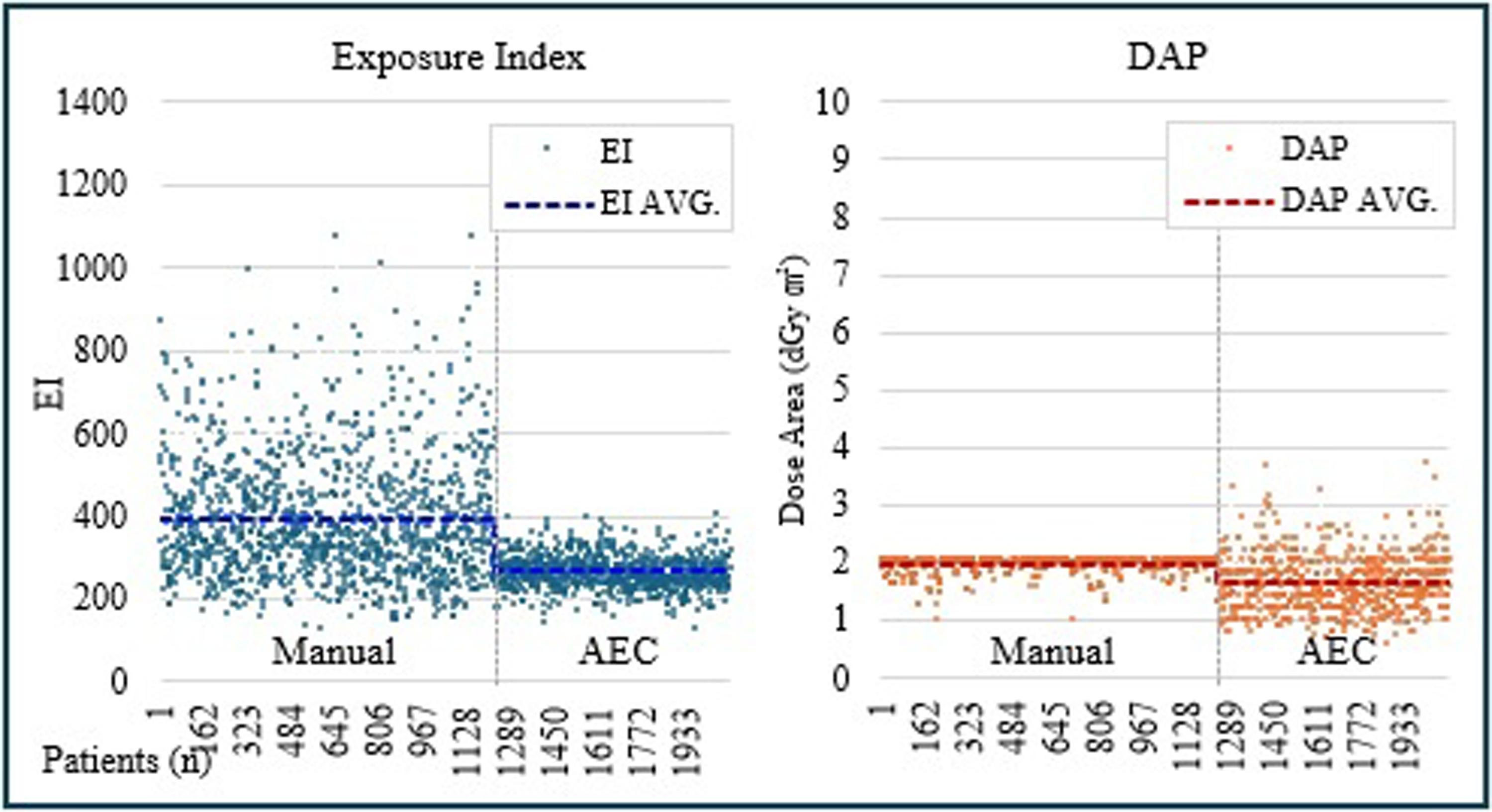
Comparison of EI and DAP in manual and AEC groups. In the AEC group, EI was reduced by 59% and DAP was reduced by 18% compared to manual mode. AEC, automatic exposure control, DAP, dose-area product; EI, exposure index.
Comparative quantitative analysis between manual and AEC groups.

Values are given as mean ± SD unless otherwise indicated.
AEC, automatic exposure control; BMI, body mass index; EI, exposure index.
Descriptive statistics for low exposure CXR examinations of manual and AEC groups.

Values are given as mean ± SD unless otherwise indicated.
AEC, automatic exposure control; BMI, body mass index; CXR, chest X-ray; DAP, dose-area product; EI, exposure index.
Observer-based analysis of image quality between the manual and AEC groups
In the observer-based analysis of average-exposure images, the VGA scores of the manual and AEC groups did not differ significantly between the readers (Table 5). Cohen's kappa value was 0.78, indicating substantial agreement. However, in lowest exposure images, AEC yielded a significantly higher VGA score compared to manual control (reader 1: manual: 12.5, AEC: 13.4; P <0.001, reader 2: manual: 12.2, AEC: 13.3; P <0.001, and reader 3: manual: 11.5, AEC: 12.5; P <0.001). In the paired comparison, the AEC group outperformed the manual group, exhibiting higher observer analysis scores (manual: 14.1, AEC: 14.3; P = 0.029) and lower DAP (manual: 1.98, AEC: 1.63; P <0.001) (Fig. 4, Table 6). The use of paired comparisons demonstrated improved consistency in exposure levels and image quality when using AEC, as reflected by lower variability in VGA scores and dose parameters across the AEC group. The Pearson correlation coefficient was 0.67, indicating a strong correlation.
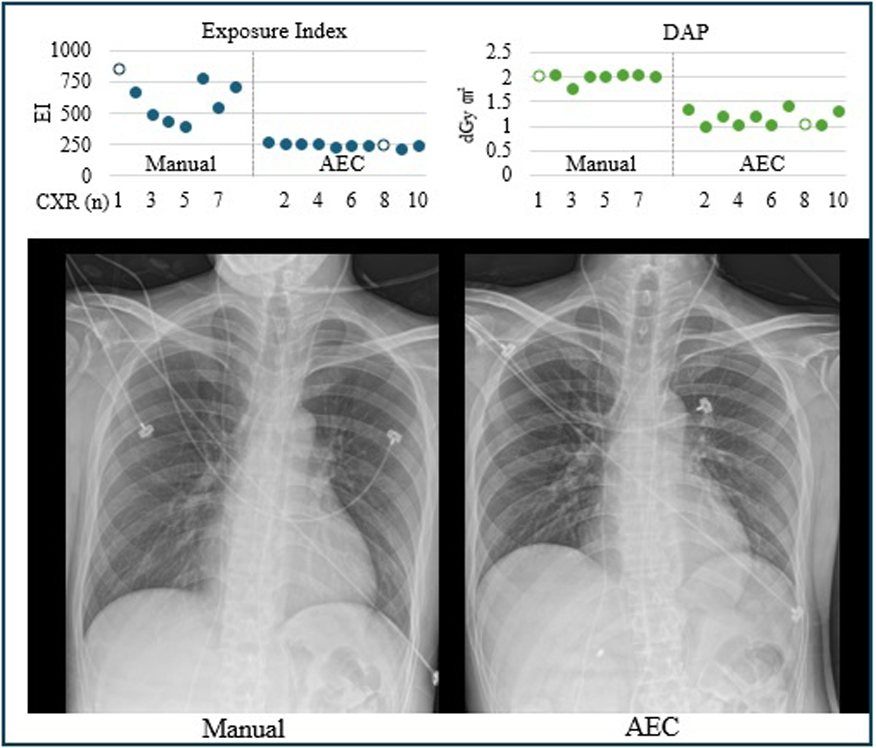
Image quality of the AEC group is similar or slightly better than that in the manual group; however, EI and DAP were decreased in the AEC group. AEC, automatic exposure control, DAP, dose-area product; EI, exposure index.
Observer-based analysis of image quality under average and low exposures in manual and AEC groups.

Values are given as mean ± SD.
AEC, automatic exposure control; VGA, visual grading analysis.
Paired comparison between manual and AEC groups.

Values are given as mean ± SD unless otherwise indicated.
AEC, automatic exposure control; DAP, dose-area product; VGA, visual grading analysis.
Discussion
In this study, the AEC of a wireless portable X-ray detector significantly decreased the radiation dose and enhanced the image quality compared to those achieved with manual control in a conventional portable X-ray device. Moreover, in the lowest exposure images, the AEC group exhibited better subjective image quality relative to the manual group.
The International Electrotechnical Commission (IEC) recommends monitoring the patient's radiation dose during X-ray examinations. The IEC standard notes that the EI may differ for each manufacturer and type of examination in clinical protocols, but the proportional relationship with the incident dose on the detector does not change (3). In clinical practice, even if an examination is performed under the same exposure, the incident dose on the detector and image quality differs depending on patient factors. Therefore, distribution of the EI values is variable and can only be used as a surrogate for dose management. In particular, it is difficult to monitor the patient dose using only the EI value in a portable X-ray system, which is not equipped with an AEC. In a fixed X-ray system, the incident dose on the CXR detector is always constant because of the AEC. Therefore, various studies have been performed to accurately measure and monitor the EI as a dose-monitoring tool in portable X-ray systems without an AEC. One study that assessed the EI as a dose-monitoring tool for AP chest X-rays of neonatal patients obtained with a portable X-ray system without AEC, reported that the EI values were in the range of 100–1600 (7). With all conditions being equal, the difference in incident dose on the detector was attributed to patient factors, such as body shape, and pathological conditions, such as the presence of pneumonia. In a study by Park et al., despite the fixed exposure conditions, EI values varied widely (range = 137.82–4924.38), largely influenced by patient body shape and size (8). In another study, when AEC was not employed, the EI value decreased as the phantom thickness increased. When the AEC was used, the EI value was the same as the phantom thickness; however, the entrance surface dose increased (9). Therefore, the build and body shape of the patient affect the EI value when an AEC system is not used.
In our study, we used a new AEC designed for a wireless portable X-ray detector, and we can expect a constant EI value, optimal radiation exposure, and consistent image quality for diagnosis similar to those of fixed X-ray systems. AEC in portable X-ray systems was applied in the film-screen radiography era, which did not require wired connections (2). The physical installation of AEC in wireless portable X-ray equipment is limited due to its usability problems, such as increased volume and weight and lack of wireless support.
To the best of our knowledge, this is the first clinical study to evaluate the efficacy of a software-based wireless AEC system implemented in portable X-ray units.
The present study has some limitations. First, this study was performed solely in the chest. It will be more meaningful to conduct additional studies at various anatomical locations and radiographic views in the future to establish the general performance of the AEC of wireless portable X-ray detectors. Second, this study was conducted in a single-institution setting without external validation. Lastly, this study did not assess diagnostic or technical image quality, which limits the generalizability of the results. A prospective study design would be needed to evaluate diagnostic accuracy and clinical utility more comprehensively.
In conclusion, the implementation of a novel, software-based AEC in wireless portable X-ray detectors improved subjective image quality while reducing radiation exposure, as substantiated by both quantitative and qualitative metrics. Furthermore, it is expected to contribute to management of patients’ radiation dose by deducing the average exposure dose.
Footnotes
Data availability
Data generated or analyzed during the study are available from the corresponding author by request.
Declaration of conflicting interests
The authors declared no potential conflicts of interest with respect to the research, authorship, and/or publication of this article.
Funding
The authors disclosed receipt of the following financial support for the research, authorship, and/or publication of this paper: This research was funded by the Future Medicine 20*30 Project of the Samsung Medical Center (grant no. SMX1240781; The Korea Health Technology R&D Project through the Korea Health Industry Development Institute (KHIDI), funded by the Ministry of Health Welfare, Republic of Korea, grant number HR21C0885.
