



Abstract
A 6-year-old, castrated male, domestic shorthair cat with progressive neurologic signs underwent magnetic resonance imaging, revealing a suprasellar mass, which resulted in euthanasia. Grossly, a tan-red tumor expanded the ventral third ventricle, compressed adjacent brain structures, and emerged ventrally at the midline. Histologically, numerous arborizing papillary formations protruded into a network of anastomosing luminal canals. Neoplastic cells immunolabeled for pan-cytokeratin, vimentin, and E-cadherin, and lacked immunolabeling for oligodendrocyte transcription factor 2 (OLIG2), glial fibrillary acidic protein (GFAP), and adrenocorticotropic hormone (ACTH). Transmission electron microscopy revealed apical microvilli, apical and lateral tight junctions, and a basal membrane. In this cat, the neuroanatomic location with ventral brain invasion was more suggestive of ependymal origin; however, there were overlapping histologic and immunohistochemical features, and ultrastructural features were more consistent with choroid plexus epithelium. Dual ependymal and choroid plexus differentiation could not be excluded. This case highlights species differences in both the occurrence and neurolocalization of intraventricular tumors in domestic animals and comparable features between papillary ependymoma and choroid plexus papilloma.
The choroid plexus is a highly vascularized papillary structure located within the ventricles of the brain. Attached to the ependymal lining, 24 the choroid plexus produces cerebrospinal fluid (CSF), which flows through the ventricular system and central canal. Embryogenic formation of the choroid plexus primordium occurs via invagination of a bilayer membrane of neuroectoderm and pia mater into the ventricles.4,18 The choroid plexus is found in 4 locations in the brain: (1) the right and left lateral ventricles, (2) the dorsal recess of the third ventricle, and (3) the fourth ventricle. 24 Specialized tight junctions (zonulae occludens) and desmosomes between the epithelial cells of the choroid plexus maintain the apical blood-CSF barrier.18,24 The choroid plexus may represent a source of neural stem cells and also plays roles in the development, homeostasis, and repair of the central nervous system (CNS). 24
Ciliated ependymal cells line the ventricles of the CNS, playing a major role in regulating CSF flow. 18 The ependyma also has homeostatic, secretory, absorptive, and metabolic functions. 18 Tanycytes are morphologically and functionally specialized ependymal cells with radially directed basal processes that extend into the neuroparenchyma and typically lack kinocilia (as opposed to non-specialized ependymal cells, which generally have kinocilia).3,24
Neoplasms of the choroid plexus and ependyma are uncommon to rare in domestic animals.18,17,20,22 In humans, choroid plexus tumors constitute 0.3% to 0.8% of all brain tumors, accounting for 2% to 4% of those occurring in young children (less than 15 years), and 10% to 20% of brain tumors in children 1 year old or less. 2 On computed tomography and magnetic resonance imaging, choroid plexus tumors are T1-weighted (T1W) isointense, T2-weighted (T2W) hyperintense, and irregular contrast-enhancing. 2 In dogs, choroid plexus tumors occur most frequently in middle aged to older animals, accounting for approximately 10% of canine primary CNS tumors. 9 Magnetic resonance imaging features of choroid plexus tumors in the dog include iso-hyperintense on T1W and T2W images; homogenously strong contrast enhancement; and signal heterogeneity with cysts, hemorrhage, necrosis, or mineralization. 23
Ependymal tumors in humans are currently categorized by molecular alterations. DNA methylation profiles are used to divide them into molecular groups across 3 anatomical compartments of the CNS (supratentorial, posterior fossa, and spine). 2 Ependymomas are rare in the dog. 16 In cats, ependymomas are more frequently described than choroid plexus tumors,11,12,18,20,22,25 with no definitive reports of choroid plexus papillomas or carcinomas to date. Magnetic resonance imaging features of ependymomas in cats include hyperintensity on T2W, T2W fluid-attenuated inversion recovery (FLAIR), proton density, and diffusion-weighted echo-planar imaging with isointensity on apparent diffusion coefficient images and subtle to strong contrast enhancement. 11 In people, ependymomas are typically T1W isointense to hypointense and T2W hyperintense, with irregular contrast enhancement and heterogeneity caused by necrosis, cysts, hemorrhage, and calcifications. 27 Herein, we describe a third ventricular neoplasm with concurrent choroid plexus and ependymal features in a cat.
A 6-year-old, castrated male, domestic shorthair cat was evaluated by the Neurology Service at the Schwarzman Animal Medical Center for progressive neurologic signs over 3 weeks. Neurologic examination revealed dull to somnolent mentation, ambulatory tetra-ataxia with head pressing, circling to the left, an absent menace response, and direct pupillary light response in the right eye. Neurologic signs were suggestive of a left supratentorial and infratentorial lesion with concern for a diencephalic lesion and subsequent brain swelling. Magnetic resonance imaging was performed with a 1.5 Tesla scanner (Philips Medical Systems) and showed a lobulated mass containing a central core of hemorrhage and/or mineralization within the third ventricle. The central core was heterogeneously hyperintense and hypointense on T1W and T2W imaging, respectively. The solid part of the mass was homogenously T1W hyperintense and T2W hypointense relative to normal brain. The tumor was variably, moderately to strongly contrast-enhancing. Ventriculomegaly, obstructive hydrocephalus, severe caudotentorial herniation, and mild cerebellar coning were noted and were presumed to be secondary to the mass. During recovery, seizures occurred. Based on these findings, euthanasia was elected, and an autopsy was performed.
Autopsy findings consisted of a soft, tan to red, 1.5 × 1.5 × 1.0 cm mass that expanded the ventral midline aspect of the brain, in the region of the hypothalamus and pituitary gland. Sectioning of the formalin-fixed brain revealed an expansile, centrally hemorrhagic midline mass located within the dilated lumen of the ventral part of the third ventricle to which it was attached (Fig. 1a, b). The mass compressed the adjacent hypothalamic structures and erupted into the ventral subarachnoid space. The dorsal aspect of the third ventricle (and its choroid plexus) was unaffected. The cerebellar vermis was elongated, indicating transforaminal herniation. Autopsy findings were suggestive of a pituitary or ependymal neoplasm, or more remotely for this intraventricular location, a choroid plexus neoplasm.

Third ventricular neoplasm, brain, cat. (a) At the level of the caudate nuclei and hippocampal fornix, a large mass with regional hemorrhages and cystic spaces fills the ventral aspect of the third ventricle and extends into the subarachnoid space. (b) Subgross view of the mass. Hematoxylin and eosin (HE). (c) Neoplastic cells are in continuity with the adjacent ependymal lining (arrow). HE. (d) Neoplastic cells form numerous, arborizing, elongated papillary projections lined by a single layer of bland cuboidal to columnar cells. HE. Inset: Small amounts of fibrous tissue are present in the intervening stroma, highlighted by the blue stain. Masson’s trichrome.
Touch imprints of the mass were stained with Wright-Giemsa and revealed cohesive, round to oval or cuboidal cells with a small amount of lightly basophilic cytoplasm and mildly pleomorphic nuclei. Histologic findings consisted of a well-demarcated, variably cellular neoplasm with occasional extension from the adjacent ependymal lining (Fig. 1c). The neoplasm consisted of numerous, arborizing, elongated papillary projections lined by a single layer of bland cuboidal to columnar neoplastic cells with scant, variably eosinophilic, lightly vascularized collagenous stroma (Fig. 1d). A prominent feature was the presence of branched, anastomosing, luminal canals (Fig. 1d) that occasionally contained eosinophilic material and the nuclei of few degenerated cells (Fig. 1d). Large cystic foci were also observed within the neoplasm, some of which contained abundant hemorrhage with cholesterol clefts and hemosiderin-laden macrophages. No rosettes or pseudorosettes were found. The mitotic count was one in 2.37 mm2 (10 FN22/40X fields). Masson’s trichrome stain revealed small amounts of fibrous connective tissue (Fig. 1d, inset) with occasional discrete central islands of collagen deposition within the tumor stroma.
Tumor sections were subjected to immunohistochemistry (IHC) for pan-cytokeratin (AE1/AE3), cytokeratin 7 (CK7), CK20, vimentin, E-cadherin, beta-catenin, S100, synaptophysin, oligodendrocyte transcription factor-2 (OLIG2), glial fibrillary acidic protein (GFAP), and adrenocorticotropic hormone (ACTH) (Table 1 and Supplemental Table S1). Neoplastic cells had diffuse, strong cytoplasmic immunolabeling for AE1/AE3 (Fig. 2a), CK7, CK20, vimentin, E-cadherin (Fig. 2a), and beta-catenin, with patchy cytoplasmic immunolabeling for S100 and synaptophysin. Neoplastic cells and stroma lacked immunolabeling for OLIG2, GFAP, and ACTH. While the tumor location (ventral third ventricle) and some of the histologic and IHC features were typical of ependymoma, other histologic features, histochemical, and IHC findings were more consistent with a choroid plexus tumor.

Abbreviations: IHC, immunohistochemistry; PanCK, pan-cytokeratin (AE1/AE3); CK7, cytokeratin 7; CK20, cytokeratin 20; E-cad, e-cadherin; B-cat, beta-catenin; GFAP, glial fibrillary acidic protein; OLIG2, oligodendrocyte transcription factor-2; EMA, epithelial membrane antigen; SYN, synaptophysin; ACTH, adrenocorticotropic hormone; CP, choroid plexus; UNK unknown; N/A, not available.
Epithelial cells, not including stroma.
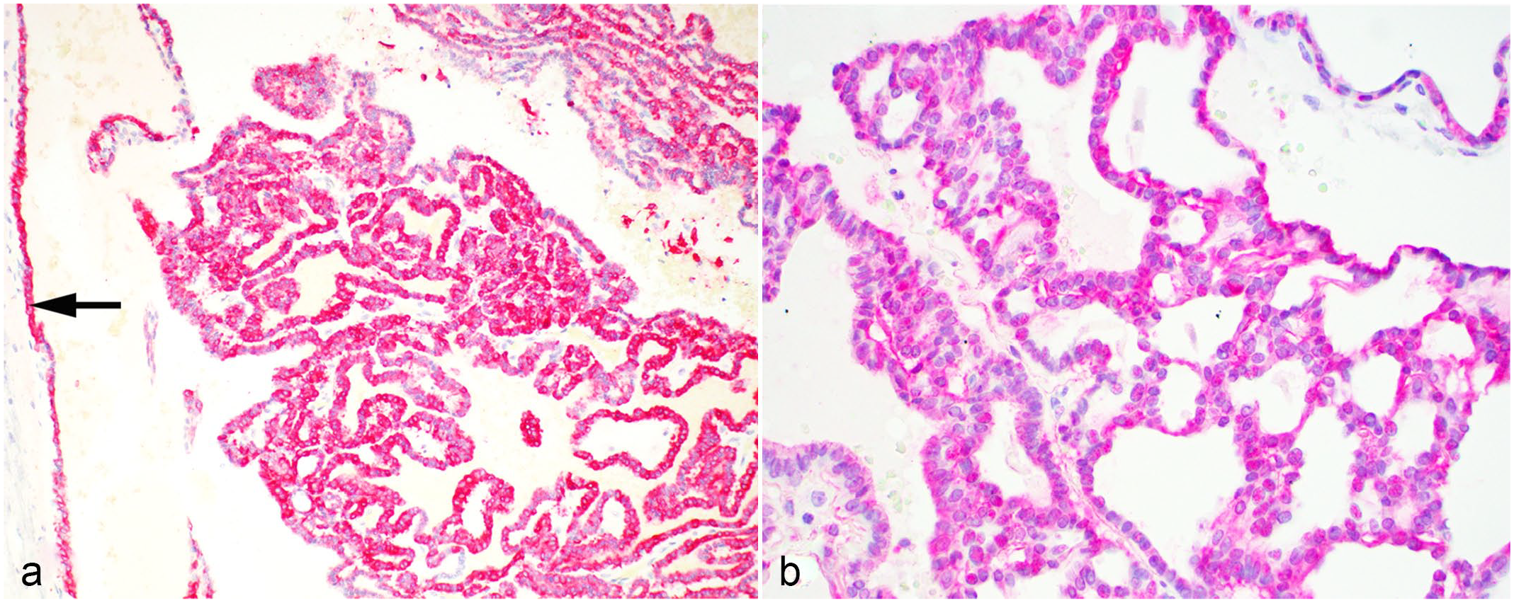
Third ventricular neoplasm, brain, cat. (a) Neoplastic cells and regional ependyma (arrow) have cytoplasmic immunolabeling for pan-cytokeratin (AE1/AE3). (b) Neoplastic cells have cytoplasmic and membranous with occasional nuclear immunolabeling for E-cadherin.
A formalin-fixed tumor sample was submitted for transmission electron microscopy. Tissues were trimmed to 1 to 2 cubic mm and post-fixed in 2.5% glutaraldehyde. Three sections were selected, processed, and resin-embedded in an automated tissue processor. Thick sections were stained with toluidine blue to identify the region of interest from which ultrathin sections were stained with uranyl acetate and lead citrate. Cuboidal neoplastic cells had an apical cellular domain characterized by tight junctions and microvilli. The lateral domain contained delicate anchoring (desmosomal) junctions and plasma membrane infolding forming interdigitations. The basal domain had a basement membrane (Fig. 3). The cytoplasm had mild to moderate numbers of round mitochondria (Fig. 3), small numbers of short endoplasmic reticulum, and vesicles located predominantly at the apical pole of the cells. No motile cilia were found. Because of the lack of motile cilia, as well as the presence of tight junctions, microvilli, and a basement membrane, ultrastructural findings were consistent with a choroid plexus neoplasm (Table 2). 15
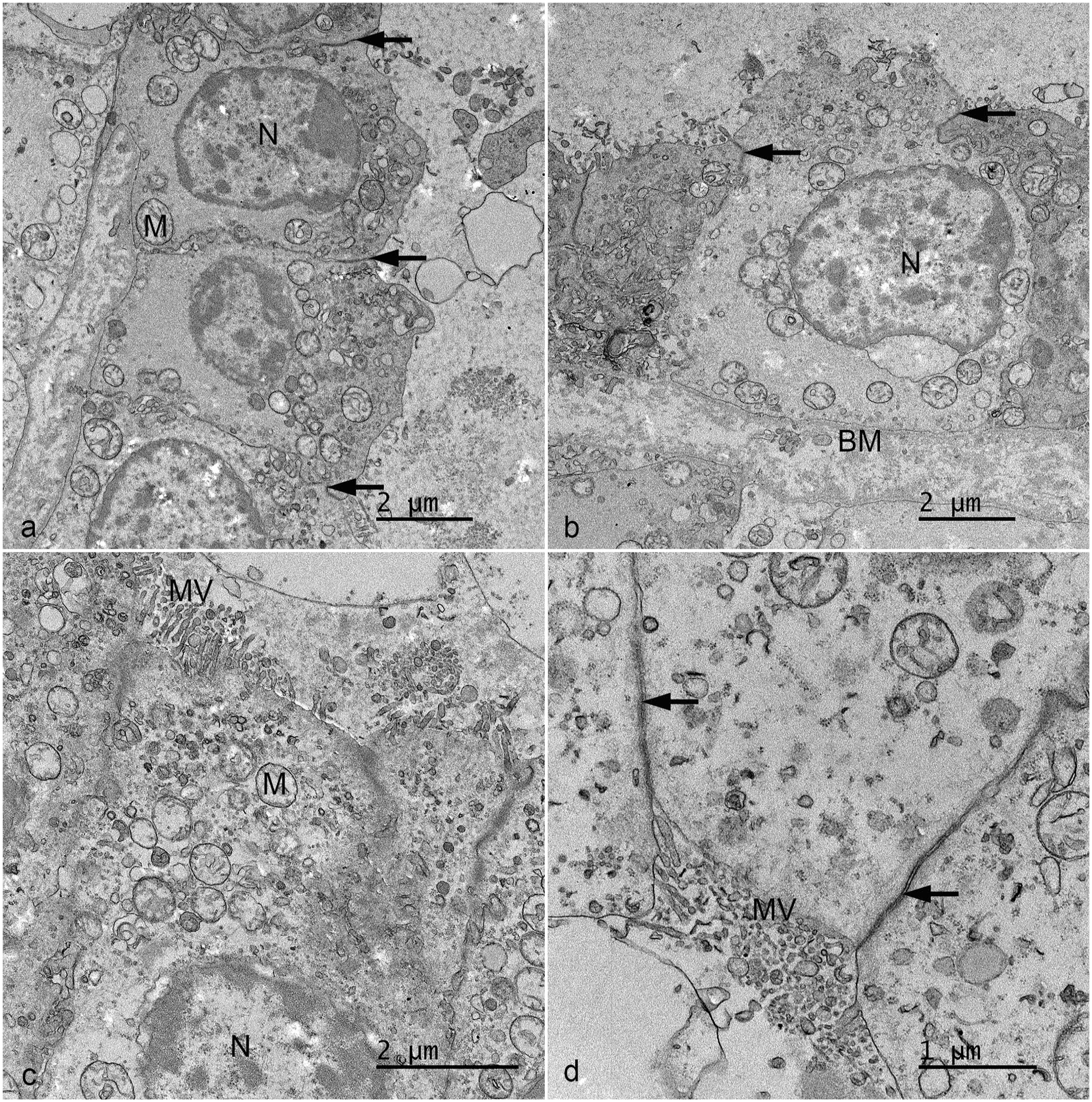
Third ventricular neoplasm, brain, cat. Transmission electron microscopy. (a) Multiple tight junctions (arrows) can be observed between the cuboidal cells. Nuclei (N) and mitochondria (M) are visible. (b) Tight junctions (arrows), a basal membrane (BM), and nucleus (N) are visible. (c) Neoplastic cells have microvilli (MV) and numerous mitochondria. (d) Tight junctions (arrows) and microvilli (MV) are present.
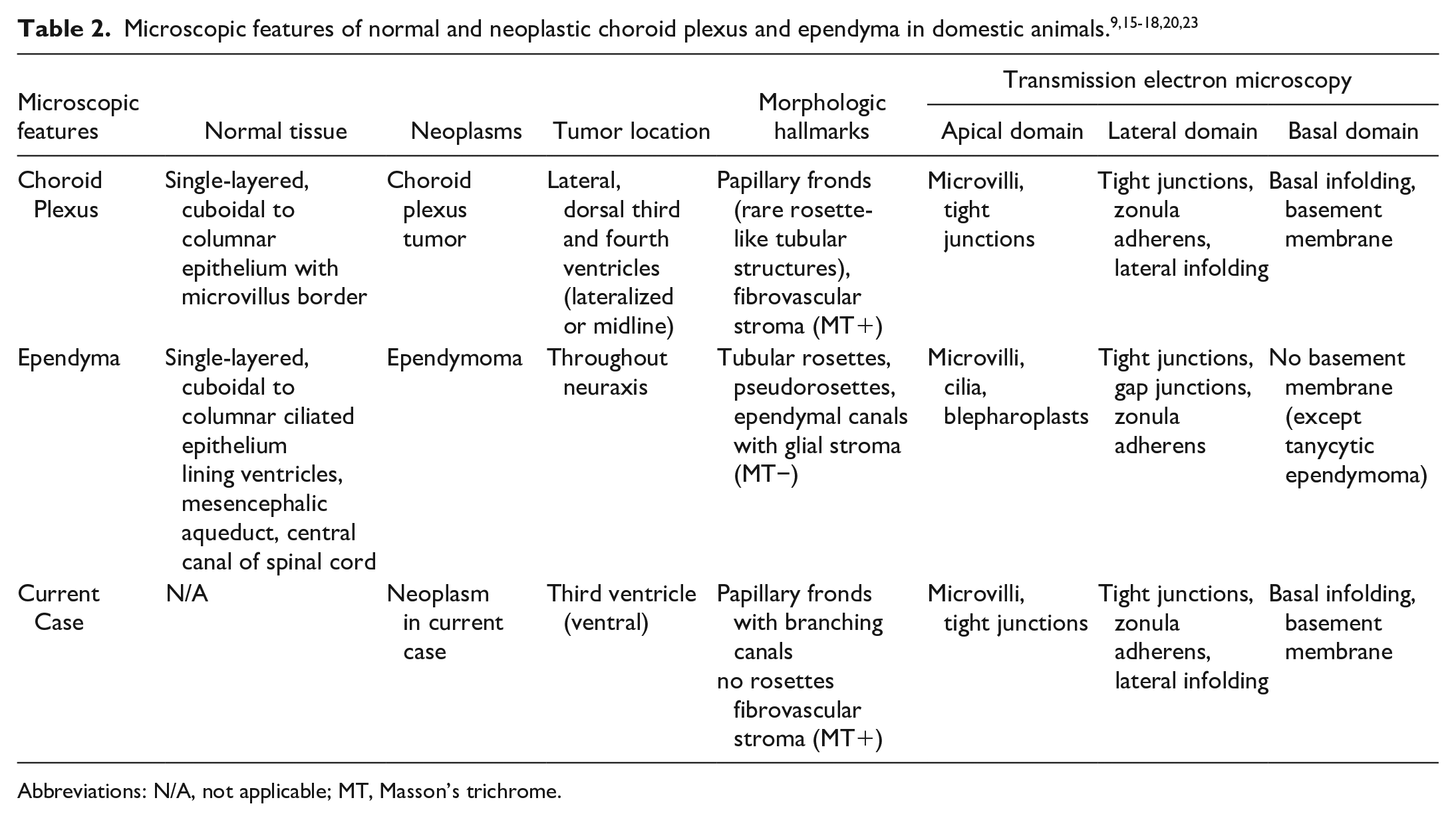
Abbreviations: N/A, not applicable; MT, Masson’s trichrome.
This case underscores the difficulty in accurately classifying primary brain tumors solely based on morphology and IHC, as cell types can exhibit hybrid phenotypes. The gross appearance (smooth surface, without granularity) and lack of tumor lateralization, which commonly occur in choroid plexus tumors of the lateral and fourth ventricles, were considered atypical for a choroid plexus neoplasm. The histomorphology and histochemical findings were interpreted differently by different pathologists, some of whom favored a diagnosis of choroid plexus papilloma, and some of whom favored a diagnosis of papillary ependymoma. In particular, the formation of branched, tubular luminal canals and large thin-walled cysts were considered more supportive of ependymoma by some of the authors, but others have observed these features in choroid plexus tumors of dogs (D.R.R., personal communication). The immunohistochemical findings were contradictory, with patterns of expression expected in both cell types. It should be noted that expression patterns may vary within a neoplasm, both due to the variability within tumors as well as differences between normal and neoplastic tissues. The ultrastructural findings were considered to be more consistent with a choroid plexus neoplasm.
Cells of the ependyma and choroid plexus are closely related; 4 thus, neoplastic choroid plexus and ependymal populations may have common morphologic features. Choroid plexus tumors and ependymomas genetically demonstrate similar methylation patterns. 13 Ambiguous or overlapping histologic and IHC findings with blurring of the boundary between choroid plexus and ependymal differentiation have been reported in the human literature,4,13 and it is well recognized that tumors of the CNS may have dual features. 7 Choroid plexus and ependymal tumors of humans are considered to be derived from multipotent neuroepithelial cells. 13 Ependymal differentiation in choroid plexus tumors of humans has ranged from changes in IHC markers without morphologic changes to observable histologic differentiation. 13 Mesenchymal tissue has also been reported in choroid plexus and ependymal tumors, possibly due to the multipotential nature of cells derived from circumventricular organs or invagination of mesenchymal tissue during development. 13 Synchronous/mixed choroid plexus and ependymal tumors in humans have morphologically distinct phenotypes of choroid plexus and ependymal cells in the same tumor. 4 These different components may represent a synchronous (mixed) tumor or a single tumor arising from a common progenitor cell with divergent differentiation (biphenotypic synchronous tumor). 4 Neural stem cells were recently confirmed to be present in the feline brain, and feline ependymomas and subependymomas were found to have a neural stem cell-like immunophenotype. 26 Ectopic choroid plexus papillomas have been reported in the human and veterinary literature without any intraventricular connection (unlike this case).1,8,21 Development from ectopic neuroectodermal remnants or metaplasia of ependymal remains has also been hypothesized as a mechanism for formation of ectopic choroid plexus tumors. 1
In veterinary medicine, choroid plexus tumors are most commonly observed in the lateral and fourth ventricles and are reported predominantly in dogs, with rare reports in horses and cattle.17,20 Histologically, these tumors form papillary (frond-like) structures supported by a core of fibrovascular stroma. Histologic features of choroid plexus tumors can overlap with papillary ependymoma, including the formation of cords, ribbons, papilliform projections, and structures that resemble tubular rosettes and pseudorosettes, or mimic metastatic carcinomas, including tubular or acinar-like structures and sheet-like growth of cuboidal epithelial cells.17,19 Choroid plexus tumors can be immunolabeled for AE1/AE3, E-cadherin, beta-catenin, and rarely GFAP. Feline choroid plexus tumors have been rarely reported, but it is unclear whether this neoplasm truly occurs in domestic cats. A case report of a dorsal third ventricle mass was suspected to represent a feline choroid plexus tumor, but a definitive diagnosis was not obtained. 14 Another report within a case series described a left lateral ventricle mass from a cat as a choroid plexus tumor, but the diagnostic details were not provided. 22 In addition, an intradural, extramedullary, ectopic choroid plexus tumor was reported in the spinal cord of a cat. 21 Neoplastic cells were immunolabeled for pan-cytokeratin and E-cadherin, and lacked synaptophysin, thyroglobulin, chromogranin A, and GFAP immunolabeling. However, none of these IHCs are confirmatory for a choroid plexus neoplasm. 21 A choroid plexus oncocytoma was reported at the left cerebellopontine angle of a cat. 8 Ultrastructural evaluation has not been previously reported for a feline choroid plexus neoplasm.
Feline ependymomas are glial tumors arising from germinal cells in the ventricular ependymal lining in the brain or spinal cord. Ependymal cells are thought to represent terminally differentiated cells that arise from either radial glial cells or multiprogenitor stem cells in the subventricular zone. 12 These neoplasms most commonly manifest as intraventricular masses of the lateral or third ventricles.12,25 While ependymomas are typically well demarcated and extend into the ventricular lumen, higher-grade tumors can infiltrate the adjacent neuroparenchyma. Characteristic histologic features include the formation of tubular (ependymal) rosettes and ependymal canals, as well as pseudorosettes, although these are not always present. In addition, morphologic variants of ependymoma can also occur, including papillary, tanycytic, clear cell, and anaplastic subtypes.12,25
A papillary ependymoma has been reported in the horse at a similar location in the third ventricle, 5 and feline ependymomas located in the ventral compartment of the third ventricle and invading the brain ventrally (as in this case) have been previously observed by some authors (BAS, personal communication). Feline third ventricle ependymomas in the dorsal compartment are rarely reported. 11 Tubular rosettes and pseudorosettes, a hallmark of many ependymomas, were lacking in our case and in the reported equine case. 5 It is proposed that papillary ependymomas have unique histologic and ultrastructural characteristics, which differ from conventional ependymomas. 19 Papillary ependymomas in domestic animals have been reported to have an astrocytic core, as opposed to the fibrovascular core expected with choroid plexus neoplasms. 25 In our case, the supporting (core) stroma did not immunolabel for GFAP and had few regions of blue staining with Masson’s trichrome (Fig. 1d, inset).
In cats, neoplastic ependymal cells are typically immunolabeled for pan-cytokeratin, GFAP, and OLIG2.12,26 The histologic features of normal and neoplastic choroid plexus and ependymal cells in domestic animals are summarized in Table 2, whereas the immunohistochemical features of choroid plexus tumors and ependymomas in domestic animals and humans and normal feline choroid plexus and ependyma are summarized in Table 1. The IHC features do not allow differentiation between the 2 possible diagnoses. In people, choroid plexus tumors are typically CK7+ and CK20−. Our case was positive for both markers, and the normal feline choroid plexus and ependyma were negative for both CK7 and CK20. Human choroid plexus and ependymal tumors are typically positive for vimentin.2,6 Our case was positive for vimentin, while the normal choroid plexus was negative and the normal ependyma was positive.
Imaging features in this case were neither discriminatory nor characteristic of any primary brain tumor reported in cats. In domestic animals and humans, both choroid plexus tumors and ependymal tumors tend to be T1W isointense, T2W hyperintense, and contrast-enhancing with cysts, hemorrhage, or mineralization. In our case, the neoplasm was characterized as T2W hyperintense (heterogeneous), contrast-enhancing with hemorrhage or mineralization. 23 However, in the solid portion of the mass, the homogenous hyperintense (T1W) and hypointense (T2W) imaging features are rare, and consistent with malignant melanoma in most species, including cats. 10
Diagnostic pathologists encounter cases in which their investigations yield ambiguous or contradictory findings. Choices are made by weighing all the available information. Our case represents a challenging diagnosis due to variable interpretation of the morphologic features and overlap between choroid plexus tumors and papillary ependymomas. Important concepts to consider include species differences in tumor predilection, common tumor locations, and histologic findings employed for diagnostic guidance. Comprehensive IHC and ultrastructural evaluation of further candidate papillary ependymomas may reveal additional cases with hybrid features. Unequivocal reports of feline choroid plexus neoplasms matching those in the dog are exceedingly rare; thus, in addition to ependymoma, other options for structurally similar intraventricular tumors include astroblastic, pituitary, or metastatic neoplasms.
Here, we highlight important points for radiologists, neurologists, and pathologists to consider when evaluating intraventricular brain neoplasms of cats. Ependymomas occur at many points along the neuraxis, whereas choroid plexus tumors are typically constrained to sites where a normal choroid plexus is found. In cats, ependymomas are well known, whereas choroid plexus tumors are not. Some of the morphologic, IHC, and ultrastructural features were most consistent with a choroid plexus tumor. However, other morphologic features, as well as the gross appearance and the atypical ventral tumor location, made a definitive diagnosis difficult and carried considerable weight in precluding interpretation as a choroid plexus neoplasm for some authors, who favored a diagnosis of papillary ependymoma. Contradictory immunohistochemical and ultrastructural features further confounded the ability to reach consensus or definitive diagnosis. These features highlight the limitations of diagnosing tumors solely based on phenotype and/or immunohistochemistry. Prospective development of additional diagnostic criteria for animal tumors will likely involve the incorporation of molecular information, focusing on mutational and methylation landscapes. Cases such as this one underscore the importance of recording and collecting data from rare or atypical tumors from multiple institutions to better understand the broader tumor landscapes in domestic animals.
Supplemental Material
sj-pdf-1-vet-10.1177_03009858251324636 – Supplemental material for Diagnostic dilemma: Papillary third ventricular neoplasm with concurrent choroid plexus and ependymal features in a cat
Supplemental material, sj-pdf-1-vet-10.1177_03009858251324636 for Diagnostic dilemma: Papillary third ventricular neoplasm with concurrent choroid plexus and ependymal features in a cat by Taryn A. Donovan, Andrew Miller, Anibal G. Armien, Brian Alan Summers, Robin Lampron, Chad West and Daniel R. Rissi in Veterinary Pathology
Footnotes
Acknowledgements
We thank Dr Melissa Nashat for providing references and Ms Amanda Ramkissoon for excellent technical assistance. We acknowledge the technical expertise of the histology laboratory at the New York State Animal Health Diagnostic Center in performing the immunohistochemistry stains.
Declaration of Conflicting Interests
The author(s) declared no potential conflicts of interest with respect to the research, authorship, and/or publication of this article.
Funding
The author(s) received no financial support for the research, authorship, and/or publication of this article.

Supplemental material for this article is available online.
References
Supplementary Material
Please find the following supplemental material available below.
For Open Access articles published under a Creative Commons License, all supplemental material carries the same license as the article it is associated with.
For non-Open Access articles published, all supplemental material carries a non-exclusive license, and permission requests for re-use of supplemental material or any part of supplemental material shall be sent directly to the copyright owner as specified in the copyright notice associated with the article.
