
Abstract
The widespread use of digital image-processing software to prepare images for publication is a matter of growing unease among journal editors, particularly in the biosciences. Concerned not so much with intentional fraud, but rather with routine and ‘innocent’ yet inappropriate alteration of digital images, several high-profile science journals have recently introduced guidelines for authors regarding image manipulation, and are implementing in-house forensic procedures for screening submitted images. Such interventions can be seen as an attempt to ‘draw a line’ for the scientific community regarding acceptable and unacceptable practices in image production. However, in trying to define simple best-practice guidelines for digital image processing, these journals raise – perhaps inadvertently – a number of longstanding ambiguities concerning the role of images in the production and communication of scientific knowledge. This paper draws on recent image-processing guidelines and journal commentaries to analyse four key tensions relating to the production, circulation and interpretation of digital images in scientific publications. By examining where and how journal editors are drawing lines with respect to image-making practices, this case study explores how trust, the distribution of authority and accountability, and the nature of objectivity are being (re-)negotiated in the digital age.
Let’s celebrate real data – wrinkles, warts and all. We want to publish gritty documentary movies, not digitally beautified yarns! (Editorial, 2006d: 203)
Over the past decade, editors of several leading science journals have expressed growing concern about the use of digital image-processing software in preparing illustrations for publication, particularly in sub-disciplines of biology including cell biology, molecular biology and genetics. The ubiquity of software such as Photoshop now means that digital images of experimental results (obtained, for example, through microscopy) can be ‘cleaned up’, ‘beautified’ or otherwise transformed with ‘a few clicks of the mouse’ (Pearson, 2005: 952). Notwithstanding its ability to yield aesthetically pleasing images, some journal editors seem to view the rise of digital image processing as posing a clear threat to the credibility of images in research papers. Expressing concern not so much with intentional fraud, but rather with ‘innocent’ and routine alteration of digital images, several high-profile science journals – including Science, Journal of Cell Biology (JCB), PLoS Biology and the Nature family of journals – have recently introduced guidelines for authors concerning image manipulation, 1 and are implementing in-house procedures for examining submitted images, including the hiring or training of ‘forensic experts’ to detect inappropriate image manipulation (Couzin, 2006b; Pearson, 2005).
In attempting to define simple best-practice guidelines for digital image processing, these journals are raising a number of complex issues regarding the role of images in the production and communication of scientific knowledge. Editors are concerned with what they describe as a crisis of trust in scientific images, nominally brought on by new technology, and their guidelines are presented as an intervention to help to restore this trust (Editorial, 2006a: 892). Apparent within their actions and writings are a number of (sometimes conflicting) views about how scientific images are made and used, and what information they convey about the natural world. Through the establishment of guidelines, I propose that journal editors have – perhaps inadvertently – exposed several fundamental tensions in the way images are used in scientific journal articles. Their interventions can be seen as an attempt to redress perceived imbalances and to ‘draw a line’ for the scientific community regarding acceptable and unacceptable practices in image production. But where and how are journal editors drawing this line, and with what rationale? This paper draws on recent image-processing guidelines and associated commentaries from Science, JCB and the Nature family of journals to explore changing practices and understandings of visual representation in relation to digital processing technologies. 2
Guidelines, spot-checks and forensic analysis
The vast majority of contemporary research articles in the natural sciences contain visual displays, in the form of tables, maps, diagrams, graphs, charts, photographs, micrographs, and so on. Such illustrations are treated as essential for the communication of knowledge claims in scientific publications, providing ‘external’ references that complement the written text and help to focus the reader’s attention on those aspects of the natural world that the author is trying to make visible. The idea that images in a journal article allow readers to ‘witness’ natural phenomena at a distance can be traced back to the origins of the scientific publication in the mid-17th century. In his analysis of Robert Boyle’s experimental programme, Steven Shapin coins the term ‘virtual witnessing’ to describe the process by which scientific claims were extended from small groups of in situ observers to members of a wider and dispersed ‘scientific public’. This was largely achieved through writing scientific reports in such a way as to produce in the reader’s mind ‘such an image of an experimental scene as obviates the necessity for either its direct witness or its replication’ (Shapin, 1984: 491, emphasis added). 3
Not all types of published images serve identical functions – they present different types of information and are associated with different conventions for reading and interpretation (Myers, 1990; Rudwick, 1976). It is images produced using techniques involving photography and scanning that are the primary focus of the recent journal guidelines, with particular attention being paid to research in cell and molecular biosciences. The journals Science and Nature publish research from across the spectrum of natural science disciplines, and present their recent guidelines as pertaining to image processing in general. In practice, the images that are currently being singled out for scrutiny are of gel electrophoretic and immunolabelled blots, together with photographs acquired through microscopy (Editorial, 2006d: 203). Notably, debates concerning the preparation of photographs and their use as evidence go back as far as the origins of photography itself (see, for example, Daston and Galison, 2007: 133; Golan, 2004; Tucker, 1997). Furthermore, STS analyses have shown that the use of digital image-processing software and tools has been commonplace since the 1980s in more mathematically grounded disciplines such as astronomy (Lynch and Edgerton, 1988), and that the adjustment and colouring of digital images in preparation for publication or presentation is now routine in medical specialities such as radiology (Burri, 2008: 49–50). Given this context, how might recent concerns about the digital realm, and particularly the focus on biological sciences, be accounted for?
At least three general factors have some role in this respect. The first relates to the increasing availability of image-processing software such as Adobe Photoshop. Originally developed in the late 1980s, Photoshop is now standard, off-the-shelf software for image preparation in bioscience laboratories that make use of scanned or digitally photographed images. As well as Photoshop, a large and rapidly growing suite of more- or less-specialist (and more- or less-validated) software tools continue to be developed for image-processing across the full range of science, engineering and medical disciplines. Second, recent years have seen increasing reports of misconduct relating to image manipulation in high-profile bioscience publications, including most infamously a 2005 Science paper by the South Korean stem-cell researcher Woo-Suk Hwang in which phase-contrast photographs claiming to indicate different stem-cell lines were in fact overlapping images of the same colony of cells, and duplicate images were presented as distinct negative control experiments (Hwang et al., 2005; see also Couzin, 2006a: 24; Rossner, 2007: 131). The US Office for Research Integrity (ORI) has noted a significant increase in the number of allegations involving questionable scientific images: in 2007–2008, 68 percent of all of the cases the ORI opened regarding research misconduct involved image manipulation, compared with only 2.5 percent in 1989–1990 (Krueger, 2009; Parrish and Noonan, 2009; Pearson, 2005: 952). 4
The growing awareness of image manipulation is related in part to the third contextual factor: the switch by most journal publishers to electronic workflows over the past decade. Until the late 1990s and even into the 2000s, scientific manuscripts were typically submitted to journals in paper form, including original or printed hard copies of images. The majority of science journals have since implemented electronic workflows; 5 today, virtually all research articles are submitted to journals via online manuscript tracking systems. Manuscript figures are prepared by authors and uploaded as digital files, in one of a number of accepted formats. This change in the mobility and circulation of images makes them more accessible to editorial scrutiny. In the digital realm, images remain associated with underlying pixel data. Digital images can be enlarged onscreen, contrast levels adjusted, and other manipulations performed in order to assess the extent of image processing (Rossner, 2006). In this way, the ability of journal editors to scrutinize or ‘police’ submitted images is catching up with practices of image adjustment. A partial answer to the question of why images are being subjected to increased scrutiny by journals might thus be because it is now technically possible to do so. Journal guidelines regarding acceptable digital image manipulation emerged fairly swiftly after the switch to electronic workflows, and are explicitly linked to the increasing ability of journals to detect adjustments made to images by authors (see Council of Science Editors, 2009: 53; Rossner, 2002: 1151). As Mike Rossner, former managing editor of JCB, points out: ‘image data is only one of many types of data we publish. But by their very nature, digital images can be easily examined for evidence of manipulation’ (Rossner, 2007: 132).
Guidelines for authors
Guidelines relating to digital image manipulation can be accessed from the journal websites, as part of the ‘Information for authors’ section. Both JCB and the Nature journals distinguish guidelines relating to digital image alteration from other guidelines relating to ‘figure preparation’. Whereas ‘figure preparation’ guidelines include information about accepted file formats, resolutions and labelling conventions for images, image manipulation is treated as part of ‘editorial policies’. For example, the image-processing guidelines for the Nature family of journals are provided on a webpage with the title ‘Image integrity and standards’. 6 At least rhetorically, this separation of content serves to distinguish image processing from more ‘technical’ aspects of manuscript preparation, instead linking the guidelines to standards of behaviour and ethics.
The guidelines essentially comprise written lists of digital image manipulations that are deemed to be essential, desirable, acceptable or unacceptable. They do not provide detailed protocols or step-by-step instructions for how to process images; rather, a stated aim is ‘to clarify boundaries of acceptability in preparing images for publication’ (Editorial, 2006b: 237). JCB (published by Rockefeller University Press) has been one of the more proactive journals in establishing image-processing guidelines, and their summary recommendations have been used as a starting point for several other journals:
No specific feature within an image may be enhanced, obscured, moved, removed, or introduced. The grouping of images from different parts of the same gel, or from different gels, fields, or exposures must be made explicit by the arrangement of the figure (e.g., using dividing lines) and in the text of the figure legend. Adjustments of brightness, contrast, or color balance are acceptable if they are applied to the whole image and as long as they do not obscure or eliminate any information present in the original. Nonlinear adjustments (e.g., changes to gamma settings) must be disclosed in the figure legend. (Rossner and Yamada, 2004: 12)
Across the journals examined here, most of the guidelines that have been developed touch on four main points regarding image processing. The first relates to the scope of manipulations. Any adjustments made using digital processing software must be applied to the whole image, not selectively to discrete parts of the image. Furthermore, no global adjustment should be undertaken if it hides or removes information present in the original image. Practices such as adjusting contrast levels on images of gels or blots in order to eliminate faint (and, perhaps according to the author, spurious or irrelevant) bands are therefore not acceptable (see Figure 1). Nor, according to this rule, is it acceptable to remove discrete artefacts from an image – for example, a speck of dust, or a routine cosmetic defect arising from a known imperfection with the scientific equipment being used (for examples of such adjustments, see Figure 2; Lynch and Edgerton, 1988: 205–209).
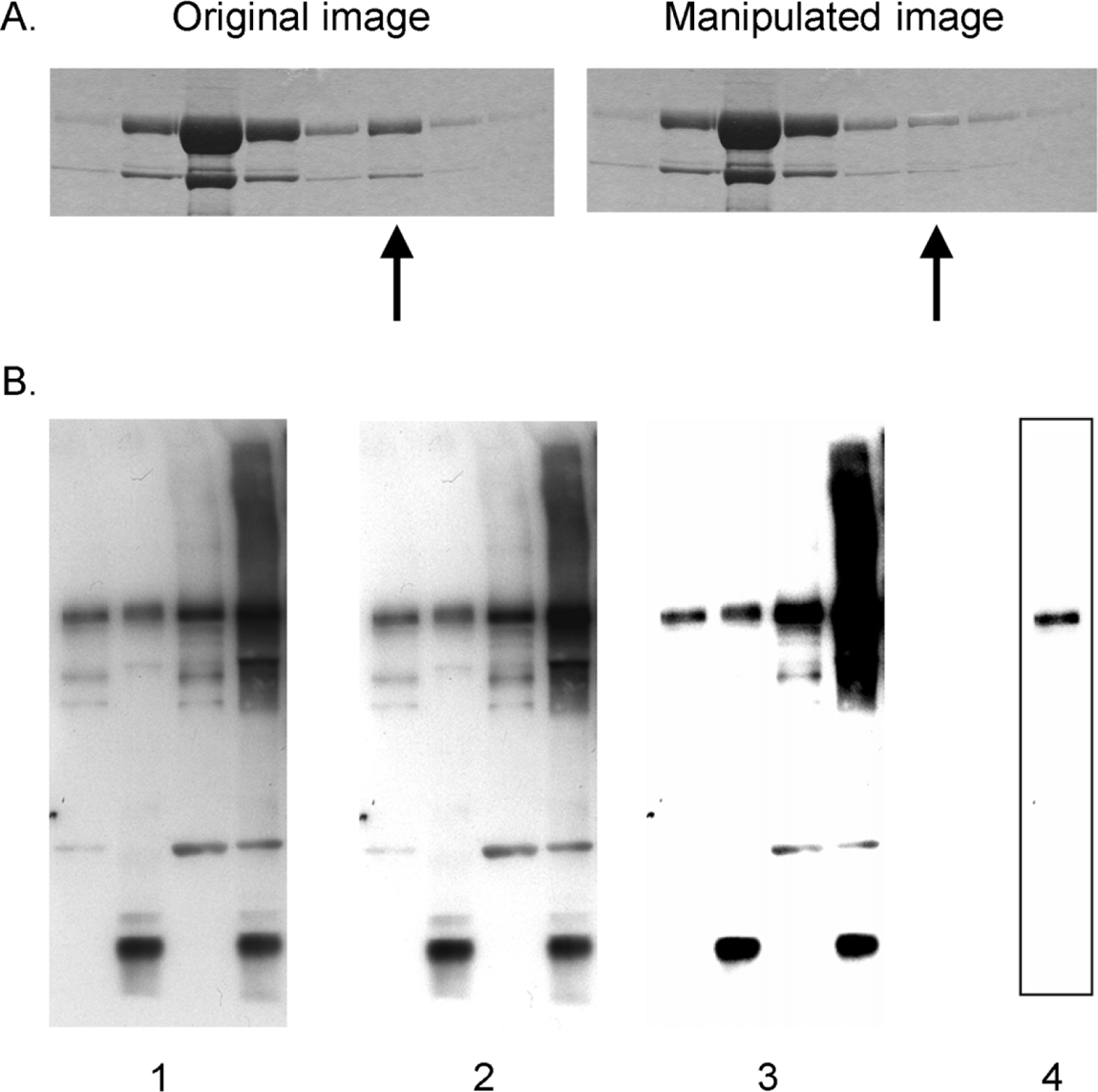
Example of inappropriate brightness and contrast adjustment of blots (reported in Rossner and Yamada, 2004: 13). The original image contains the following caption: ‘Manipulation of blots: brightness and contrast adjustments. (A) Adjusting the intensity of a single band (arrow). (B) Adjustments of contrast. Images 1, 2, and 3 show sequentially more severe adjustments of contrast. Although the adjustment from 1 to 2 is acceptable because it does not obscure any of the bands, the adjustment from 2 to 3 is unacceptable because several bands are eliminated. Cutting out a strip of a blot with the contrast adjusted provides the false impression of a very clean result (image 4 was derived from a heavily adjusted version of the left lane of image 1).’ ©2004, Rockefeller University Press. Originally published in The Journal of Cell Biology 166:11-15 doi:10.1083/jcb.200406019.
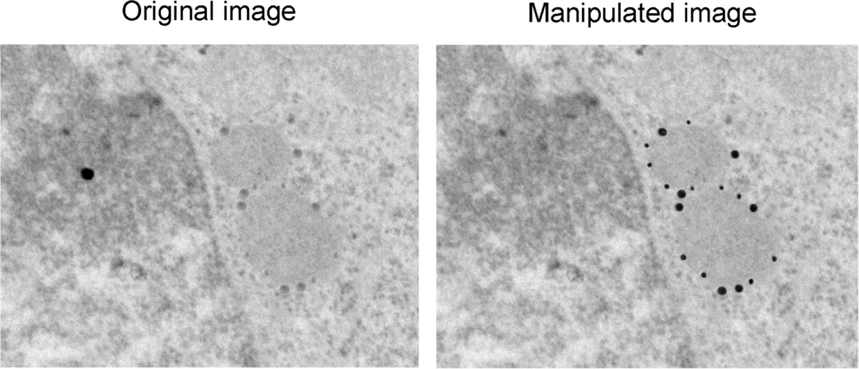
Example of selective adjustment of images (reported in Rossner and Yamada, 2004: 14). The original image contains the following caption: ‘Misrepresentation of immunogold data. The gold particles, which were actually present in the original (left), have been enhanced in the manipulated image (right). Note also that the background dot in the original data has been removed in the manipulated image.’ ©2004, Rockefeller University Press. Originally published in The Journal of Cell Biology 166:11–15 doi:10.1083/jcb.200406019.
The second point relates to cutting and pasting, or manipulations involving the rearrangement or grouping of images. According to journal guidelines, producing composite images by cutting and pasting together (selected portions of) images can be acceptable, provided that the various sub-images are explicitly delineated on the figure and the composite nature of the figure is described in the accompanying figure legend. An example deemed to have involved inappropriate image manipulation in this regard relates to two articles on cell signalling published in Nature Cell Biology in 2003 (Sawada et al., 2003a,b), in which the first author – one of the laboratory’s postdoctoral researchers – used Photoshop to make composite western blot images by cutting and pasting together bands from several different experiments (Pearson, 2005: 953). A formal investigation by the journal editors concluded that, although the interpretation of the research findings was not affected by the image manipulation, ‘the frequency and severity of the manipulations’ undertaken necessitated full retraction of the papers (Editorial, 2007b: 355; Sawada et al., 2007).
The third point emphasized in the journal guidelines is transparency in process. Just as details regarding the arrangement of composite images should be provided in the relevant figure caption (see above), so too should other aspects of image preparation be detailed. For example, the guidelines produced by the Nature journals stipulate that ‘authors should list all image acquisition tools and image processing software packages used’, as well as ‘document key image-gathering settings and processing manipulations’. 7 For images obtained using microscopy, this information includes the make and model of the microscope and lens used, together with a list of the instrument settings used for image capture, a description of the experimental sample, and details of any post-acquisition adjustments. As well as providing greater context for image interpretation and promoting increased transparency in the reporting of findings, the documenting of such information is described as a useful mechanism for raising awareness among scientists about processes of image preparation. 8
The fourth key issue mentioned in most of the journal guidelines is a requirement to keep all original data relating to images. This stipulation applies less to the manipulation of digital images themselves than to practices of data storage and record-keeping by scientists. The consequences of failure to comply with this requirement are potentially serious; for example, JCB editorial policies state that the journal may request to see original data ‘for comparison to the prepared figures’. Regardless of whether inappropriate image manipulation has occurred, ‘if the original data cannot be produced, the acceptance of the manuscript may be revoked’. 9
In-house policies and procedures
In parallel with establishing normative guidelines for digital image manipulation by authors, journals are also developing in-house policies and procedures for handling and evaluating digital images, and for guiding action in cases of suspected misconduct. The Council of Science Editors has also become involved with summarising recent developments concerning image integrity, and suggesting best-practice strategies for journals (Council of Science Editors, 2009: 53–55). A number of editorials mention the reluctance of their journal staff to act as ‘data police’ (see Editorial, 2007a: 215; Editorial, 2007b: 355), but they nonetheless identify a need for procedures designed to detect and manage cases of misconduct in image manipulation. Several journals have thus implemented procedures for screening images once a manuscript has been provisionally accepted for publication. 10 For example, JCB instituted a comprehensive screening programme in 2002, which employs an in-house ‘forensic expert’ to systematically screen all digital images in manuscripts that have been accepted for publication (Pearson, 2005: 952; Rossner, 2002: 1151). Since then, Science and the Nature family of journals have also instituted systems for ‘spot-checking’ digital images in papers that have been provisionally accepted – these systems variously involve choosing papers at random, or selecting articles thought to be at ‘high risk’ of image manipulation. 11
Editorial staff at JCB estimate that 25 percent of the manuscripts submitted to their journal contain what they deem to be inappropriately manipulated images. 12 In most cases, the editors determine that the manipulations do not affect data interpretation; if authors are able to supply new figures that comply with the journal guidelines, the manuscript is published. However, 1 percent of manuscripts contain image manipulations that are deemed to have ‘crossed the line’ and affect data interpretation – in such cases, the manuscript is rejected, and the case may be formally reported to bodies concerned with scientific integrity.
Drawing a line: Questions and tensions
The advent of new technologies and methods for the digital capture, transformation and adjustment of images raises a number of questions about the epistemic virtues, practices and ethos of scientific image production (Daston and Galison, 2007). The recent development of guidelines for image processing can be read as an intervention by journal editors intended to influence community practices of image-making. However, I suggest that far from being a straightforward, practical intervention, these guidelines raise (perhaps inadvertently) a number of longstanding and complex questions regarding visual representation in scientific communication and practice. Although focused on image preparation for the purposes of publication (the point at which journals are most obviously able to exert influence), the reach of these guidelines arguably extends deep into the methods, morals and metaphysics of representation in scientific practice (Daston and Galison, 1992: 84). Implicit in the guidelines (and sometimes explicit in the accompanying journal editorials) are particular understandings of the practices of image production, and the role(s) of images for communicating scientific knowledge claims. I propose that through these guidelines, the journals raise and must negotiate four interrelated ambiguities relevant to published images: the relationship between image production and image processing; the line between ‘innocent’ and fraudulent image alteration; the relationship between the authors and readers of journal articles; and the meaning of objectivity in the digital age. None of these issues is a fundamentally new concern with respect to visual representation in science, but each is re-cast and recalibrated as new imaging technologies and ‘ways of seeing’ are developed. In what follows, I consider how the (guide)lines being drawn by journal editors negotiate these concerns while trying to develop practical, working solutions to the perceived problem of digital image alteration in biology. 13
The line between benchtop and desktop: Skill and practice in image production
Journal editors describe the use of digital image acquisition and processing tools as facilitating the production of ‘clean’ and aesthetically pleasing images, and indeed as having ‘removed the physical impairments to perfect images’ (Editorial, 2006a: 892). The increasingly widespread availability of desktop image-processing software is identified as problematic, because it has the potential to disrupt a long-held supposition: that the quality of an image tells us something about the skill of the scientist who made it. Pretty pictures are highly valued in scientific work; for example, having one’s image published as the cover artwork for a high-profile scientific journal is a coveted honour. Furthermore, a high-quality image, suggest editors of journals including JCB, Nature and Nature Immunology should reflect ‘effort’, ‘skill’, ‘expertise’ and even ‘technical mastery’ on the part of scientists (Editorial, 2006a: 892; Editorial, 2007a: 215; Rossner and Yamada, 2004: 11). The advent of software such as Photoshop threatens to sever this relationship, as care and skill at the laboratory bench are no longer necessary to generate an image of publishable quality. Instead, to cite an extreme example, it becomes possible ‘to transform a featureless black microscope snap into a starry vista littered with labelled proteins’ while sitting in front of a desktop computer (Pearson, 2005: 952).
This quotation introduces a second and related concern on the part of journal editors. Technologies such as Photoshop do not simply offer a shortcut or a substitute for careful work at the laboratory benchtop, but furthermore can result in the generation of images that do not ‘accurately’ represent the experimental data obtained. Again, the editors suggest, this development represents a departure from longstanding practice, in which ‘data acquired at the bench were almost identical to the data published, blemishes and all’ (Editorial, 2007a: 215). Journal editors frown upon the use of image-processing software to remove such blemishes, or to otherwise tidy, clean up or prettify images in preparation for publication. Nature in particular has adopted a strong stance in this regard, stating that ‘beautification is a form of misrepresentation. Slightly dirty images reflect the real world’ (Editorial, 2006a: 892). Digital image processing is thus identified as problematic not only because it can yield images that provide a false impression of how skilled an individual scientist is, but because it might result in misrepresentation of the original data, and thus mislead the readers as to what the phenomenon under investigation ‘looks like’. The possible misrepresentation of data through image processing will be discussed further below. The point I wish to make here is that statements such as ‘beautification is a form of misrepresentation’ effectively serve to draw a line between ‘scientific’ and ‘aesthetic’ practices in image production. According to journal editors, the production of beautiful images for publication should rely on scientific skill as opposed to aesthetic dexterity; practices guided by aesthetic considerations have the potential to misrepresent data, and to mislead reviewers and readers alike.
The guidelines for image preparation being developed by journals might thus be read as an intervention designed to limit actions in the name of beauty or aesthetics, and to safeguard the scientific skill involved in image production. In practice, these guidelines define a boundary between the scientific and the aesthetic by drawing a line between practices of image-making at the laboratory benchtop, and adjustments made at the computer terminal or desktop. The focus is predominantly on limiting the use of software for processing images after they have been captured in a digital format. This separation of image acquisition from image processing, and of ‘scientific’ from ‘aesthetic’ practices, is arguably a pragmatic move on the part of journals. However, this move simultaneously exposes ambiguities that have been recognized through science studies research on visual representation. Two key points might be singled out in this regard. First, detailed ethnographic studies such as those carried out by Latour and Woolgar (1979), Lynch (1985a,b), Knorr-Cetina and Amann (1990) and Myers (2008) reveal difficulties in distinguishing clearly between scientific and aesthetic practices in image production; indeed, image-making is often portrayed as something akin to a skilled craft or a design process (Knorr-Cetina and Amann, 1990: 280). In their study of digital image-processing in astronomy, Lynch and Edgerton identify aesthetic practices and judgments as deeply embedded within practices of image production. Such aesthetic considerations are not discussed or deployed by scientists in the name of creativity or beauty, 14 but are typically directed towards achieving a certain ‘representational realism’ (Lynch and Edgerton, 1988: 200), of ‘composing visible coherences, discriminating differences, consolidating entities, and establishing evident relations’ (p. 212). Framed this way, the incorporation of aesthetic judgment might be considered necessary for the production of meaningful scientific images, with an absolute separation of the scientific and aesthetic being difficult to maintain in practice. Indeed, Carusi (2008: 248) suggests that a ‘compelling’ scientific image has both epistemic and aesthetic qualities.
Second, the recent journal guidelines concentrate on digital image-processing for the purpose of publication. Perhaps as a consequence of this pragmatic focus, images are discussed primarily as products or outputs of scientific research, and as representational devices for communicating findings. Again, ethnographic studies reveal possible difficulties in constraining the scope of journal guidelines to this stage of image-making. Images do not come into being at the point of preparing a manuscript for publication (however, images destined for publication are typically singled out for particular scrutiny and preparation – see Knorr-Cetina and Amann (1990) and Lynch (1985a: 94–98)). Rather, as the scientific community is well aware, visual representations are key and constitutive parts of the knowledge production process, and are actively constructed, transformed and rendered throughout scientific practice. For natural phenomena operating at scales not accessible to direct, unmediated observation with the human eye, such transformations may be necessary to make these phenomena ‘visible’ in the first place: ‘Researchers cannot directly observe living brain cells, ribosomes, strands of DNA, or bird migration routes without making use of complex procedures for technically visualizing these phenomena as picturable, graphable, mappable, or measurable configurations’ (Lynch, 1991: 208). 15 In such instances, reality cannot be distinguished from visual representation: ‘there is no way to compare a representation of a … phenomenon to the “real” thing, since the thing becomes coherently visible only as a function of representational work’ (p. 208). The reliability of the tools and work practices underpinning the entire observation process thus becomes crucial to the credibility and representational accuracy of knowledge claims being made. This point has particular salience for digital imaging, as new, increasingly intricate and interactive configurations of equipment and practice (requiring increasingly specialized knowledge and judgment) are used to render phenomena visible and meaningful (see, for example, Burri and Dumit, 2008: 303; Cambrosio and Keating, 2000; Editorial, 2005; Myers, 2008).
Although focusing primarily on images as illustrations or end-products, journal editors do acknowledge that the generation and acquisition of images are important parts of scientific practice, and some of their recommendations extend into the realm of benchtop practice. For example, image-processing guidelines for the Nature family of journals (arguably the most detailed of all the journal guidelines analysed here) state that ‘positive and negative controls, as well as molecular size markers, should be included on each gel and blot’. 16 Such requirements have consequences for the design of individual experiments. For the most part, however, statements in the guidelines regarding benchtop practice remain quite general, such as ‘authors must … take care to exercise prudence during data acquisition, where misrepresentation must equally be avoided’. 17 On the whole, alterations to the form and composition of images are deemed preferable if performed at the laboratory bench as opposed to with post-acquisition image-processing software. This rhetorical line drawn between benchtop and desktop results in some seemingly contradictory guidelines. For example, when discussing the preparation of figures containing blots and gels, the editors of JCB would prefer authors to ‘perform multiple exposures to get the bands at the density you want, without having to overadjust digitally the brightness and contrast of the scanned image’ (Rossner and Yamada, 2004: 13). In their study of image preparation in a molecular genetics laboratory, Knorr-Cetina and Amann (1990: 279–280) detail seven manipulations of an autoradiograph blot that were considered by scientists when they were preparing what they deemed to be an image of publishable quality: these included cutting the image so as to retain only those data of interest, changing the exposure time of the x-ray film (to highlight or reduce the intensity of particular bands), and running the experiment again under different conditions designed to yield an appropriate image for publication. According to recent image-processing guidelines, each of these possibilities would be deemed preferable to adjusting the contrast levels on a digital image of the autoradiograph. Such advice may seem contradictory in terms of curbing the possibility for intervention in the final, published image, but it becomes a necessary consequence of ‘drawing a line’ between image acquisition and image processing. 18
The line between acceptable and unacceptable manipulation
When developing image-processing guidelines, journal editors also attempt to define a boundary between acceptable and fraudulent image alteration (Council of Science Editors, 2009: 53). Again, drawing a clear line here is difficult.
19
On the one hand, editors acknowledge that image-processing software can be used for the complete fabrication of results. On the other hand (and related to the discussion above), they acknowledge that banning all digital image manipulation is not only unnecessary and undesirable, it also would be impossible. As suggested in a news feature from Nature:
No one wants to ban image manipulation outright. In cell-biology experiments, for example, researchers often have to adjust the relative intensities of red, green and blue fluorescent markers in order to show all three in a single image. Even drastic changes are sometimes considered tolerable if scientists spell out exactly what they did. But it is tough to draw a precise line between acceptable and unacceptable image manipulation. (Pearson, 2005: 953)
Their guidelines aim not to outlaw the use of digital image-processing software, but to limit it to instances where it might be determined ‘essential’, with the general stipulation that ‘the final image must correctly represent the original data and conform to community standards’. 20 Journal editors identify a spectrum of problematic image manipulations, ranging from honest mistakes or ‘innocent embellishment’, through to ‘scientific misconduct’ and deliberate fraud (Editorial, 2006b: 237; see also Council of Science Editors, 2009: 53). Despite the use of terms such as ‘innocent’ or ‘deliberate’, editors do not distinguish the acceptability of specific image adjustments on the basis of intent alone. They do not treat a lack of understanding or training in how to use image-processing software as an excuse for image beautification – as an editorial in Nature Methods states, ‘good intent does not make all practices acceptable’ (Editorial, 2006b: 237). Rather, journal editors distinguish between acceptable, inappropriate and fraudulent image adjustments largely on the basis of whether they affect the interpretation of data (see Council of Science Editors, 2009: 54). However, this criterion is itself far from straightforward; one might ask whether it is possible to determine a single, ‘correct’ interpretation of an image against which manipulations should be compared, given that, in principle, the interpretation of data is always context-dependent and open to re-evaluation (Lynch, 1991: 201). 21 The reference point according to the journal guidelines seems to be the interpretation that the authors offer in their manuscript to account for the presented data. Authors are thus granted authority to present a written interpretation of the image they have prepared for publication, but simultaneously bear the responsibility for ensuring that the interpretation offered is appropriate to the image manipulations performed (and relatively independent of any image adjustments made for primarily aesthetic purposes).
Although clear and unambiguous images are held up as an ideal, journal editors acknowledge the context-dependence of data, and furthermore emphasize that interpretations of images may change over time: ‘some observations that do not appear to make sense in the context of the current body of knowledge may turn out to be logical once the biology of the system is understood’ (Editorial, 2006b: 237). They warn authors against unnecessary image alterations, as ‘removing … peripheral information from images today will lead to contradictions tomorrow’ (p. 237). Their editorials associate extensive alteration of images with misrepresentation, and in turn link misrepresentation with possible misinterpretation of images by readers. For example:
innocent efforts to smarten or prettify images [can] end up with unintended consequences. At the very least, biologists risk erasing potentially valuable information, such as low levels of fluorescently labelled protein swilling around a cell’s cytoplasm. At worst, such manipulations can lead researchers to the wrong scientific conclusions. (Pearson, 2005: 952–953)
Altering selective parts of images – to remove artefacts or ‘unnecessary clutter’ – should be avoided for similar reasons: ‘it is not acceptable … to remove ugly, unexplainable or confusing areas of gels/blots for cosmetic reasons. Not only can it mislead the editor, referee, and ultimately the reader, but it can also hide important information pointing to real biological insight’ (Editorial, 2004: 275). 22
Sensitive to this complexity, in practice the guidelines treat image manipulations as ‘acceptable’ if they conform to the published guidelines and do not affect data interpretation. ‘Inappropriate’ adjustments are those that violate the guidelines, but are not seen to affect the particular interpretation or conclusions that the authors propose. Most of the problematic adjustments identified through editorial scrutiny of submitted images are described as belonging to this category. Such alterations can be quite extensive, as long as the resulting images do not alter the conclusions of the paper. For example, they can include ‘adjustments of brightness/contrast to a gel image that completely eliminate the background … or that obscure background smears or faint background bands’, and ‘the splicing of images from different microscope fields into a single image that appears to be a single field’ (Council of Science Editors, 2009: 54). Image manipulations that fall into the ‘fraudulent’ category are those that do affect data interpretation, for example by ‘deleting a band from a gel to “fix” a negative control that did not work or adding a band to a gel to indicate the presence of a product that was not really there’ (Council of Science Editors, 2009: 54).
Cases of outright fraud are discussed as being fairly rare, and in any case are difficult to safeguard against through the use of guidelines alone. 23 Rather, the guidelines are presented with the aim of promoting integrity in ‘normal’ or routine scientific practice. Journal editors suggest that much inappropriate image manipulation is done unknowingly, that scientists are often ‘unaware that their efforts to achieve the cleanest images for publication have crossed the line of acceptability’ (Pearson, 2005: 952). The introduction of guidelines for image preparation shines a spotlight on this process, and in this way may start to open up issues surrounding practices of image-making that have hitherto not received explicit attention. Furthermore, the guidelines go beyond awareness-raising by detailing a number of criteria that publishable images must comply with. They avow that the need for guidance to shape best practice in biological sciences (as opposed to other scientific sub-disciplines) is particularly acute, owing to a perceived lack of training for young researchers with respect to good practice in image preparation: ‘graduate school curricula typically do not offer systematic instruction in microscopy or image formation, with the result that most biology graduate students rely on ad-hoc training by more senior students or postdocs’ (Peterson, 2005: 881). 24 Image-processing programmes such as Photoshop offer simple interfaces for adjusting images, and journal editors are concerned that biologists can be tempted or even ‘duped’ into inappropriate image manipulation owing to the availability and ease of using such software (Editorial, 2006c: 101). 25
The development of journal guidelines serves as a basis from which to evaluate existing circumstances and to develop new standards or community norms. Consistent with the training function of guidelines, new briefing materials, training courses and online tools are being developed in direct response to the image-processing guidelines. 26 By encouraging training and discussion regarding image-making practices, the guidelines aim to promote norms of transparency and integrity in scientific practice. Procedural requirements such as noting down instrument settings, detailing any image adjustments performed and retaining all original images are seen as practices that should help to make visible, and render more accountable, the process of image preparation.
To summarize, the image-processing guidelines being developed by journals must grapple with the complicated issue of what counts as appropriate or inappropriate image adjustment. Definitions of research misconduct necessarily enter this discussion, and journal editors have drawn a line at the point where image alteration affects data interpretation (in itself an ambiguous point). However, they are concerned primarily with shaping and guiding normal scientific practice in light of technological advances in image-processing capabilities. This ambition is consistent with a broader shift in the 1990s from initiatives designed to prevent research misconduct to those promoting research integrity (see Montgomery and Oliver, 2009). The production of image-processing guidelines is an example of the role that journals can have as sites or institutions active in continually defining and refining norms of research integrity, both with and for the scientific community.
The relationship between author and reader
The publication of guidelines for digital image processing raises questions about the relationship between the author and the reader of a research paper. In general terms, such papers might be understood as a means of advancing knowledge claims to the scientific community for the purpose of evaluation. Authors try to convince readers of the validity and importance of the claims being made by demonstrating logically sound reasoning, skilled execution of work, and a clear display of the phenomenon under investigation. In a practical sense, images are treated as central components of scientific manuscripts; it is through images that readers may see for themselves what has been achieved, and judge whether the links made between the author’s observations and knowledge claims seem reasonable. By introducing guidelines that require authors to disclose details of the experimental setup for capturing images and to list any image manipulations performed, journals are promoting an ethic of transparency focused on tracing the histories of presented images, and providing clearer correspondence between image and experiment. 27
In addition to promoting transparency, some of the guidelines attempt to impose broad limits on author intervention in image production; for example, the first sentence of the Nature journals’ guidelines states that ‘images submitted with a manuscript for review should be minimally processed’. 28 By using guidelines to shape author practices, journals are arguably attempting to safeguard a degree of power and autonomy for the reader, protecting his or her ability to witness from afar. Although, in principle, readers are granted authority to judge for themselves the meaning of a scientific image, it is the author who creates and selects the image(s) to present. The process of selecting, refining and framing data is central to the presentation of scientific knowledge claims, but the availability of image-processing software such as Photoshop potentially grants the author unprecedented control over the appearance of images. Authors routinely identify growing pressure to publish beautiful images, not least linked to the fierce competition for publication in high-impact-factor journals (Franzen et al., 2007). 29 For a paper to catch the eye of discerning editors and reviewers, the belief is that it must contain positive and ‘significant’ findings, and provide a compelling visual demonstration of the phenomenon under question. Journal editors acknowledge the resulting pressure on authors, and state a desire to ‘end the fetish of the perfect image’ (Editorial, 2006a: 892). In practice, though, images are often chosen to represent the ‘best’ data and experiments out of many possible examples in order to persuade editors and readers of the importance and validity of a particular knowledge claim. These data might not reflect the average of all of the experiments performed. 30 The selection of representative images or averaging data for the purpose of publication clearly differs from the ideal of performing an experiment directly in front of observers or witnesses, where there is no opportunity to select the most appropriate results to present.
Particularly for highly interdisciplinary journals such as Science and Nature (which appeal to a broad scientific audience), one might ask whether images in contemporary scientific papers are deployed predominantly as tools for ostension or for witnessing. 31 If readers are assumed to unquestioningly accept knowledge claims made in publications, a pedagogical or idealized rendering of images may be acceptable. If, however, a publication is understood to be a basis for evaluating or testing a particular set of claims, then representational accuracy and completeness become more salient. 32 The journal guidelines seem to advocate the latter understanding, asserting expertise and authority on the part of the reader for validating knowledge claims.
Importantly, despite being able to control the selection and presentation of images, ultimately authors cannot determine how these images are understood or used by readers – not least because ‘an image usually carries information beyond the specific point being made’ (Rossner and Yamada, 2004: 11). A recent initiative pioneered by JCB editors affords readers increased control over data access and image interpretation: in December 2008, the journal launched JCB DataViewer, an application that allows readers of an article published in JCB to download the original image data and its associated metadata in order to perform their own image analysis. The JCB DataViewer is designed particularly for microscopy image datasets and time-lapse videos, but can also display other images (including gels and blots) captured in a variety of file formats. JCB editors emphasize that ‘the JCB DataViewer interface makes any published image as accessible to readers as if they had acquired it’ themselves, and importantly, that it grants readers access to ‘a maximum amount of information from published images, far more than can be gleaned from a single, two-dimensional optical slice’ (Hill, 2008: 969). Through the JCB DataViewer, the interactive, digital format of captured images is maintained for readers. With this tool, readers are granted the ability to probe beneath the ‘face value’ of a printed image (Coopmans, 2011), and through this, are thought able to assume more of a ‘witnessing’ and evaluative role than previously possible. Indeed, this development might be seen to blur the relationship between authors and readers of manuscripts.
The JCB DataViewer is also presented as a tool for ensuring ‘transparency’ and ‘data integrity’ in published articles (Hill, 2008: 970). Both authors and readers are suggested to benefit – authors because they can ‘better showcase their data and … better substantiate the conclusions drawn’, and readers because they have ‘all of the information necessary to evaluate authors’ interpretations’ (p. 970). 33 In requiring original data files, the JCB DataViewer repository indirectly serves as a mechanism to limit the degree of processing that authors undertake in preparing their images for publication: ‘We are not just asking to see a larger section of a microscope field or a larger piece of a blot, but the actual data files acquired by authors (that have not had a whiff of Photoshop!)’ (p. 970). At present, uploading of image files to the JCB DataViewer is optional; JCB editors note that as of September 2010, just over 20 percent of published papers were accompanied by images in this interactive archive, which is now being accessed by about 15,000 users each month (deCathelineau et al., 2010).
In summary, at least two aspects of recent journal interventions address the relationship between authors and readers of scientific images. By requiring increased methodological transparency associated with individual images (through written disclosure of image acquisition and processing steps), and by developing software tools that allow researchers (authors and readers) to access ‘raw’ image data and metadata, the guidelines try to limit the scope for image processing and interpretation by the author, and assert a greater role for the reader in evaluating the claims being put forward. These actions effectively serve as an attempt to reduce or constrain what might be seen as an imbalance between author and reader (one that is potentially exacerbated in the face of digital image-processing technologies), by trying to ensure comparable access to digital image data and thus equal authority on the part of authors and readers to propose and challenge interpretations of observed phenomena.
Defining objectivity in the digital age
Broadly speaking, the image-processing guidelines being advanced by journals can be read as attempts to define what counts as visual evidence and good representational practice in contemporary molecular biosciences (and nominally science more generally). This promotion of guidelines might be seen as part of a broader movement to assert a form of procedural objectivity in science, and to promote trust through ‘apparatuses of surveillance, control, and institutional discipline’ (Shapin, 2008: xvii). This push to articulate guidelines highlights a fundamental tension regarding the role of images in scientific publications: on the one hand, images should convey information that convincingly supports a particular knowledge claim, and on the other they should provide an objective representation of some aspect of the natural world. These ideals are not mutually exclusive, but the tension between them has ramifications for scientific practice, influencing the degree to which authors can acceptably intervene in framing and shaping the content of published images. By attempting to define (or refine) the position of this line, journal editors inevitably address the nature of objectivity in scientific practice. Indeed, the very development of written guidelines and conventions for image preparation might be thought of as a symptom or a reflection of a new moment in the trajectory of objectivity as a guiding value in science. 34 The growing ease with which images can be digitally produced, transformed and manipulated re-poses questions about the epistemic virtues, practices and ethics of image production (Daston and Galison, 1992, 2007). What, then, might objectivity mean in the digital age, and how might it be best upheld?
The journals’ push to limit author intervention in image preparation is not limited to concerns regarding a role for the reader in evaluating evidence. The rhetoric of journal editors treats the authentic or unadulterated image as somehow a ‘better’ or more accurate reflection of the natural world than a digitally manipulated one: ‘slightly dirty images reflect the real world’ (Editorial, 2006a: 892). The current guidelines appeal to an understanding of objectivity similar to what Daston and Galison refer to as mechanical objectivity in their historical account of objectivity as performed through atlas images. They situate the emergence of mechanical objectivity in the 19th century, and stress its emphasis on scientific and ethical self-control; mechanical objectivity is about resisting ‘the temptations of aesthetics … the desire to schematize, beautify, simplify’ (Daston and Galison, 2007: 120). This form of objectivity is manifest through ‘the insistent drive to repress the willful intervention of the artist–author, and to put in its stead a set of procedures that would, as it were, move nature to the page through a strict protocol, if not automatically’ (Daston and Galison, 2007: 121). The development of journal guidelines, together with the emphasis in their accompanying editorials on distinguishing between ‘aesthetic’ and ‘scientific’ practices in image preparation, promotes a view of objectivity consistent with this description. This view plays out in the details of the guidelines too, for example through the stipulation that authors should not selectively remove known artefacts from images. 35 In this respect, the guidelines treat the authenticity of an image as key to its objectivity, and as critical for engendering trust in the image. This link between trust and abstaining from intervention is not an inevitable one; for example, Lynch and Edgerton’s (1988: 206–208) study of digital image processing in astronomy identifies the ‘cleaning up’ and removal of artefacts from images as a possible (although not universally accepted) strategy for demonstrating competence among scientific colleagues. A key difference between these two examples may lie in the perceived expertise of the author in using image-processing tools – with Photoshop in particular being singled out as problematic in encouraging authors to ‘black-box’ the underlying mathematics of digital image manipulation (Greene, 2005: 143). Questions of whether it is better to leave in or to remove noise from an image, or about whether data manipulation enhances or detracts from objectivity, are intimately bound up with particular disciplinary tools, conventions and judgments about what counts as good scientific practice. 36
Daston and Galison’s account of mechanical objectivity in the 19th century describes an increasing reliance on machines (such as the camera) to uphold objectivity where the scientific self might falter. Contemporary debates about image preparation are unfolding in the context of a pervasive (and yet fairly recent) digital medium for image capture and processing, and in an era of highly specialized scientific and medical research that relies on increasingly complicated configurations of machines and software for making natural phenomena visible in the first place. Imaging software and practices can be used simultaneously as tools for controlling apparatus, analysing data and representing phenomena. Cambrosio and Keating (2000) suggest that the scope of these functions effectively collapses the distinction between representing and intervening – a distinction that is actively promoted through the ideal of mechanical objectivity. Although the assertion of mechanical objectivity is a common response when expertise is challenged and distrust is high (Porter, 1995), one might ask whether reinforcing this ideal is realistic (or even desirable) in light of the pervasive switch to digital media in scientific practice.
Conclusion
The scientific community is increasingly identifying digital image processing as a site of concern in scientific practice, one that has few explicitly articulated norms and conventions. The widespread use of such image processing across the natural sciences, and particularly its rise in molecular biosciences, is being associated with problems relating to the trustworthiness of the published image. Several journals are trying to intervene and help redress this crisis of trust by developing guidelines for image processing. While affirming the importance of images in the communication and validation of scientific knowledge, these efforts also expose a number of underlying tensions and ambiguities.
The impetus behind the development of journal guidelines is nominally the increasing use of digital image acquisition and software tools. Such technologies certainly offer new possibilities for image capture and adjustment; it is clear that software such as Photoshop can be used inappropriately, and in extreme cases can support the complete fabrication of experimental data. Yet it is less clear that digital image-processing technologies as a class present a unique and fundamental threat to the integrity of the published image, compared with their analogue equivalents. Despite statements such as ‘slightly dirty images reflect the real world’, the current concerns of journal editors revolve less around determining the so-called truth or falsity of digital images and are more around setting norms for image production as a means of safeguarding trust in the published image. By developing guidelines, journal editors are trying to draw lines for the scientific community that define best practice in digital image preparation. However, their interventions reveal a deep and longstanding tension between the theory and practice of representation, one that is continually challenged as new imaging technologies and practices emerge. In this paper I have suggested that, while trying to develop practical, working solutions to the perceived problem of digital image alteration in biology, journal editors must negotiate four complex and interrelated concerns: the relationship between image production and image processing; the line between appropriate and inappropriate image alteration; the relationship between authors and readers of journal articles; and the meaning of objectivity in the digital age. Through analysing their guidelines and commentaries, we see how difficult it is for journal editors to uphold a simple and internally consistent understanding of scientific images; indeed, the ‘hybrid’ character and function of images (Lynch, 1990: 171) becomes increasingly apparent as attempts are made to set guidelines for practice.
These recently proposed guidelines expose a number of longstanding ambiguities inherent to practices of visual representation, but do not resolve them. Rather, and as earlier STS studies of guidelines in different domains have suggested, these efforts might more appropriately be understood in terms of structuring relations of accountability in an attempt to manage the distribution and eventual resolution of these ambiguities (Lee, 1999; Rappert, 2001). Journal editors are faced with the challenge of wanting to provide clear guidance for the scientific community yet not being in a position to prescribe in minute detail exactly how image preparation should be performed for all possible configurations of research questions, technologies and practices. Their guidelines are intended to be generalisable and applicable to all digital image processing; as such they offer general rules but few detailed prescriptions for what may or may not be done to prepare a given digital image for publication. They do draw certain lines (for example, between image processing at the benchtop and the desktop), but they cannot justify these solely on the basis of technical arguments about contemporary imaging practices or the properties of resulting images, and also make appeals to values such as transparency, objectivity and trust.
As presented, the journal guidelines retain considerable definitional ambiguity, for example, in relation to the boundary between ‘appropriate’ and ‘inappropriate’ manipulation. The resolution of this ambiguity gets devolved to case-by-case assessments of the practices associated with individual images, and requires that the specific context and interpretation of a given image must be taken into account. In this way, the journal guidelines do not resolve inherent tensions in image production, but rather attempt to manage this ambiguity by asserting general practices that should be followed while deferring the actual moment of resolution to individual, future cases (see Lee, 1999). Decisions about exactly which image-processing steps to pursue are left to individual authors, but in being required to adhere to the guidelines authors also are made explicitly accountable to journal editors and readers. To try to enforce this accountability, journal editors are implementing procedures for checking images and mechanisms for sanctioning authors who do not comply. In this respect, this case study offers a clear example of the influence that journals attempt to exert on community norms and scientific knowledge production. Consistent with Brian Rappert’s study of guidelines surrounding the use of ‘non-lethal’ weapons, we see that the provision of appropriate training (in this case, for researchers using digital image-processing tools) becomes an important community issue accompanying the development of guidelines (Rappert, 2001: 579).
In sum, the recent image-processing guidelines, despite their internal contradictions about the role of images in knowledge production and their ambiguities concerning questions of acceptable and unacceptable practices, can be understood as tools that help to (re)structure and make explicit relationships of accountability among authors, editors and readers of journal articles when it comes to making, interpreting and validating images in publications. However, and particularly given the complexities surrounding contemporary image-making practices, a number of questions warrant continued empirical investigation. These include whether the journal guidelines, as proposed, become accepted as legitimate by the scientific community. They also concern the extent to which the guidelines influence image-making practices. 37 Ultimately, these questions concern whether the guidelines help to restore the perceived crisis of trust in published images. Pursuing these lines of enquiry should, in turn, deepen our understanding of whether and how objectivity may be undergoing recalibration in the digital age.