



Abstract
Gonadotropin-releasing hormone (GnRH) agonists, such as leuprorelin, are recommended in the patients with pedophilia at highest risk of offending. However, the cerebral mechanisms of the effects of these testosterone-decreasing drugs are poorly known. This study aimed to identify changes caused by leuprorelin in a pedophilic patient’s brain responses to pictures representing children. Clinical, endocrine, and fMRI investigations were done of a man with pedophilia before leuprorelin therapy and 5 months into leuprorelin therapy. Patient was compared with an age-matched healthy control also assessed 5 months apart. Before therapy, pictures of boys elicited activation in the left calcarine fissure, left insula, anterior cingulate cortex, and left cerebellar vermis. Five months into therapy, all the above-mentioned activations had disappeared. No such activations and, consequently, no such decreases occurred in the healthy control. The results of this pilot study suggest that leuprorelin decreased activity in regions known to mediate the perceptual, motivational, and affective responses to visual sexual stimuli.
Introduction
Pedophilia is a severe public health problem, with about 25% of girls and 16% of boys being victims of sexual abuse in the United States (American Psychiatric Association, 2000; Dube et al., 2005). Long-term consequences of sexual abuse are highly problematic: In a large study of same-sex twin pairs, increased risk for a wide range of psychiatric disorders was observed in women and in men reporting a history of childhood sexual abuse (Nelson et al., 2002). Prevention of such outcomes depends not only on the judicial system but also on improved medical management of men with pedophilia. In men at high risk of offending, gonadotropin-releasing hormone (GnRH) agonists may be indicated in association with psychotherapy. While receiving GnRH agonists and psychotherapy, men with pedophilia reported a dramatic decrease in deviant sexual desires and behaviors (Rösler & Witztum, 1998). However, the side effects of GnRH agonists, for example, osteoporosis, make many practitioners wary of the medical responsibility associated with the prescription of powerful testosterone-decreasing drugs that have not been approved by the U.S. Food and Drug Administration for this indication. In addition to providing new basic knowledge on the cerebral mechanisms of the therapeutic effectiveness of GnRH agonists, identifying changes in cerebral activity associated with the clinical effects of GnRH agonists could clarify the rationale for their usage.
Aim
The aim of this pilot study was to investigate whether, in a pedophilic man attracted to boys, brain responses to pictures representing boys would be changed by leuprorelin, a GnRH agonist. In aiming to identify brain regions whose responses might change under treatment with leuprorelin, we tried to specify regions mediating sexual desire felt before treatment and the cerebral mechanisms whereby leuprorelin might exert its effects. Based on our previous work on the cerebral correlates of sexual arousal and their modulation by testosterone, we hypothesized that leuprorelin-induced decreased plasma testosterone would be associated with decreased activation of brain areas involved in processing visual sexual stimuli, namely, the inferior and middle occipital gyri, the lateral part of the right orbitofrontal cortex, the anterior cingulate cortex (ACC), the insulae, and the cerebellar vermis (Mouras et al., 2008; Redouté et al., 2005).
Method
We report the case of a 46-year-old right-handed man diagnosed with pedophilia (American Psychiatric Association, 2000) who had been charged with eight sexual offenses against boys. He was compared to a 47-year-old right-handed heterosexual male healthy control. The local research ethics committee approved the study and both participants gave their written informed consent after complete description of the study.
Both subjects were assessed through a medical examination, hormonal assays, the Intensity of Sexual Desire and Symptoms Scale (Rösler & Witztum, 1998), which we revised to include ratings based on explicit criteria and ranging from 1 to 18, the Composite International Diagnostic Interview (Organisation Mondiale de la Santé, 1996) to identify psychiatric comorbid conditions, the Symptom Checklist-90–Revised (SCL-90R; Derogatis, 1977) to evaluate the extent of associated psychopathological disturbances, an abridged form of the Wechsler Adult Intelligence Scale III (Axelrod, Ryan, & Ward, 2001), and functional magnetic resonance imaging (fMRI) to assess brain responses to various categories of pictures (see below).
Assessments were first conducted before initiation of therapy, then 5 months into therapy (3.75 mg of leuprorelin every 4 weeks). Treatment was administered in a double-blind fashion within a randomized clinical trial (ClinicalTrials.gov Identifier: NCT00601276) from which this pilot study was drawn. Leuprorelin was associated with individual supportive psychotherapy and mianserin (30 mg/day), an antidepressant agent. The healthy control did not receive treatment and was also assessed on two occasions separated by 5 months.
Main Outcome Measure
fMRI data were acquired on a 1.5-Tesla Philips Intera Achieva scanner (Best, Netherlands). Five categories of photographs were presented: male children in swimsuit (MCS), female children in swimsuit (FCS), ordinarily dressed female children (FCD), female adults in swimsuit (FAS), and ordinarily dressed female adults (FAD). No pornographic photographs were used. The run comprised three 35-s-long blocks of each of the five categories. On the second assessment, the same categories of pictures were presented in the same order as on the first assessment. However, to prevent activation of brain areas involved in memory processes and to maintain the same degree of novelty, we used two different sets of photographs on the first and second sessions. During the fMRI runs, the penile responses were measured through a volumetric penile plethysmograph (Mouras et al., 2008). After the end of the run, single-item scales, ranging from 1 (totally absent or extremely low) to 9 (extremely high), were presented to assess perceptions experienced during the run in response to each category of photographs, including interest for pictures. Functional data preprocessing steps and statistical analyses were performed using the SPM5 software package (http://www.fil.ion.ucl.ac.uk/spm). The main analyzed statistical contrast, (MCS-FCS)time1 – (MCS-FCS)time2, was meant to identify brain areas (a) showing higher responses to boys than to girls before treatment and (b) exhibiting smaller differences of activation under treatment. To identify areas that showed such decreased activation in the patient, but not in the control, the patient’s brain activation map yielded by the above contrast was masked with the activation map derived from the same contrast applied to the control subject’s brain responses. The statistical threshold of this exclusive mask was set low (p < .05, uncorrected for multiple comparisons) to ensure that regions showing decreased activation in the patient were not exhibiting decreased activation in the control subject.
Supplementary analyses were performed using FCD or FAD as control conditions instead of FCS. FCD and FAD had the advantage of representing more adequate neutral control conditions in the case the patient had felt a bisexual attraction toward children. Except the nature of the control conditions, all the steps of these analyses were the same as previously.
In addition, to investigate whether areas responding to visual stimuli showed functional connectivity, we studied statistical correlations among responses of distinct areas. To do so, we used an analysis of the psychophysiological interactions, a method that allowed to identify areas, As, where the Blood-Oxygenation-Level Dependent (BOLD) signal was differentially correlated with the BOLD signal in other areas, Bs, as a function of the psychological context C (Gitelman, Penny, Ashburner, & Friston, 2003). Here, among brain regions (Bs) that responded differentially under therapy, we sought to identify those areas (As) whose functional correlation with these brain regions was different as a function of the gender of children on the presented pictures.
Results
The patient was single, had 14 years of education, and worked as an employee. On initial assessment, the patient was obsessed with his pedophilic desires. “They were so powerful that I did not hesitate to put myself in danger and to ruin the life of others,” he declared. He spent hours on illegal Internet sites, felt very guilty, thought of committing suicide, and had thrown away two computers containing illegal pictures of children.
The patient’s and the control subject’s IQs were 121 and 136, respectively (Axelrod et al., 2001). Before initiation of the treatment, the patient suffered from major depressive disorder (American Psychiatric Association, 2000; Organisation Mondiale de la Santé, 1996). On the SCL-90R (Derogatis, 1977), before treatment initiation, then 5 months into treatment, the patient’s scores were (a) Global Severity Index: 1.21, then 0.2 (M ± SD in nonpatient reference sample: 0.60 ± 0.44; control: 0.03); (b) depression scale: 2.38, then 0.62 (M ± SD in nonpatient reference sample: 0.73 ± 0.55; control: 0.00). Both participants were physically healthy.
Previous psychotherapeutic treatment had not enabled the patient to control his attraction to children. After 5 months of leuprorelin therapy combined with individual supportive psychotherapy and mianserin, he stated that this treatment had “opened a new life” for him. He reported no more sexual fantasies, desires, and masturbatory behaviors related to children and had ceased visiting illegal sites and committing offenses. He felt content with the decrease of sexual desires. Self-esteem was restored. Guilt feelings and suicidal thoughts had disappeared. On the Intensity of Sexual Desire and Symptoms Scale–Revised, the patient’s score related to children decreased from 11.7, a very high score, to 1.0, the minimum possible score. On the two assessments, the healthy control’s scores were 1.0.
Under leuprorelin therapy, the plasma testosterone level fell from 18.44 mmol/L (normal level) to 0.69 mmol/L (hypogonadal level). On the scale of interest for pictures of boys, he gave a rating of 8 (very strong) on the first fMRI session, and of 3 (low) on the second. The time courses of the penile plethysmographic signal recorded before and under treatment demonstrate the disappearance of penile responses to pictures of boys on the second session (Figure 1). Before treatment, the plethysmographic signal showed penile responses also in reaction to pictures of female children in swimsuit. These responses were smaller than those in reaction to male children in swimsuit and were not observed under treatment.
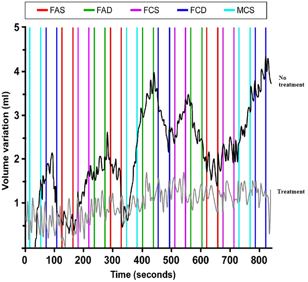
Penile volumetric variations recorded in the patient during the fMRI runs
Before treatment, areas showing activation in response to pictures of boys in the patient’s brain, but not in the control’s, were the left calcarine fissure (p < .001, corrected for multiple comparisons), the left anterior insula (p < .05, corrected), the caudal ACC (x = 0, p = .001, corrected), and the left cerebellar vermis (p < .01, corrected). Under therapy, all four activations disappeared. Decreased activation was statistically significant in all four regions (Figure 2). The activations in the left calcarine fissure and in the ACC were correlated, that is, exhibited functional connectivity (p < .001, uncorrected). Because of the occurrence of weak penile responses in response to the pictures of female children in swimsuit, the activation pattern in the MCS condition was also compared with the brain responses to the pictures of ordinarily dressed female adults [(MCS-FAD)time1 – (MCS-FAD)time2] and of ordinarily dressed female children [(MCS-FCD)time1 – (MCS-FCD)time2] in the patient and in the healthy subject. Then, for each of these contrasts, the patient’s brain activation map was masked with the control subject’s activation map. In these analyses, the calcarine fissure and the cerebellar vermis showed decreased activation under treatment.
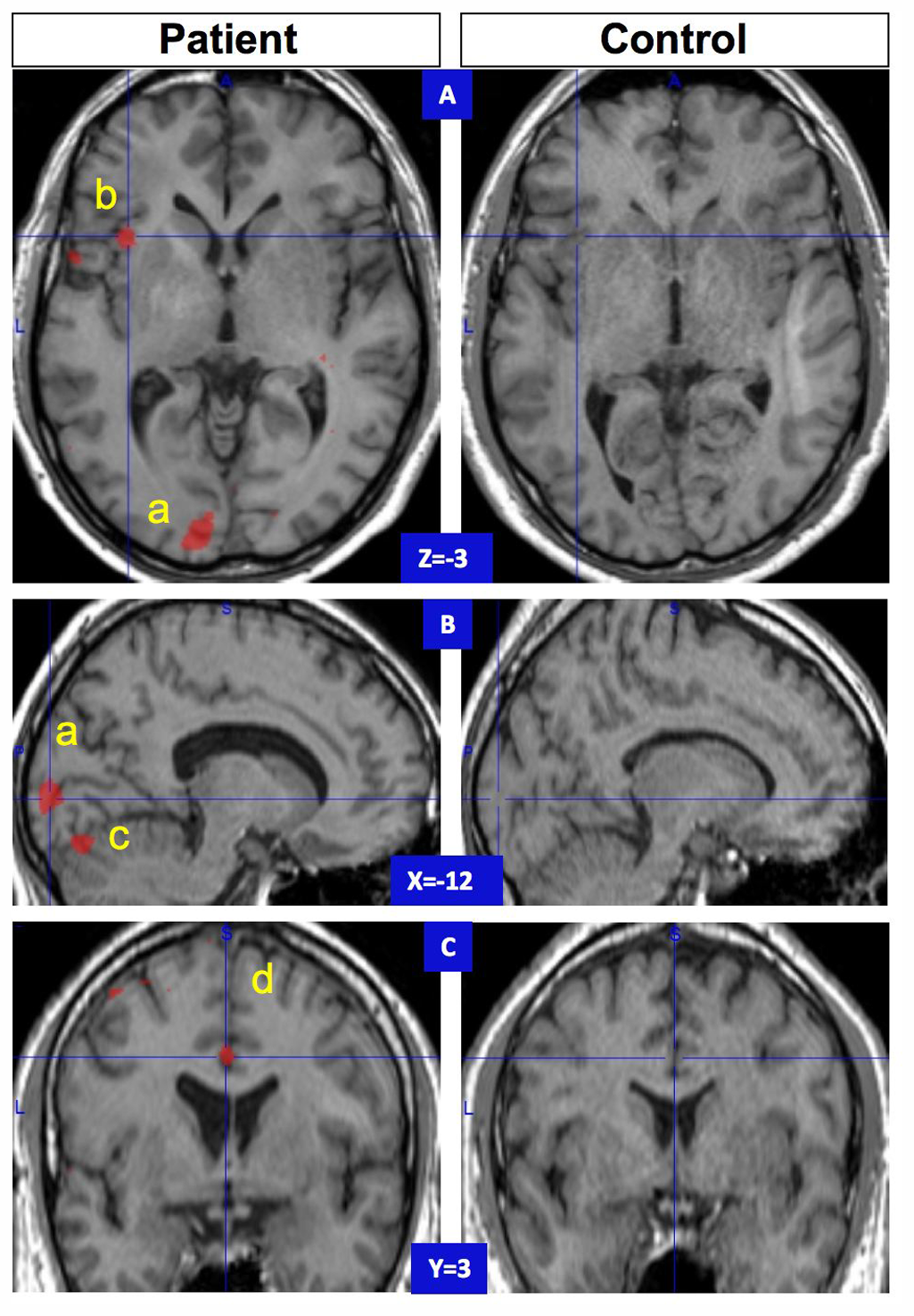
Brain areas where activation in response to pictures representing boys decreased under leuprorelin therapy
Discussion
This is the first fMRI study contrasting the brain activation pattern in response to sexually relevant visual stimuli in a man with pedophilia before and during GnRH agonist treatment. It could be a first step toward the demonstration of the mechanisms of the efficiency of leuprorelin in this indication.
The responses of four brain regions, namely the left calcarine fissure, the left anterior insula, the caudal ACC, and the cerebellar vermis, were modified under leuprorelin therapy. These changes are likely to represent mechanisms whereby the decrease of plasma testosterone is associated with decreased sexual interest and desire. The higher activation in the primary visual area in response to photographs of boys is striking as it suggests that the first step of this patient’s visual perception was already influenced by sexual attraction to boys. The functional connectivity analysis (i.e., activations in the left calcarine fissure and in the ACC being correlated) suggests that this visual cortex activation resulted from top–down modulation of calcarine activation by the ACC rather than from a direct effect of visual stimuli. An activation of the ACC, including its caudal part, has been reported in studies of the neuroanatomical correlates of sexual arousal in healthy men (Stoléru & Mouras, 2007) and in men attracted to boys (Schiffer et al., 2008a). The specific role of the caudal ACC in motor function is similar to the role of premotor area cortices (Dum, 1993). Thus, in monkeys, stimulation of the ACC elicits genital manipulation of a masturbatory character (Robinson & Mishkin, 1968). The caudal ACC plays a crucial role in the motivation and the initiation of goal-directed behavior in general (Devinsky, Morrell, & Vogt, 1995) and of sexual behavior in particular (Stoléru & Mouras, 2007).
The insula has been consistently found activated by sexual stimuli (Stoléru & Mouras, 2007). Furthermore, the level of activation in the anterior insula is correlated with the level of penile tumescence (Mouras et al., 2008), with variations of insular BOLD signal level leading those of penile volume with a 20-s delay. This part of the anterior insula (Mouras et al., 2008) is very close to the cluster found activated in our patient before treatment and deactivated under therapy.
Despite the traditional view confining the role of the cerebellum to motor coordination, more recent evidence indicates its role in emotional and motivational processes (Parsons et al., 2000). Cerebellar activation in response to visual stimuli and/or a correlation of cerebellar activation with penile erection has been reported (Mouras et al., 2008). In nonhuman male primates, the cerebellum showed a positive BOLD response to periovulatory odors and a negative BOLD response to odors of ovariectomized females (Ferris et al., 2004).
The pattern of distributed brain activation in response to pictures of boys reported above is similar to the pattern in heterosexual men presented with pictures of women. In the patient, this pattern changed dramatically under treatment. The decreased activation of the left insula is consistent with the modulation of the activity in this area by plasma testosterone levels (Redouté et al., 2005). Similarly, in hypogonadal patients, blood perfusion in the midcingulate cortex is enhanced by testosterone administration (Azad, Pitale, Barnes, & Friedman, 2003).
A recent fMRI study (Schiffer, Gizewski, & Kruger, 2009) of a heterosexual pedophile under leuprorelin treatment found no activation in the hypothalamus, thalamus, amygdala, insula, substantia nigra, hippocampus, and the rostral part of the ACC in response to pictures of undressed female children, regions that the same group had previously found activated in a previous study of unmedicated patients (Schiffer et al., 2008b). Our findings are thus partially consistent with this previous study (Schiffer et al., 2009). Lack of complete agreement is not surprising as both studies are single-case studies, and one focuses on a patient attracted to boys, the other on a patient attracted to girls.
The decreased activations in the four areas mentioned are unlikely to be related to the effect of the repetition of fMRI acquisitions as no such changes were observed in the matched healthy control, even with a statistical threshold set very low. As the patient received mianserin for major depression, one may ask whether the changes in the brain activation pattern were related to the antidepressant effect or to a general psychological improvement. However, in a comparison between depressed and nondepressed men presented with visual sexual stimuli, no between-groups differences were found in the responses of the four regions mentioned above (Yang, 2004). In a similar comparison between depressed and nondepressed female subjects, healthy women did show higher responses in middle occipital gyrus, insula, and anterior cingulate gyrus (Yang et al., 2008). However, whereas these higher brain activations were consistent with self-reported higher sexual arousal in healthy than in depressed women, in our patient the alleviation of depression was on the contrary accompanied by self-report and phallometric evidence of decreased sexual arousal as well as by decreased activation in these brain areas. If changes in brain responses observed in our patient had been caused by mianserin-induced alleviation of depression, one would have expected to observe increased responses in these areas, not decreased responses as recorded in our patient. Alternatively, one could ask whether mianserin caused sexual side effects, such as decreased desire. However, mianserin, rather than a selective serotonin reuptake inhibitor (SSRI) antidepressant, was selected to treat this patient’s depression because we wanted to discriminate the effects of leuprorelin from the side effects of antidepressant drugs. Sexual side effects of mianserin are considered as less marked than those of SSRI drugs so that mianserin has been recommended as a treatment of the side effects of SSRI drugs (Baldwin & Birtwistle, 1998). It can also be argued that psychotherapy, and/or the interaction between pharmacological treatments and psychotherapy, was the cause for changes in brain and in behavioral responses. As another limitation, given the patient’s attraction to boys, a homosexual control would have been more adequate than a heterosexual one and the presentation of pictures of men (instead of women), both ordinarily dressed and in swimming suits, would have made possible further analyses.
Conclusion
In spite of these limitations, this pilot study suggests that leuprorelin decreased activity in regions known to mediate the perceptual, motivational, and affective responses to visual sexual stimuli. We submit that this observation provides a valid basis for controlled studies of the effects of GnRH agonists on functional brain responses in pedophilia. In addition, given the questionable reliability of self-reports in this clinical area, if studies on large enough samples confirm our findings, fMRI could become a tool to objectively monitor effectiveness of pharmacologic and other therapeutic approaches of pedophilia.
Footnotes
The authors declared no conflicts of interests with respect to the authorship and/or publication of this article.
This research has been sponsored by Assistance Publique-Hôpitaux de Paris and has been supported by the DGS at the French Ministry of Health, the DAP at the French Ministry of Justice, the DGAS at the French Ministry of Labour, Social Relations, Family and Solidarity, and by the French Ministry of Research. We thank D. Glutron, B. Grandjean and R. Rouxel for technical assistance and T. Similovski, J. J. Hauw, and P. Bouchard for suggestions about a previous version of the manuscript.