



Abstract
Background
Axillary lymph node metastasis (ALNM) serves as a critical prognostic determinant in breast cancer, yet the molecular drivers governing lymphatic dissemination remain poorly characterized. Integrating single-cell transcriptomic profiling with Mendelian-randomization (MR)-based genetic prioritization may help reveal cell type-specific mechanisms underlying metastatic progression.
Methods
We analyzed the GSE195861 single-cell RNA sequencing dataset encompassing six invasive ductal carcinoma (IDC) samples and paired ALNM specimens. t-distributed Stochastic Neighbor Embedding-based clustering and SingleR annotation delineated cellular heterogeneity, while differential expression analysis identified metastasis-associated genes in epithelial compartments. MR analysis employing five robust methods (inverse variance-weighted, weighted median, MR-Egger, simple/weighted mode) integrated genome-wide association study data (GCST90018799) to establish causal gene-breast cancer associations. CellChat reconstructed ligand-receptor networks across nine annotated cell types.
Results
Unsupervised clustering resolved 27 cell clusters into nine lineages, revealing ALNM-specific expansion of monocytes, pre-B cells, and CD34+ hematopoietic stem cells (HSCs). Epithelial cells exhibited 2421 differentially expressed genes (DEGs) between IDC and ALNM, including 12 genes whose genetically predicted expression showed significant associations with breast cancer risk in MR analysis (P < 0.05). CD53 (odds ratio (OR) = 1.110, 95% confidence interval (CI) = 1.019–1.209, P = 0.017) and TCDD-inducible poly-ADP-ribose polymerase (TIPARP) (OR = 1.153, 95% CI = 1.032–1.288, P = 0.012) were prioritized as candidate genes, as their genetically predicted expression was associated with increased breast cancer risk in weighted median MR. Cell–cell communication analysis implicated macrophage-derived midkine-nucleolin signaling and B-cell-orchestrated macrophage migration inhibitory factor-(CD74 + CXCR4) axis in metastatic crosstalk. Functional enrichment linked DEGs to extracellular matrix remodeling and MAPK/PI3K-Akt activation.
Conclusion
This multi-omics integration prioritizes CD53 and TIPARP as ALNM-associated candidate genes with genetically supported associations with breast cancer risk, with macrophage-epithelial and B-cell-HSC interactions serving as potential therapeutic targets. Our findings provide a roadmap for developing metastasis-interceptive strategies through precision targeting of the ALNM-associated tumor microenvironment.
Keywords
Introduction
Breast cancer (BC) remains the most prevalent malignancy in women globally, with axillary lymph node metastasis (ALNM) serving as a pivotal determinant of disease staging, therapeutic decision-making, and patient survival.1–3 Despite advancements in multimodal therapies, approximately 20–30% of patients with early-stage invasive ductal carcinoma (IDC) develop lymphatic metastases, as evidenced by large-scale clinical cohorts, underscoring persistent gaps in understanding the molecular drivers of metastatic dissemination.4,5 The metastatic cascade—from local invasion to lymphovascular infiltration and colonization of distant niches—involves intricate crosstalk between neoplastic epithelial cells and stromal/immune components within the tumor microenvironment (TME). 6 However, traditional bulk transcriptomic approaches obscure cellular heterogeneity, limiting insights into metastatic cell states and intercellular signaling networks.
The emergence of single-cell RNA sequencing (scRNA-seq) has significantly advanced our ability to dissect the TME at single-cell resolution, uncovering previously unrecognized epithelial cell subpopulations with metastatic potential and complex immune–stromal interactions.7–9 In BC, recent scRNA-seq studies have identified epithelial cell subsets exhibiting hybrid epithelial–mesenchymal transition (EMT) features, as well as immunosuppressive myeloid cells that contribute to the formation of metastatic niches.10–12 However, a key knowledge gap remains: the mechanisms by which spatially and temporally regulated cell–cell communication within primary tumors promotes the selection and expansion of tumor cell populations with a predisposition for lymph node metastasis are still poorly understood.
Mendelian randomization (MR), a genetic instrumental variable approach, offers a powerful framework to infer causal relationships between molecular traits and disease outcomes while minimizing confounding biases.13,14 When synergized with scRNA-seq-derived cell type-specific expression profiles, MR can help prioritize candidate genes identified from single-cell analyses by providing orthogonal genetic support for their relevance to BC biology. 15
Here, we present a multi-omics investigation that integrates scRNA-seq of paired IDC and ALNM samples with MR analysis of genome-wide association study (GWAS) data to dissect the cellular and genetic landscape of BC lymphatic metastasis. Focusing on epithelial cells—the central drivers of metastatic spread—we explore how the TME is remodeled during lymph node colonization, unravel ligand–receptor signaling networks that mediate metastatic communication, and identify causal gene-disease associations through MR analysis. By bridging single-cell biology with genetic causal inference, our study offers novel insights and potential therapeutic targets for intercepting lymphatic metastasis.
Methods
Data source
The single-cell dataset GSE195861 was downloaded from the Gene Expression Omnibus (GEO) database (https://www.ncbi.nlm.nih.gov/geo). This dataset includes samples from seven cases of ductal carcinoma in situ (DCIS), six cases of IDC, six cases of ALNM, and one normal breast tissue sample. According to the GEO record, each of the six IDC cases was accompanied by a corresponding lymph node metastasis specimen collected from the same patient (Supplementary Table 1). Therefore, the comparative analyses in this study were based on six matched IDC–ALNM pairs. Gene-specific eQTL exposure datasets were obtained from the IEU OpenGWAS eqtl-a series, corresponding to the eQTLGen resource. According to the source database, these datasets represent blood-derived/whole-blood gene expression data from individuals of predominantly European ancestry and are aligned to HG19/GRCh37. The eQTLGen resource includes up to 31,684 samples from 37 cohorts, although the effective sample size may vary across specific gene-level exposure datasets available in OpenGWAS. BC GWAS summary statistics (ebi-a-GCST90018799) were used as the outcome dataset. 16
scRNA-seq data processing and normalization
scRNA-seq is a transformative technology that provides insights into intercellular heterogeneity, high-resolution disease mechanisms, and potential clinical applications. 17 In GSE195861, the Seurat package was used to create a Seurat object for scRNA-seq data. 18 The PercentageFeatureSet function was applied to calculate the percentage of mitochondrial genes. Cells with fewer than 200 detected genes and genes expressed in fewer than three cells were filtered out. The data were then normalized, and genes with the highest cell-to-cell variation coefficients were selected for downstream analysis.
Dimensionality reduction, unsupervised clustering, and visualization
PC analysis was employed for dimensionality reduction of the integrated dataset. 19 The variance explained by each principal component (PC) was ranked, and the top 20 PCs (dims = 20) were selected for downstream analysis. Graph-based clustering was performed using modularity optimization, and t-distributed Stochastic Neighbor Embedding (t-SNE) was used for visualization. 20 The SingleR package was utilized for cell type annotation, and the Monocle package was used for cell trajectory analysis.
Cell–cell communication analysis
Cell–cell communication was evaluated by quantifying ligand-receptor interactions between different cell types. The CellChat R package (www.cellchat.org) was used to infer intercellular communication, leveraging the CellChatDB database. 21 This approach integrates expression data with known ligand-receptor interactions, including cofactors, to model cell–cell communication networks.
Identification of differentially expressed genes
Differential expression analysis between IDC and axillary lymph node metastatic epithelial cells was conducted using the FindMarkers function in the Seurat package. The filtering criteria for differentially expressed genes (DEGs) were adjusted P-value (P.adj) < 0.05 and |log2 fold change (log2FC)| > 1.
Functional enrichment analysis
To explore the functional roles of DEGs, Gene Ontology (GO) and Kyoto Encyclopedia of Genes and Genomes (KEGG) pathway enrichment analyses were performed.22,23 These analyses identified common biological functions and pathways associated with the DEGs.
MR analysis
To genetically prioritize epithelial DEGs identified from the IDC-versus-ALNM comparison, we performed two-sample MR using eQTLs as instrumental variables and BC risk (ebi-a-GCST90018799) as the outcome. Single nucleotide polymorphisms (SNPs) associated with gene expression at P < 5 × 10⁻⁸ were selected as candidate instruments. To ensure independence among instruments, linkage disequilibrium clumping was performed using a window of 10,000 kb and an r2 threshold of 0.001. Instrument strength was assessed by calculating R2 and F-statistics, and SNPs with F ≤ 10 were excluded as weak instruments. Exposure and outcome datasets were harmonized using the standard procedure implemented in the TwoSampleMR package. Palindromic SNPs were handled according to allele frequency information, and ambiguous variants were excluded when strand alignment could not be reliably determined. The inverse variance-weighted (IVW) method was used as the primary analysis, complemented by weighted median, MR-Egger, simple mode, and weighted mode methods. Cochran's Q test was used to assess heterogeneity, the MR-Egger intercept was used to assess directional pleiotropy, leave-one-out analysis was used to evaluate robustness, and MR-PRESSO was additionally performed when the number of retained instrumental variables was sufficient. A two-sided P value < 0.05 was considered statistically significant 24 (Supplementary Tables 2 and 3).
Gene–gene interaction network
GeneMANIA (https://genemania.org/) was used to construct a gene–gene interaction network to identify genes functionally related to the identified biomarkers. 25 This tool integrates publicly available biological datasets to infer gene relationships.
Statistical analysis
MR results are reported as odds ratios (ORs) with 95% confidence intervals (CIs). After harmonization of exposure and outcome datasets, the IVW method was used as the primary MR analysis, complemented by weighted median, MR-Egger, simple mode, and weighted mode approaches. Cochran's Q test was used to assess heterogeneity among instrumental variables. Directional pleiotropy was evaluated using the MR-Egger intercept, with funnel plots used as a visual aid to inspect potential asymmetry. Leave-one-out analysis was performed to determine whether the observed associations were driven by any single SNP. A two-sided P value < 0.05 was considered statistically significant. Group comparisons were conducted using the Mann–Whitney U test.
Results
Identification of cell clusters and composition in IDC and metastatic axillary lymph nodes
In this study, we analyzed six IDC samples and their paired six axillary lymph node metastases from the GSE195861 single-cell dataset. t-SNE clustering identified 27 distinct cell clusters, which were further classified into nine major cell types using SingleR annotation: monocytes, pre-B cells (CD34−), epithelial cells, granulocyte colony-stimulating factor-mobilized hematopoietic stem cells (G-CSF-HSC), macrophages, bone marrow (BM) cells, CD34+ hematopoietic stem cells (HSC CD34+), neutrophils, and stromal cells (Figure 1(a) and (b)).
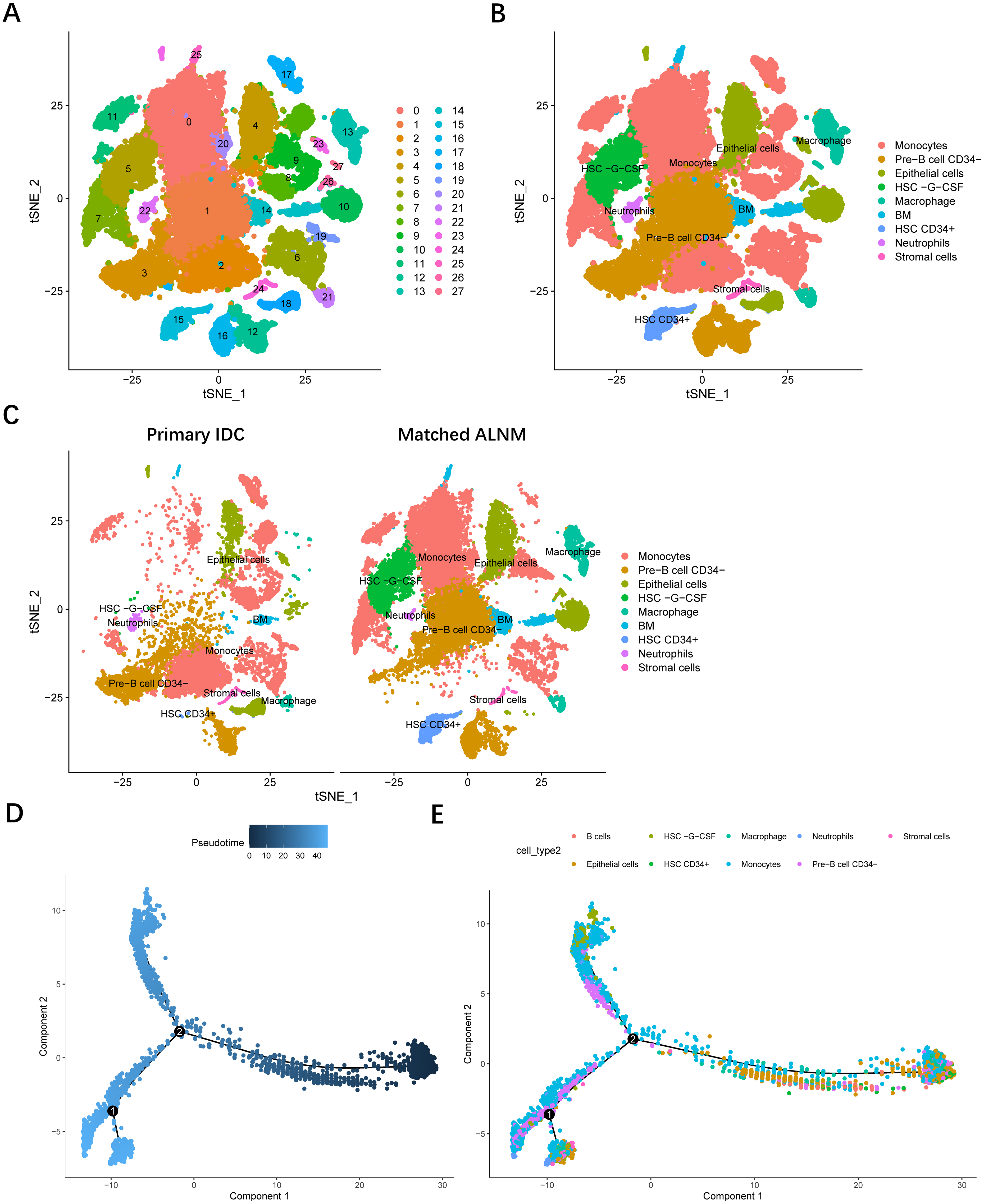
Cell clustering and trajectory analysis of single-cell RNA-Seq data. (a) t-SNE plot showing cell clustering of all samples in the single-cell RNA-Seq dataset. Each color represents a distinct cell cluster. (b) Annotation of cell types using the SingleR package, identifying nine major cell types. (c) Comparison of cell type distributions between IDC and matched metastatic axillary lymph nodes. (d) and (e) Cell trajectory analysis using Monocle. Black curves indicate inferred differentiation trajectories, and numbered nodes (1, 2) represent potential critical transition states. Pseudotime denotes the inferred temporal progression of cellular states.
Compared to the IDC group, the metastatic lymph node group exhibited a significantly higher proportion of monocytes, pre-B cells, epithelial cells, G-CSF-HSC, macrophages, bone marrow cells, and CD34+ hematopoietic stem cells (Figure 1(c)). Cell trajectory analysis revealed dynamic changes in the tumor microenvironment, with black curves representing differentiation trajectories and numbered nodes (1, 2) indicating potential critical transition states (Figure 1(d) and (e)). The overall trend suggested that epithelial cells might undergo specific differentiation or phenotypic changes during ALNM, which could be closely related to tumor progression and metastatic potential.
Cell–cell communication in IDC and metastatic axillary lymph nodes
To investigate intercellular communication, we performed cell–cell interaction analysis on the nine identified cell types. The results showed that macrophages and B cells had the highest number of ligand-receptor interactions, while monocytes and B cells exhibited the strongest communication intensity (Supplementary Figure 1).
At the signaling pathway level, we constructed an interaction network to depict cell–cell communications. Bubble plots indicated that the macrophage migration inhibitory factor (MIF)-(CD74 + CXCR4) interaction was enriched between B cells and G-CSF-HSC, whereas the MIF-(CD74 + CD44) interaction was predominant between B cells and monocytes. Additionally, the MDK–NCL interaction between macrophages and epithelial cells was particularly strong, suggesting that these interactions may play a crucial role in ALNM in BC (Figure 2).
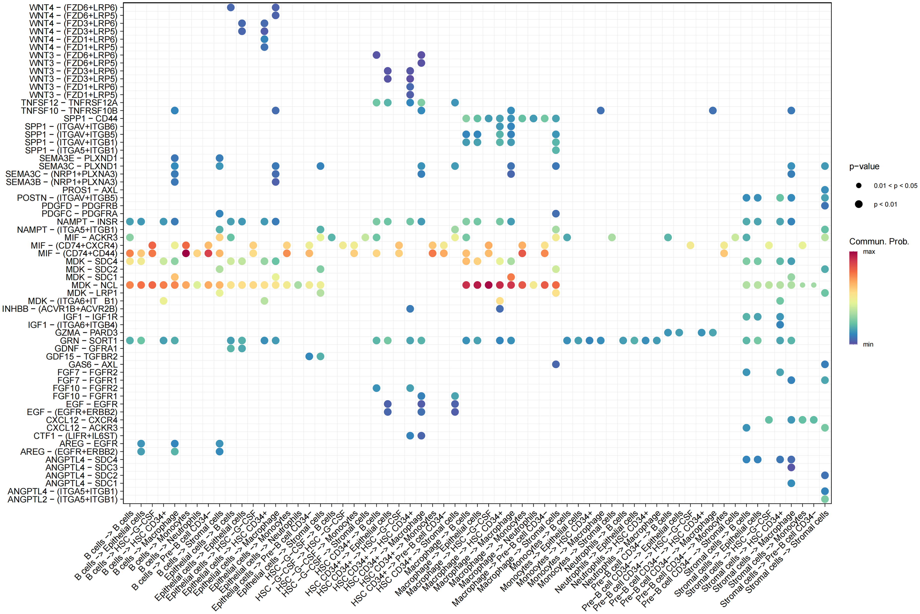
Bubble plot of cell–cell communication at the signaling pathway level. A bubble plot was constructed to visualize the intercellular communication network at the signaling pathway level. Each bubble represents a ligand–receptor interaction between specific cell types, with bubble size indicating interaction strength and color denoting pathway enrichment.
Identification of key differentially expressed genes and MR analysis
Given that epithelial cells are the primary source of tumor cells and are likely to be closely associated with ALNM, we conducted a differential gene expression analysis between epithelial cells from IDC and their paired metastatic lymph nodes. A total of 2421 DEGs were identified.
To further explore the role of these DEGs, we obtained gene-specific eQTL instruments for these DEGs from the IEU OpenGWAS eqtl-a resource and performed MR analysis using BC (ebi-a-GCST90018799) as the outcome. Twelve DEGs were significantly associated with BC (p < 0.05), including ACAP1, APOL6, ASRGL1, CD53, FSCN1, ISG15, PI3, PLEKHO1, RNF144B, SPART, TCDD-inducible poly-ADP-ribose polymerase (TIPARP), and TTC23 (Supplementary Tables 2 and 3, Figure 3).
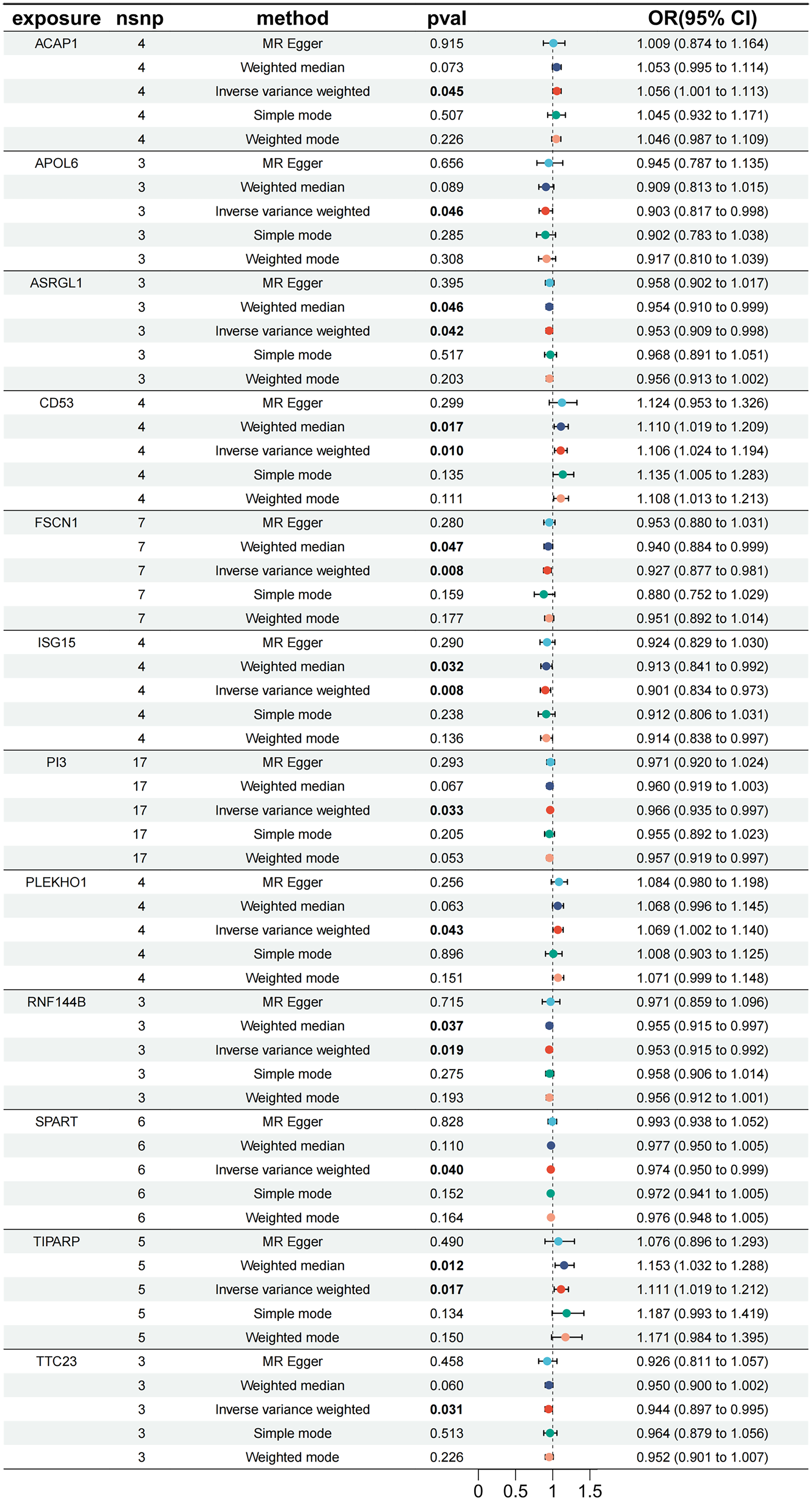
Forest plot of expression quantitative trait loci (eQTL)-based MR results for ALNM-associated epithelial DEGs using breast cancer (ebi-a-GCST90018799) as the outcome. eQTLs of candidate central genes were extracted from GWAS summary statistics and analyzed using Mendelian randomization (MR), with breast cancer (GCST90018799) as the outcome. Sensitivity analysis was performed by comparing causal effect estimates across multiple MR methods, including MR-Egger, penalized weighted median, simple mode, inverse variance weighted (IVW), and weighted mode. A P-value < 0.05 was considered statistically significant.
Among them, ACAP1, CD53, PLEKHO1, and TIPARP showed positive associations between genetically predicted expression and BC risk (OR > 1), while the remaining genes appeared to have protective effects. The best causal estimates showed that CD53 (OR (95% CI) = 1.110 (1.019–1.209), P = 0.017, weighted median) and TIPARP (OR (95% CI) = 1.153 (1.032–1.288), P = 0.012, weighted median) were significantly associated with an increased risk of BC. Leave-one-out sensitivity analysis demonstrated the robustness of these findings, supporting the potential relevance of these genes in BC biology and their possible involvement in ALNM-associated molecular alterations (Figure 4).
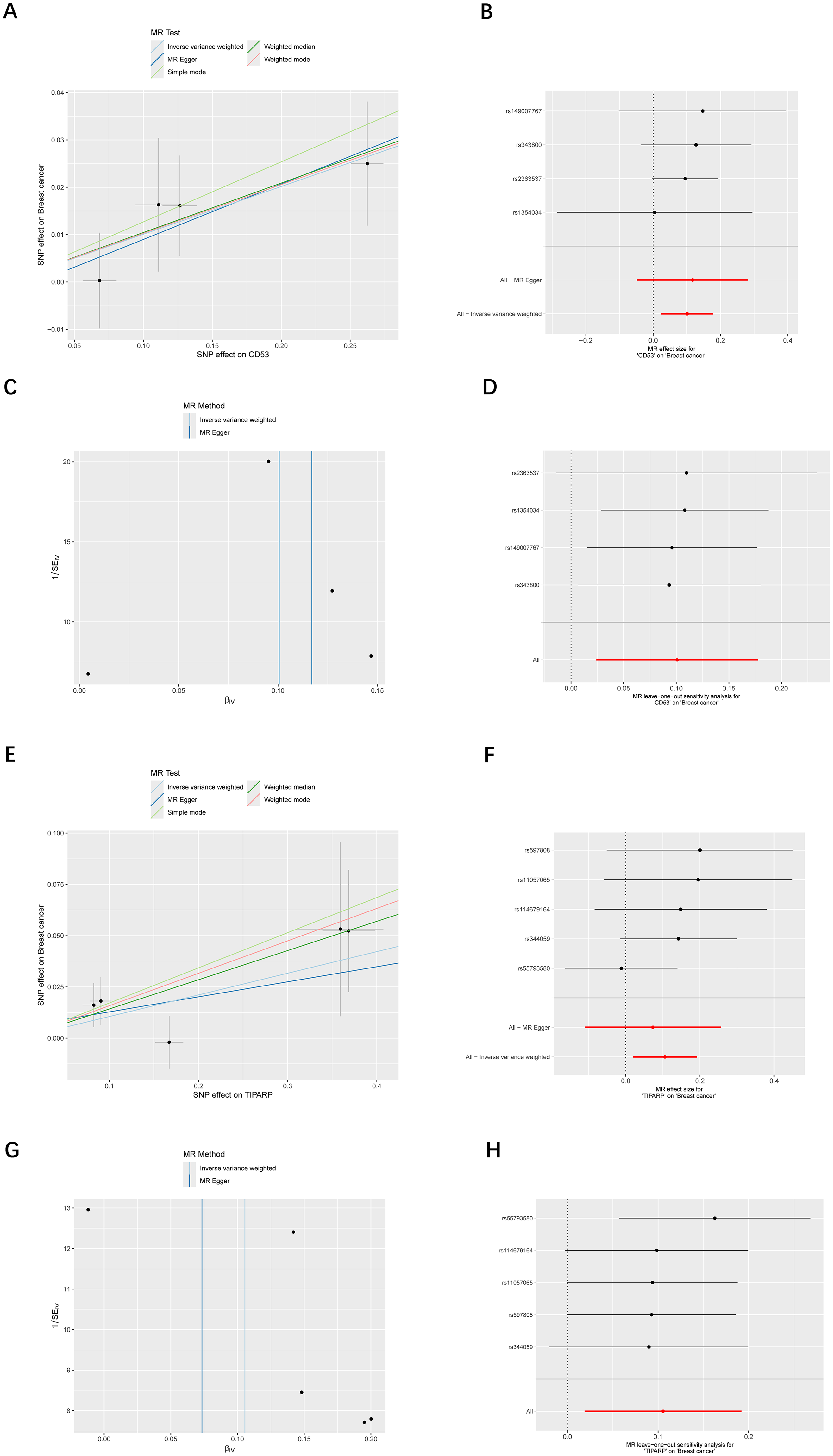
Mendelian randomization analyses of CD53 and TIPARP, two candidate genes prioritized from ALNM-associated epithelial DEGs, using breast cancer risk as the outcome. (a) and (e) Scatter plots showing the associations between genetically predicted expression of CD53 and TIPARP and breast cancer risk. (b) and (f) Forest plots showing effect estimates from multiple Mendelian randomization (MR) methods for CD53 and TIPARP. (c) and (g) Funnel plots assessing potential asymmetry suggestive of directional pleiotropy in the MR analyses of CD53 and TIPARP. (d) and (h) Leave-one-out sensitivity analyses evaluating the robustness of the MR results for CD53 and TIPARP.
Functional enrichment and pathway analysis of key genes
Using GeneMANIA, we predicted 32 functionally related genes associated with the 12 identified DEGs, with RNF144B showing the highest correlation. The protein–protein interaction (PPI) network revealed various types of associations, including co-expression, co-localization, and genetic interactions.
GO enrichment analysis indicated that these genes were involved in gland development, response to lipopolysaccharide, and collagen-containing extracellular matrix organization. KEGG pathway analysis showed significant enrichment in MAPK signaling and PI3K-Akt signaling pathways, suggesting their potential involvement in the molecular mechanisms underlying BC ALNM (Figure 5).

Functional enrichment and pathway analysis of key genes. (a) Gene–gene interaction network of identified biomarker genes constructed using the GeneMANIA database. (b) and (c) Gene Ontology (GO) enrichment analysis of key genes, including biological processes, cellular components, and molecular functions. (d) Kyoto Encyclopedia of Genes and Genomes (KEGG) pathway analysis of key genes.
Discussion
In this study, we employed a comprehensive approach combining scRNA-seq, functional enrichment analysis, and MR to investigate the molecular mechanisms driving ALNM in BC. The integration of scRNA-seq data with clinical and genetic information provided deep insights into the cellular dynamics and gene interactions involved in BC metastasis, highlighting several key findings that could shape future diagnostic and therapeutic strategies.
Our scRNA-seq atlas of IDC and matched ALNM samples reveals a profound reorganization of the TME during lymphatic dissemination. The enrichment of monocytes, G-CSF-HSC, and CD34+ hematopoietic progenitors in ALNM aligns with emerging evidence that metastatic niches recruit BM-derived cells to support colonization.26–31 Notably, the expansion of pre-B cells in ALNM echoes recent reports of B cell-mediated immune tolerance in metastatic sites, where regulatory B cells (Bregs) may suppress anti-tumor T-cell responses through interleukin-10 and transforming growth factor-beta (TGF-β) secretion.32,33 The observed epithelial cell trajectory bifurcation (nodes 1→2) suggests a metastable transitional state—possibly reflecting EMT-like plasticity—that warrants further investigation using pseudotime-velocity analyses.
The dominance of macrophage-epithelial interactions via MDK–NCL signaling provides mechanistic insights into niche preparation. MDK, a heparin-binding growth factor overexpressed in BC, promotes metastasis through PI3K/Akt activation and vascular leakiness, while its receptor nucleolin facilitates extracellular MDK internalization to drive pro-survival signaling.34,35 Similarly, the MIF-(CD74 + CXCR4) axis between B cells and HSCs may orchestrate stemness programs in metastatic epithelial cells, as MIF is known to enhance cancer stem cell properties via CXCR4-mediated Wnt/β-catenin activation.36,37 These findings position macrophage- and B cell-derived ligands as microenvironmental “fertilizers” for metastatic outgrowth.
Our MR analysis provided genetic support for prioritizing CD53 and TIPARP as candidate genes associated with BC risk, and these genes may also be relevant to ALNM-associated molecular alterations. CD53, a tetraspanin regulating immune synapse formation in lymphocytes, paradoxically emerges as a metastasis promoter—a duality potentially explained by its role in stabilizing integrin clusters that enhance epithelial cell adhesion to lymphatic endothelia.38–40 TIPARP (TCDD-inducible poly-ADP-ribose polymerase), traditionally studied in xenobiotic metabolism, may foster metastasis through poly-ADP-ribosylation-dependent modulation of hypoxia-inducible factor 1-alpha (HIF-1α) stability under hypoxic stress.41,42 The protective association of APOL6 and ISG15 aligns with their roles in ER stress-induced apoptosis and interferon-mediated immune surveillance, respectively, suggesting their loss in metastasis enables immune evasion.43–46
The convergence of DEGs on MAPK and PI3K-Akt pathways underscores their dual roles in BC progression: while MAPK signaling (e.g. via ERK1/2 phosphorylation) drives EMT and matrix remodeling, PI3K-Akt activation promotes survival in detached circulating tumor cells.47–49 Pharmacologically targeting these pathways has shown limited success due to compensatory feedback loops; however, our cell-type-specific expression data suggest that combinatorial inhibition of macrophage-derived MDK and epithelial Akt may disrupt metastatic resilience. Additionally, the association of RNF144B—an E3 ubiquitin ligase regulating epidermal growth factor receptor degradation—with extracellular matrix organization implies a novel link between growth factor receptor turnover and metastatic niche formation.50–52
Despite these advances, our study has limitations. The modest cohort size (n = 6 pairs) necessitates validation in larger scRNA-seq datasets. Spatial transcriptomics could further resolve ligand-receptor interactions within microanatomical niches, while CRISPR-based perturbation of CD53/TIPARP in organoid models would clarify their functional roles. Future MR analyses should incorporate cis-eQTLs with pleiotropy-robust methods to refine causal estimates.
In conclusion, this study provides novel insights into the cellular and molecular mechanisms of ALNM in BC. The identification of key genes, cellular interactions, and signaling pathways opens new avenues for the development of targeted therapies aimed at preventing or treating lymph node metastasis. Future studies should focus on validating these findings and exploring the potential therapeutic implications of targeting these molecular drivers in clinical practice.
Supplemental Material
sj-docx-1-jbm-10.1177_03936155261443650 - Supplemental material for Exploring key biomarkers associated with axillary lymph node metastasis in breast cancer using single-cell RNA sequencing and Mendelian randomization
Supplemental material, sj-docx-1-jbm-10.1177_03936155261443650 for Exploring key biomarkers associated with axillary lymph node metastasis in breast cancer using single-cell RNA sequencing and Mendelian randomization by Limeng Qu, Jinyang Li, Shirong Ding, Qian Long and Wenjun Yi in The International Journal of Biological Markers
Footnotes
Abbreviations
Acknowledgments
Thanks to the anonymous peer-reviewers for their insightful suggestions and careful reading of the manuscript.

Ethics approval
Not applicable.
Consent to participate
Not applicable.
Author contributions
Q-LM and L-JY: Writing—original draft. D-SR: Writing—original draft. L-Q: Writing—review & editing, Supervision. Y-WJ: Writing—review & editing, Conceptualization.
Funding
This study was supported by the National Natural Science Foundation of China [grant number: 82303526 and 82403073],the Natural Science Foundation of the Hunan Province of China [grant number: 2023JJ40842 and 2024JJ6590], the Innovation Platform and Talent Plan of Hunan Province [grant number: 2023SK4019], the Natural Science Foundation of Changsha City [grant number: kq2208309], the China Postdoctoral Science Foundation [grant number: 2023M733955 and 2023M743946], Open Funds of State Key Laboratory of Oncology in South China [grant number: HN2024-04, NH2024-07], and the Scientific Research Launch Project for new employees of the Second Xiangya Hospital of Central South University [grant number: QH20230256 and QH20230268].
Declaration of conflicting interests
The authors declared no potential conflicts of interest with respect to the research, authorship, and/or publication of this article.
Supplemental material
Supplemental material for this article is available online.
References
Supplementary Material
Please find the following supplemental material available below.
For Open Access articles published under a Creative Commons License, all supplemental material carries the same license as the article it is associated with.
For non-Open Access articles published, all supplemental material carries a non-exclusive license, and permission requests for re-use of supplemental material or any part of supplemental material shall be sent directly to the copyright owner as specified in the copyright notice associated with the article.
