



Abstract
Objective:
Real-time assessment of calyceal and tissue temperatures during laser lithotripsy.
Methods:
Patients eligible for endoscopic combined intrarenal surgery were assigned to Soltive 60 W SuperPulsed thulium fiber laser (TFL; Olympus, Inc.) or holmium:YAG (Ho:YAG; Lumenis Pulse™ 120H) based on availability. After translocation of stones, a 1.5-mm multipoint thermal sensor needle measuring temperatures 5, 15, 25, and 35 mm from the tip was placed percutaneously with ultrasound guidance. The 5-mm mark recorded calyceal fluid temperature and the others parenchymal temperatures for 2 minutes during fragmentation (1 J/2 Hz TFL and 0.5 J/5 Hz Ho:YAG, 2–2.5 W), dusting (0.3 J/50 Hz, 15 W), and pop-dusting (0.5 J/80 Hz, 40 W). If tissue temperature exceeded 43°C, lasering was paused. After measurements, tract dilation and percutaneous nephrolithotomy were performed. Categorical data were compared using chi-square or Fisher’s exact tests and continuous variables via t-tests.
Results:
The 20 patients (10 TFL and 10 Ho:YAG) had similar baseline characteristics. During fragmentation, calyceal (Tcalyx) and parenchymal (Ttissue) temperatures never exceeded 41°C. During dusting, Tcalyx exceeded 43°C in four patients for each laser and Ttissue exceeded 43°C in one Ho:YAG patient (49°C after 60 seconds). Maximum calyceal temperature (Tmax) in TFL and Ho:YAG patients was 51°C and 56°C (p = 0.18), respectively. During pop-dusting, Tcalyx exceeded 43°C in 10 TFL and 8 Ho:YAG patients, and average maximum temperature (avgTmax) was significantly higher for TFL (52.9°C vs 45.1°C, p = 0.027). The tissue Tmax was 47°C and 49°C (p = 0.96), respectively, with 43°C exceeded in three TFL and three Ho:YAG patients.
Conclusion:
During high-powered lasering, temperatures can exceed 43°C in as little as 15 seconds. This is concerning since temperatures >43°C may be associated with permanent tissue injury.
Introduction
Retrograde intrarenal surgery (RIRS) using holmium:YAG (Ho:YAG) and thulium fiber laser (TFL) systems for lithotripsy has become the most common surgical treatment for kidney stones. 1 Although advancements in laser technology, particularly the development of high-power modalities, have enhanced stone ablation efficiency, concerns have been raised regarding the potential for thermal injury to surrounding urothelial tissues. 2
Energy emitted by a laser used in ureteroscopy (URS) may dissipate, in the form of heat, into surrounding tissues, potentially causing thermal damage such as coagulation, carbonization, and protein denaturation.
Existing literature highlights the importance of modifiable surgical parameters, including irrigation temperature and flow rate, utilization of ureteral access sheaths (UASs), laser settings (power, pulse duration, and frequency), and operator duty cycle, in lowering intrarenal temperatures and thus mitigating potential thermal damage during URS.3–5
However, no human studies, to date, have been done to measure both renal parenchymal and calyceal temperatures during RIRS with lithotripsy with both pulse-modulated Ho:YAG (Lumenis Pulse™ 120H) and SuperPulsed TFL (Olympus, Inc.) systems. The primary objective of this study was to measure human intracalyceal and renal parenchymal temperature changes in real time during endoscopic stone surgery with two high-power laser systems. Secondary objectives included correlating irrigation fluid temperatures with parenchymal temperatures in real time during endoscopic stone and determining which modifiable surgical factors influence renal temperature changes during endoscopic stone surgery with high-power laser systems. Another secondary objective included comparing calyceal and renal parenchymal temperature changes between the pulse-modulated Ho:YAG and SuperPulsed TFL systems.
Methods
Study design
An Institutional Review Board approved (IRB STUDY-23-00046) from December 2023 to July 2024 recruited patients with unilateral stone burden requiring a combined approach (endoscopic combined intrarenal surgery [ECIRS]) at Mount Sinai Medical Center. Although surgeon preference plays a role, the primary indications for ECIRS included a large stone burden (≥2 cm) and multiple stones in different calyces, in an effort to prevent the need for a multiaccess percutaneous nephrolithotomy (PCNL). Patients were then screened for the following inclusion criteria: minimum age of 18 years; nonpregnant and able to provide informed consent; preoperative creatinine ≤1.3 mg/dL; negative preoperative urine culture; and preoperative CT scan within 60 days of surgery. Exclusion criteria included nephrolithiasis procedure (i.e., shockwave lithotripsy, URS/laser lithotripsy [LLT], or PCNL) within the previous 90 days; preoperative indwelling urinary drain (ureteral stent and percutaneous nephrostomy tube); history of urinary diversion; congenital kidney anomalies, solitary or atrophic kidney; and preexisting urinary tract obstruction or ureteral stricture disease.
Procedure
Stone burden was defined as the sum of the single longest diameters of all stones, as measured in the coronal dimension. For a more precise measurement, total stone volume was calculated by summing the volumes of individual stones. Maximal width and length were measured on axial views and height on coronal views, all under ≥8× magnification. Using these dimensions, individual volumes were determined via manual calculation with the European Association of Urology ellipsoid formula: V = 0.167 × π × L × W × H.
Patients were assigned to receive LLT with either a TFL (60 W Soltive™ SuperPulsed) or Ho:YAG (Lumenis Pulse™ 120H) laser system based on the availability of the laser system.
All patients were positioned in the modified Barts flank-free position followed by standard sterile preparation and draping. Cystoscopy was performed followed by flexible URS (Flex X2, Karl Storz) under direct vision into the renal collecting system. The procedure was performed without the use of a UAS. A comprehensive pyeloscopy was performed to identify and characterize the stone burden. As is our standard procedure with ECIRS, all stones were translocated using a basket to a favorable calyx for LLT followed by percutaneous evacuation of all stones, stone particles, and dust. All patients received gravity irrigation at room temperature with saline irrigant bags placed 80 cm above the patient’s abdomen (distance from top of the abdomen to the bottom of the irrigant bag). Although the precise volume of infused fluid was not measured, our standard protocol for URS involves using no more than a single 1-L saline bag.
After translocation of stones, a 1.5-mm multipoint thermal sensor (MTS) needle (Boston Scientific) was utilized to get percutaneous access to the favorable renal calyx in place of using a 18 g access needle (Fig. 1). The MTS needle was inserted into the collecting system under ultrasound and fluoroscopic guidance. Moreover, with the flexible ureteroscope in position, the needle penetration was observed in real time. After the needle was positioned properly with the distal most sensor in the calyx, it was calibrated according to protocol to ensure accurate functionality.
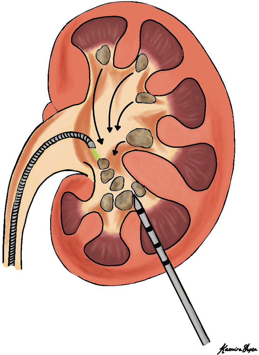
Diagram of translocation of stones to a favorable calyx and percutaneous placement of multipoint thermal sensor with calyceal (5 mm) and parenchymal (15 mm [1 cm], 25 mm [2 cm], and 35 mm [3 cm]) marks.
The Soltive 60 W SuperPulsed TFL (Olympus, Inc.) or Lumenis P120 Ho:YAG laser with MOSES™ technology (Boston Scientific) systems was used for LLT with a 200-µm laser fiber. Temperature data were collected from each patient during LLT using three commonly used laser settings. Fragmentation was performed with the following settings: 1 J/2 Hz (TFL) and 0.5 J/5 Hz (Ho:YAG), 2–2.5 W with the aim to break stones into large pieces. Dusting was performed with the following settings: 0.3 J/50 Hz, 15 W with the aim to pulverize stone into fine powder in a continuous “painting” motion over the stone’s surface. Pop-dusting was performed with the following settings: 0.5 J/80 Hz, 40 W with the aim to rapidly fragment kidney stones into extremely fine, powder-like pieces. Each laser mode was used for 2 minutes, with intracalyceal and parenchymal tissue temperatures measured in real time. If tissue temperature exceeded 43°C, lasering was paused. Before commencing a new laser setting, the temperature of the caliceal fluid and renal parenchyma was allowed to return to baseline body temperature prior to initiating a new laser setting.
During LLT, temperature measurements were recorded using the MTS needle at 5 mm (calyceal), 15 mm (1 cm), 25 mm (2 cm), and 35 mm (3 cm) from the probe’s tip. The 5-mm sensor was located just within the collecting system, whereas the other sensors measured temperatures along the parenchyma (medulla and cortex) in real time. At each site along the MTS needle, we monitored for temperatures over 43°C and recorded the duration for which this threshold was surpassed. The operating surgeon assessed and graded any laser-induced damage intraoperatively, immediately following the completion of lithotripsy. The laser damage was graded with the following criteria: Grade 0: none, Grade 1: mucosal, Grade 2: submucosal, and Grade 3: full thickness. 6
Following temperature measurements, a fascial dilator was placed over the MTS needle under direct vision, to facilitate the placement of a guidewire and to perform balloon dilation of the tract to 24F followed by PCNL of all stones and fragments and dust in standard fashion. At the conclusion of the case, the surgeon decided as usual if a ureteral stent or nephrostomy tube was necessary. Categorical data were compared using chi-square or Fisher’s exact tests, whereas continuous variables were analyzed via t-tests. Normality of distributions for all comparisons was assessed via Kolmogorov–Smirnov and Shapiro–Wilk tests of normality. Given that the majority of comparisons/distributions in our study did not violate the assumption of normality, we opted to compare data between our groups using means/t-tests and standard deviations.
Results
Between December 2023 and July 2024, a total of 26 patients met the inclusion criteria for ECIRS. Six patients were preoperatively excluded according to exclusion criteria, resulting in a final analysis of 20 patients, with 10 in each treatment arm (Fig. 2).

Flow diagram.
There were no differences in demographics, stone characteristics, and clinical characteristics between both groups (Table 1). Total operative time, operator duty cycle, and frequency of visible laser damage were similar between both groups. Nine out of 20 (45%) were done totally tubeless, without stents or nephrostomy tubes. The remaining 11 had stents but no nephrostomy tubes.
Baseline Characteristics and Operative Information Between Ho:YAG and TFL Groups
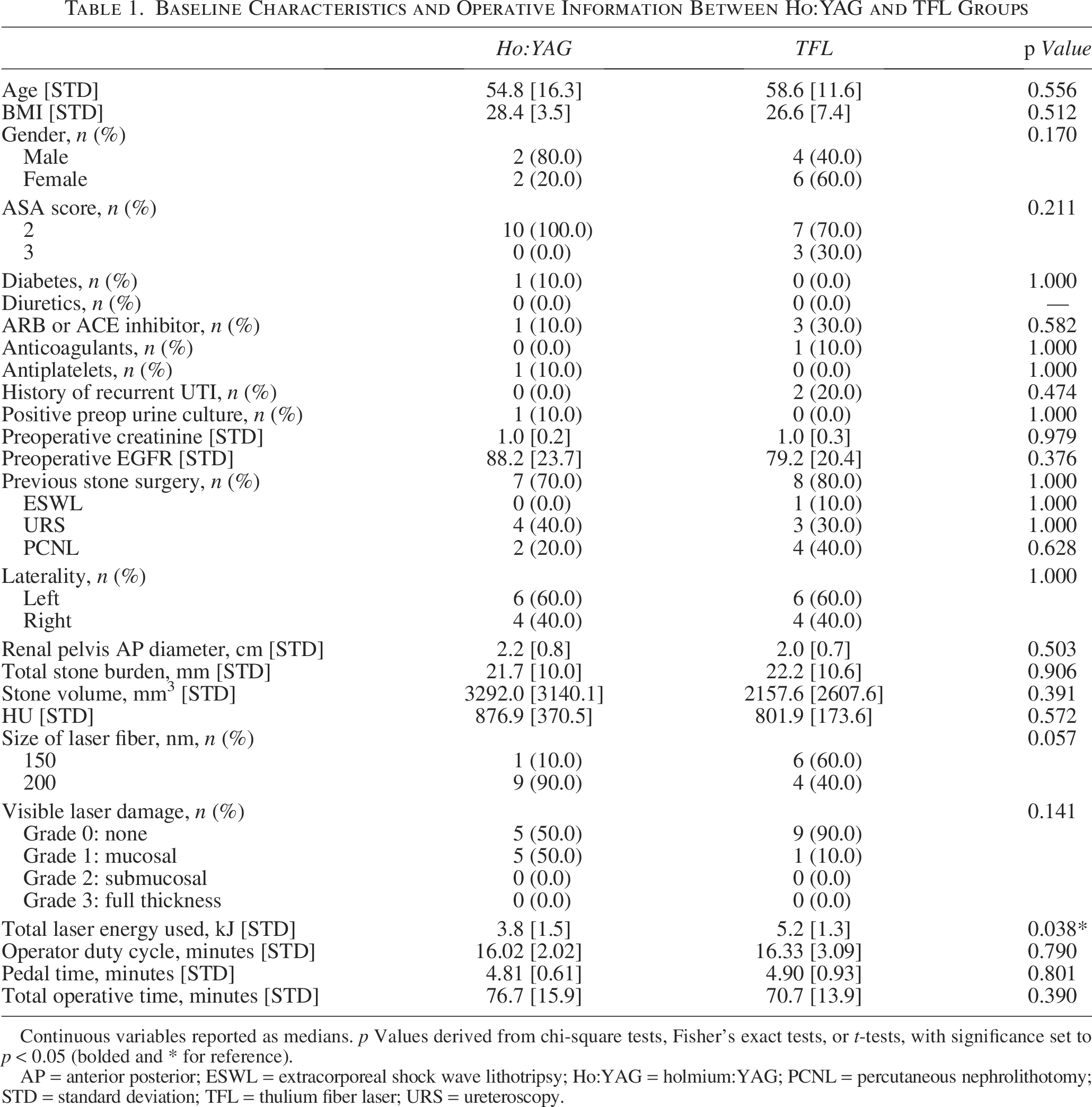
Continuous variables reported as medians. p Values derived from chi-square tests, Fisher’s exact tests, or t-tests, with significance set to p < 0.05 (bolded and * for reference).
AP = anterior posterior; ESWL = extracorporeal shock wave lithotripsy; Ho:YAG = holmium:YAG; PCNL = percutaneous nephrolithotomy; STD = standard deviation; TFL = thulium fiber laser; URS = ureteroscopy.
Mean baseline calyceal temperatures (Tcalyx) for Ho:YAG and TFL were below 33°C across all time points for both groups (Table 2). During fragmentation, 1 J and 2 Hz, 2 W, mean calyceal temperatures for Ho:YAG and TFL were 31.8°C vs 32.5°C at 0 seconds, 33.4°C vs 33.3°C at 60 seconds, and 34.0°C vs 33.4°C at 120 seconds (p = 0.404, p = 0.941, p = 0.699; Fig. 3). During dusting, 0.3 J and 50 Hz, 15 W, mean Tcalyx for Ho:YAG and TFL was 32.5°C vs 31.9°C at 0 seconds, 39.1°C vs 42.1°C at 60 seconds, and 37.6°C vs 42.6°C at 120 seconds (p = 0.526, p = 0.289, p = 0.082). During dusting, Tcalyx exceeded 43°C in four TFL patients and four Ho:YAG patients (mean Tmax: 44.3°C vs 40.4°C, p = 0.175). Maximum calyceal temperature (Tmax) in TFL and Ho:YAG patients was 51°C and 56°C (p = 0.18), respectively. During pop-dusting, 0.5 J and 80 Hz, mean Tcalyx for Ho:YAG and TFL was 33.0°C vs 32.7°C at 0 seconds, 43.0°C vs 47.3°C at 60 seconds, and 40.0°C vs 51.2°C at 90 seconds (p = 0.754, p = 0.264, p = 0.071). The Tcalyx exceeded 43°C in 10 TFL and 8 Ho:YAG patients with a significantly higher mean Tmax in the TFL arm (TFL: 52.9°C, Ho:YAG: 45.1°C, p = 0.027).

Mean calyceal temperature by laser and setting over time. Error bars represent standard error of the mean.
Temperature Outcomes Between Ho:YAG and TFL Groups

p Values derived from t-tests, with significance set to p < 0.05 (bolded and * for reference).
Mean baseline parenchymal temperatures (Ttissue) for both groups were also comparable and remained below 36°C across all time points. During fragmentation, mean Ttissue at the 15-mm probe tip for Ho:YAG and TFL was 34.2°C vs 35.2°C at 0 seconds, 34.7°C vs 35.3°C at 60 seconds, and 34.8°C vs 32.3°C at 120 seconds (p = 0.087, p = 0.468, p = 0.430). Maximum calyceal and parenchymal temperatures averaged below 36°C and never exceeded 43°C during fragmentation (Table 3).
Temperature Outcomes Between Ho:YAG and TFL Groups
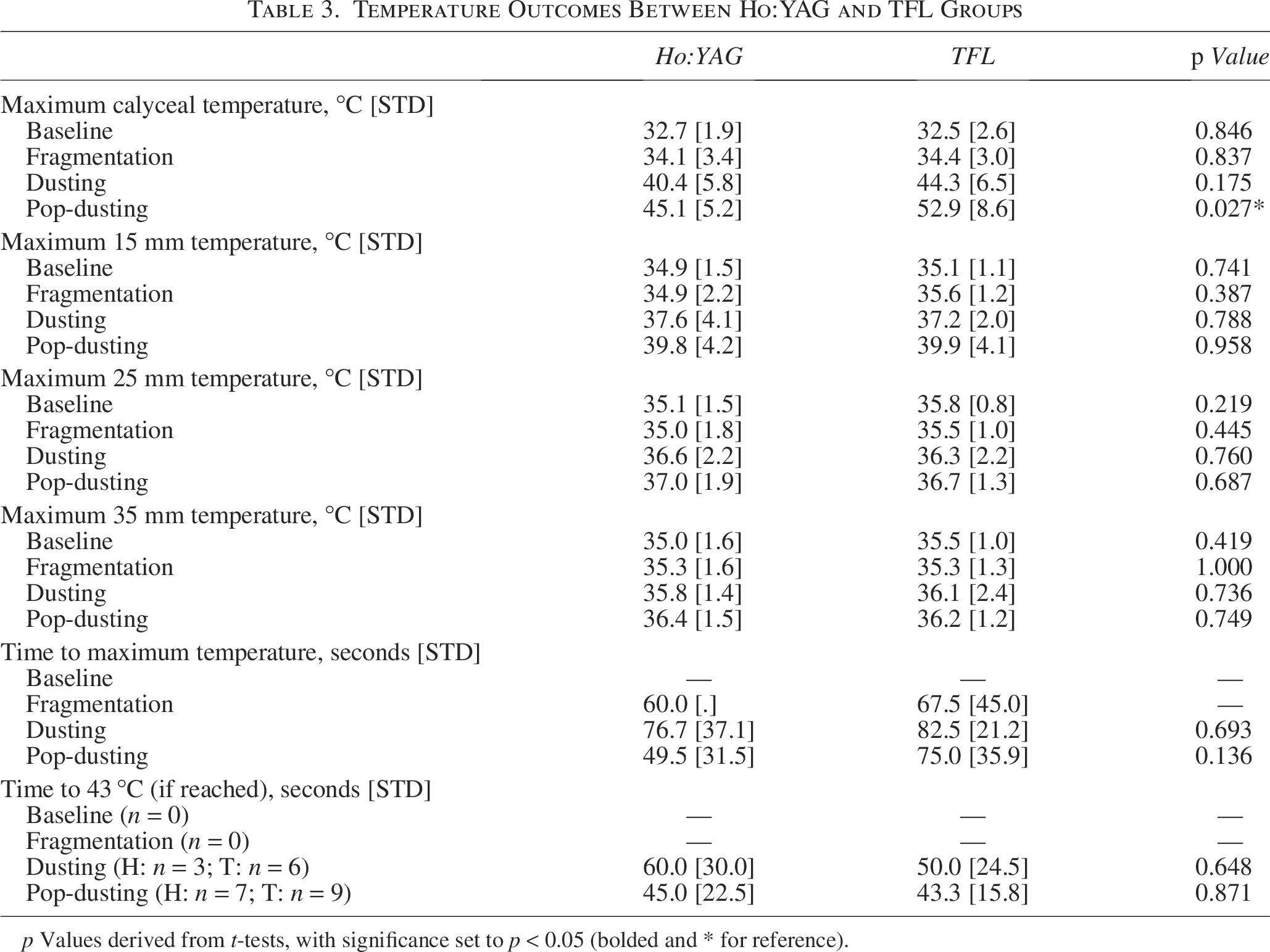
p Values derived from t-tests, with significance set to p < 0.05 (bolded and * for reference).
During dusting, mean Ttissue at the 15-mm probe tip for Ho:YAG and TFL was 35.0°C vs 35.0°C at 0 seconds, 36.9°C vs 36.5°C at 60 seconds, and 36.3°C vs 36.6°C at 120 seconds (p = 1.000, p = 0.805, p = 0.554). The mean parenchymal Tmax at 15 mm was 37.6°C and 37.2°C during dusting in the Ho:YAG and TFL groups, respectively (p = 0.788; Fig. 4). Ttissue during dusting only exceeded 43°C in one patient in the Ho:YAG cohort (49°C after 60 seconds at the 15 mm probe; Fig.5). At the 25 mm and 35 mm markers, the mean Tmax was <37°C for both Ho:YAG and TFL with no significant statistical differences between groups. During pop-dusting, mean Ttissue at the 15-mm probe tip for Ho:YAG and TFL was 34.6°C vs 35.1°C at 0 seconds, 38.4°C vs 38.3°C at 60 seconds, and 39.0°C vs 39.5°C at 90 seconds (p = 0.607, p = 0.954, p = 0.899). The mean parenchymal Tmax at 15 mm was 39.8°C and 39.9°C during pop-dusting in the Ho:YAG and TFL groups, respectively (p = 0.958). Ttissue during pop-dusting exceeded 43°C two times in the Ho:YAG cohort and three in the TFL cohort, which all occurred at the 15 mm probe (Fig. 6). At the 25-mm and 35-mm probes, mean Tmax was comparable between groups and averaged below 37°C during pop-dusting.
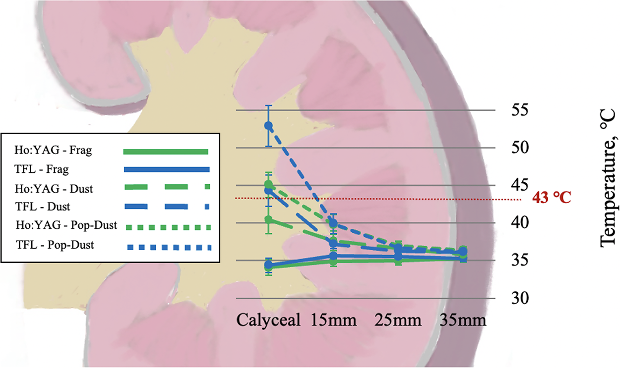
Mean Tmax at each renal landmark by laser and setting. Error bars represent standard error of the mean.
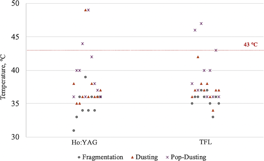
Individual maximum temperature readings at 15-mm probe for Ho:YAG and TFL across laser settings. Ho:YAG = holmium:YAG; TFL = thulium fiber laser.
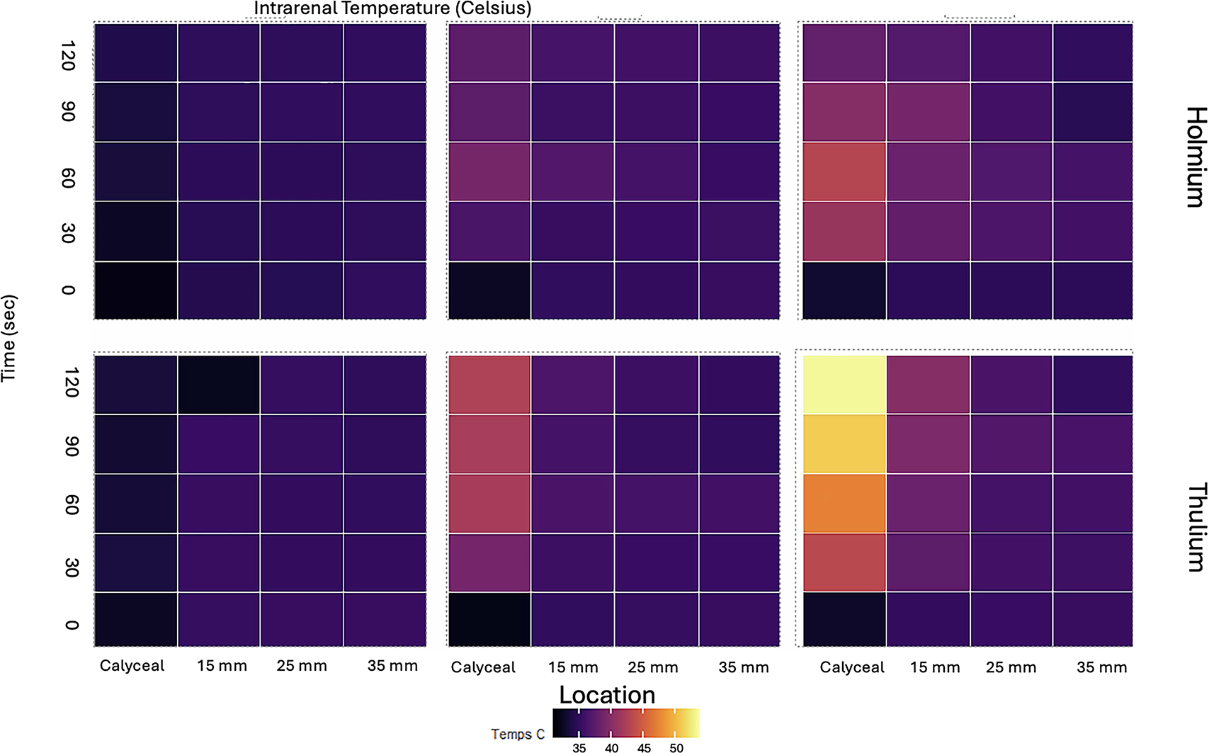
Heat map representation of calyceal and parenchymal temperatures for both pulse-modulated Ho:YAG and SuperPulsed TFL for all settings.
Discussion
High-powered lasers have demonstrated increased efficiency in LLT; however, they also pose a risk of elevating intrarenal temperatures, potentially leading to thermal injury to the kidney. This study evaluates calyceal and parenchymal temperature elevations up to 120 seconds following the activation of high-powered lasers. Additionally, we analyzed the minimum, maximum, and mean calyceal and parenchymal temperatures during laser activation using both pulse-modulated Ho:YAG and SuperPulsed TFL systems.
With the advent of high-powered lasers, several modifiable risk factors have been investigated to mitigate the risk of thermal injury, one of which is laser settings. In this study, we observed that with low-powered settings (2–2.5 W), renal temperatures—including both calyceal and parenchymal measurements—remained below 43°C for both TFL and Ho:YAG systems. In contrast, high-powered settings (>15 W) led to temperatures exceeding 43°C within as little as 15 seconds. Similarly, a study that measured calyceal fluid temperature utilizing a Type K thermocouple during retrograde LLT with a TFL laser at incremental settings reported that a temperature of 43°C was reached in 3 out of 10 patients at 20 W and in 8 out of 10 patients at 30 W, whereas it remained below this threshold at 5 W and 10 W. 7 In an in vivo porcine model, all dusting and most fragmentation settings (dusting: 0.5 J, 80 Hz, 40 W; low-power fragmentation: 1 J, 10 Hz, 10 W; high-power fragmentation: 1.5 J, 20 Hz, 30 W) resulted in calyceal temperatures reaching or exceeding 44°C. 8 All the settings >5 W in our study and in prior in vivo studies have resulted in calyceal temperatures reaching or exceeding 43°C. These findings underscore the direct relationship between laser power settings and peak calyceal fluid temperatures. During pop-dusting, the average maximum temperature (avgTmax) was significantly higher for TFL than for pulse-modulated Ho:YAG. This finding is consistent with a previous in vitro URS study using an impacted ureteral stone model with no outflow where they found that TFL produced significantly higher temperatures than Dornier and Empower HL lasers across most power settings (p < 0.001). 9 The sole exception occurred at 20 W, where no significant difference was observed between TFL and Empower HL. However, the study’s methodology presents a key limitation. Temperature was measured with a thermocouple placed in the ureteral fluid, just 2 mm from the laser fiber. When treating an impacted stone, this method is known to yield falsely elevated readings, which may not accurately reflect the true temperature of the ureteral tissue. Calyceal fluid temperatures do not necessarily indicate that renal tissue temperatures increase, and parenchymal temperature elevations may be more indicative of renal injury because of high-powered lasers.
Given the importance of parenchymal temperatures in evaluating potential tissue injury, our study measured medullary and cortical temperatures at approximately 1, 2, and 3 cm from the tip of the renal papilla. According to the thermal dose model described by Sapareto and Dewey, tissue maintained at 43°C for 120 minutes experiences an equivalent thermal dose to tissue maintained at 50°C for 56 seconds or 56°C for 1 second. This concept has been validated across multiple tissue types, including renal tissue.10,11 Thermal cell injury is known to occur progressively at sustained temperatures above 43°C. 12 Our study demonstrated that under high-powered laser settings, calyceal and parenchymal temperatures exceeded 43°C in as little as 15 seconds. Specifically, during dusting (0.3 J, 50 Hz, 15 W), parenchymal temperatures exceeded 43°C in 1/20 patients (Ho:YAG patient), reaching 49°C after 60 seconds at the 15-mm marker. At the 25-mm and 35-mm markers, the mean peak temperature remained below 37°C for both Ho:YAG and TFL, with no significant differences between the two groups. However, during pop-dusting (0.5 J, 80 Hz), parenchymal temperatures exceeded 43°C in 5/20 patients (2 Ho:YAG patients and 3 TFL patients), all occurring at the 15-mm marker. At the 25-mm and 35-mm markers, mean peak temperatures remained below 37°C across both groups. Notably, with pop-dusting, parenchymal temperatures exceeded 43°C in as little as 20 seconds.
Conversely, findings from an in vivo porcine study indicated that temperatures surpassing 43°C did not penetrate deeper within the kidney. 4 Even under the most unfavorable conditions—including prewarmed irrigation, absence of a UAS, and the use of a single-lumen ureteroscope—renal temperatures remained below 43°C. This phenomenon is likely attributable to increased ureteral peristalsis and the heat-sinking properties of porcine renal tissue. Our findings from the first in vivo assessment of human renal parenchymal temperature during RIRS with LLT highlight the importance of optimizing laser settings to minimize the risk of thermal injury while maintaining procedural efficacy. In addition, our findings of elevated parenchymal temperatures during lithotripsy, coupled with the advent of new high-powered lasers, underscore the urgent need to study the associated risk of thermal damage.
Calyceal and parenchymal temperatures are influenced not only by transmitted energy but also by the surrounding medium and flow dynamics, including renal pelvis anteroposterior diameter (APD) and irrigation/outflow characteristics. In vitro studies have demonstrated that decreasing irrigation flow rate leads to elevated calyceal temperatures. 13 Specifically, Wollin et al. reported that with no flow, all maximum temperatures exceeded 43°C, surpassing those observed with lower-energy settings of the Lumenis Pulse 100 W Ho:YAG at 0.8 J/8 Hz, 1 J/10 Hz, and 1 J/20 Hz (p < 0.05). The mechanism by which irrigation lowers calyceal temperature is attributed to heat transfer to the irrigant as it passes over the lasered stone. 14 In a similar in vitro study, the highest temperature recorded was 70.3°C, occurring with no irrigation after 60 seconds of continuous laser firing (pulse 120 W Ho:YAG, 1.0 J × 40 Hz). Notably, fluid temperature in the test tube was measured via a thermocouple positioned 5 mm from the tube’s bottom. 15 These in vitro studies employed a thermocouple within a static fluid environment (tube or water bath), lacking outflow. When extrapolating in vitro findings to in vivo conditions, the interplay between irrigation and outflow becomes critical. 8 An in vivo porcine study investigating various irrigation and outflow scenarios, including single- vs dual-lumen ureteroscopes, warmed vs room-temperature irrigation, and with/without a 14 F UAS with the SuperPulsed TFL, identified the use of a UAS as the most significant factor affecting intrarenal temperature. Temperatures reached or exceeded 44°C across all laser and irrigation settings when a UAS was not employed. 4 The present study, conducted without UAS, using room-temperature irrigation and single-lumen ureteroscopes, represents a “worst-case scenario.” Although previous research suggests an inverse correlation between intrarenal temperature and APD, this association was not observed in the current study. Although APD can reflect renal pelvic volume, it does not consistently predict renal outflow. This discrepancy may stem from variations in temperature measurement methodologies.
We acknowledge several limitations in our study. To ensure a standardized methodology across laser types, we performed RIRS without a sheath and with gravity irrigation via a single-lumen ureteroscope—which is our standard method. We acknowledge that this is a “worse-case scenario,” but RIRS is commonly done in this manner, including at our own center. Although this approach enhances comparability, it may reduce the generalizability of our findings. Despite the limited sample size, the qualitative data provide compelling evidence of clinical relevance. Despite these limitations, we believe our study is well designed and provides valuable insights into an underexplored topic such as temperature dynamics during URS, particularly within the renal parenchyma.
Conclusions
High-powered settings in both Ho:YAG and TFL raise greater concerns regarding calyceal and parenchymal heating (>43°C) and the risk of renal injury compared with low-powered settings. The duration of high-powered energy activation should be carefully considered. Our study highlights the need for further research to explore parenchymal temperatures and their possible association with renal injury.
Authors’ Contributions
K.G.: Conceptualization and study design. C.C., V.D., and K.G.: Data collection. C.C., K.G., and D.L.: Data analysis. K.G., K.G., D.L., and Z.S.: Writing. R.K., W.M.A., B.G., K.G., and M.G.: Review and editing.
Footnotes
Funding Information
This study was supported by the Ferdinand C. Valentine Fellowship Award for Research in Urology, Boston Scientific Corporation (equipment only).
Authors’ Disclosure Statement
There are no competing financial interests.