



Abstract
Purpose:
To describe our 10-year experience with EBD for primary obstructive megaureter.
Materials and Methods:
Children needing intervention for primary obstructing megaureters (POM) (based on increasing distal dilatation >1.4 cm, differential renal function [DRF] <40%, or symptoms) were counseled to undergo a trial of EBD. A 9.5Fr cystoscope and stent “pusher” were used to insert a 14’’ guidewire to allow insertion of a 4/25 mm coronary dilatation balloon that was insufflated to 15 ATM for 5 minutes. Afterwards a 4.7Fr Double-J stent was positioned for 6 weeks. Ureteral diameter, DRF, length of obstruction, and complications were registered. Success was defined as improvement of the indication without further need for a more invasive procedure.
Results:
A total of 31 dilated ureters in 28 patients (23/5 M/F) were included, with a median age of 9 months (M) (range: 1–111) and a follow-up of 41 M (range: 12–84). EBD showed a narrow ring in 29 (93.5%) and longer narrow distal ureters in 2 (6%). EBD was successful in 29/31 ureters (93.5%), with different success rates for a narrow ring (96.6%) and a longer narrow distal ureter (50%).
Conclusion:
As most POM resolve spontaneously, hard indications are needed before treatment is offered. Symptomatic patients (e.g., infections) and declining DRF with dilated ureters are strong indications. In such cases, EBD with a coronary dilatation balloon has a high success rate and provides diagnostic information regarding the length of the narrow ureteral segment. Longer narrow ureteral segments are rare but result in a higher failure rate.
Introduction
Focusing on the classification of Smith et al, 1 megaureters (dilated >7 mm) 2 can be divided in obstructing, refluxing, or non-obstructing and non-refluxing. Each of these types can be primary or secondary. In this study we focus on primary obstructing megaureters (POM).
Nowadays conservative treatment with or without antibiotic prophylaxis is standard practice because of the 60%–75% spontaneously improvement of POM in the first 2 life years.3–5 Nevertheless, intervention is required if symptoms (pain, calculi, and recurrent breakthrough pyelonephritis) are caused by POM. Other, more relative reasons for intervention are progressive dilatation of the anterior-posterior-(AP) diameter of the renal pelvis, dilatation of the distal ureter, or a declining differential renal function (DRF) during follow-up. An accurate follow-up of the dilated ureter and upper tract requires a standardized US protocol with a fixed time to scan after fluid intake. 6
Standard intervention for POM (if required) is a ureteral reimplantation. However, in younger babies (<1 year of age), often a temporary open or laparoscopic ureterostomy or a Kaefer anastomosis is preferred.2,7 However, especially in Europe, an increased interest in endoscopic balloon dilatation (EBD) is noted.8–14 In this retrospective article, an addition to a prior published poster in the Journal of Urology, 15 we review our first 10 years of experience with EBD, both as a diagnostic and therapeutic tool for POM.
Materials and Methods
Study type
We performed a retrospective cohort study to evaluate all cases of POM for which an EBD was tried over the last 10 years at our Institution. At the time of the surgical procedure, all families gave consent for the proposed surgical procedures. The results described in this article are seen as an audit of our outcomes, and as such, ethical board and institutional review board approval wasn’t needed because of the retrospective nature and anonymization of cases. However, the research principles of the Declaration of Helsinki were followed. Analyzed data sets are available from the corresponding author on reasonable request.
Patients
Inclusion
All children who required an intervention for POM based on the following criterion:
Presenting symptoms that include pain, calculi, or recurrent breakthrough pyelonephritis. An increasing and progressive dilatation of minimally 1.4 cm, or a declining of DRF <40%.
Exclusion
All secondary cases of megaureter (such as neurogenic bladder and infravesical obstruction) were excluded. Exclusion of refluxing megaureters was preformed through a voiding cystourethrogram pre-EBD.
Procedure
After general anesthesia, the patient was placed in the lithotomy position.
Further cefazolin prophylaxis was administered during the operation.
A 9.5Fr compact cystoscope with a straight 5Fr working channel (Karl Storz) was used to perform a cystoscopy and identify the orifice of the POM.
Next a 3Fr stent “pusher” was used to allow the insertion of a 14’’ guidewire in the orifice of the POM. This is the most difficult part of the procedure (Fig. 1). Each false attempt to introduce the guidewire will result in more edema, making the next attempt to introduce the guidewire more difficult and result in failure.

Intervention Picture technique.
Fluoroscopy was used to confirm the guide wire was routed into the ureter. No attempt was undertaken to run the guidewire completely up into the kidney.
Once the guidewire was introduced, a 4/25 mm coronary dilatation balloon (TREK, 4 mm in diameter, 25 mm in length) was inserted over the guidewire. The balloon was placed into the ureter, but we made sure the distal part of the balloon remained in the bladder, just outside the ureteral orifice.
With the help of an inflation device (Encore 26), the balloon was slowly and steadily filled with contrast solution until 15 atm was reached. During insufflation a fluoroscopic image was taken, and the length of obstruction was noted; this can be seen in Figure 2. If only a single line or indentation was observed in the balloon, it was classified as a narrow ring, whereas a longer area of indentation was considered a longer narrow ureter.
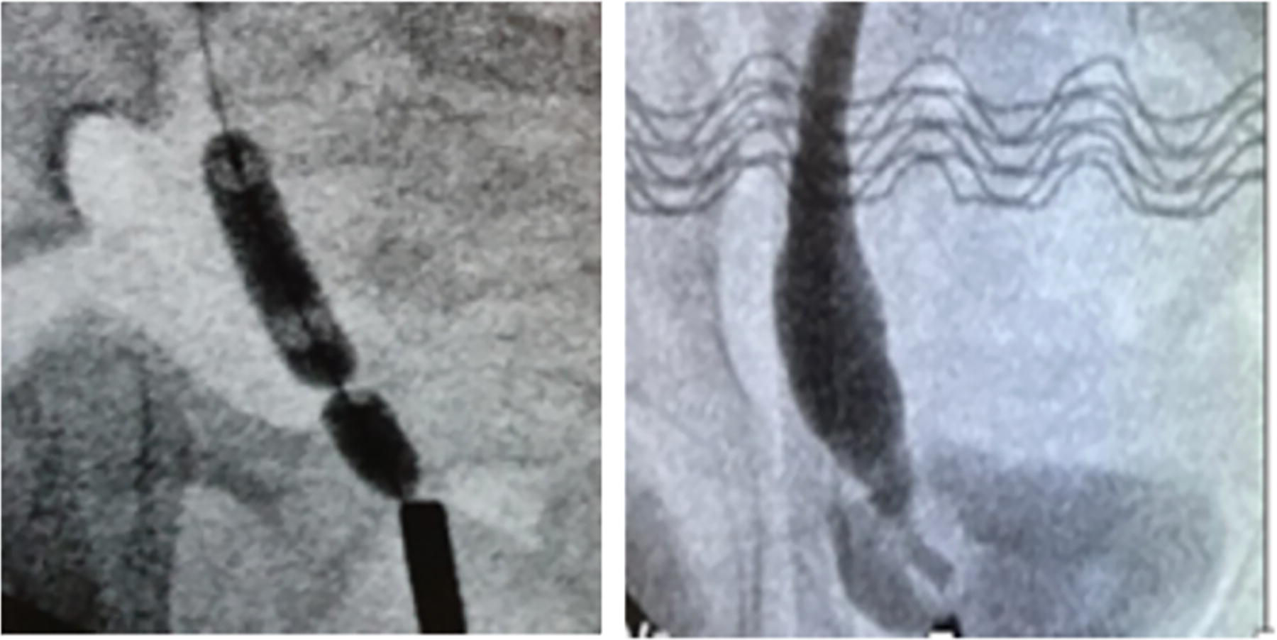
Ureteral obstruction. Narrow ring vs Narrow distal ureter.
The 15 ATM pressure was maintained for 5 minutes (min.).
A slight outward pressure was maintained on the balloon to prevent the balloon from moving into the ureter beyond the obstruction. After 5 min., another fluoroscopic image was taken to evaluate the evolution of the narrowing.
The balloon was removed together with the cystoscope.
Next, an 18” guidewire was inserted, followed by a single 4.7Fr multilength (8/20) Double-J (DJ) stent (Cook). This stent was positioned across the vesical-ureteral junction (VUJ) for a minimum of 6 weeks. No attempt was undertaken to place the DJ stent in the proximal ureter or kidney.
After 6 weeks, the DJ stent was removed cystoscopically with a short sedation. In our initial cases, a second dilatation was performed following the same protocol after 6 weeks at the time of stent retrieval. If the balloon could not be inserted, a more invasive procedure (e.g., reimplantation or ureterostomy) was performed during the same anesthesia.
Outcome Assessment
Distal ureteric diameter (DUD), DRF, and AP diameter were registered. These outcome parameters were registered both before and 6 months (M) after the procedure. Parameters were recorded following a standardized bolus drink schedule 30 min. before research. Surgical complications such as postop infections and stent migration were noted.
All cases were followed up beyond the initial 6-M period. Success was defined as improvement of the indication for therapy and no need for any further intervention. This longer follow-up allowed to assess long-term outcome, as no need for reintervention.
Statistics
Descriptive statistics were performed to get an overview of patient characteristics and results. Normally divided before–after were analyzed with a paired t test. DRF data were not normally divided, and here was a Wilcoxon-signed ranked test performed.
Results
Subjects
Thirty-one dilated ureters of 28 patients (23/5 M/F) were included with a median age of 9 M (range: 1–111 m) and a follow-up of 41 m (range: 12–84 m).
In 19 patients (67.9%), the POM was located on the left side; in 6 patients (21.4%), on the right side; and 3 patients (10.7%) had a bilateral POM. The reasons for intervention in these 28 cases are summarized in Table 1.
Cases Included

Symptomatic: 5 recurrent infections, 2 stones, and 2 stones + infection.
DRF = differential renal function; PUJ = pyelo-ureteral junction; VUJ = vesico-ureteral junction.
Feasibility
In all cases, we were able to insert a guidewire and dilate the balloon.
Afterwards a 4.7 DJ stent was placed in 28 ureters; for the other 3 ureters, a 3.7 DJ stent was placed for an average of 8 weeks.
The overall mean surgical procedure time was 88 min. (standard deviation [SD] 51min.). Divided into groups the unilateral surgical procedure was performed in a mean time of 76 min. (SD 33 min.), bilateral intervention 96 min. (SD 7 min.: SEM 5 min.) and the more difficult pyelo-ureteral junction/vesical-ureteral junction (PUJ/VUJ) disorders 164 min. (SD 109 min.).
Outcome
Radiological presentation of the dilated balloon showed a narrow ring in 29 ureters (93.5%) and a longer narrow distal ureter in 2 ureters (6%). The diameter of the distal ureter improved in 96% of the ureters at 6 M postop.
Patients were divided into groups, based on their indication for therapy (symptomatic, declining DRF <40%, and increasing dilatation >1.4 cm). The outcome parameters (DUD, AP diameter and DRF-affected kidney) for each indication were compared before and after EBD within a follow-up period of 6 M. The results are summarized and shown in Figures 2–5.

AP-diameter: preop vs postop by group. X-as: blue before EBD, red 6 months after EBD; Y-as: AP-diameter in millimeters. AP = anterior-posterior; EBD = endoscopic balloon dilatation.
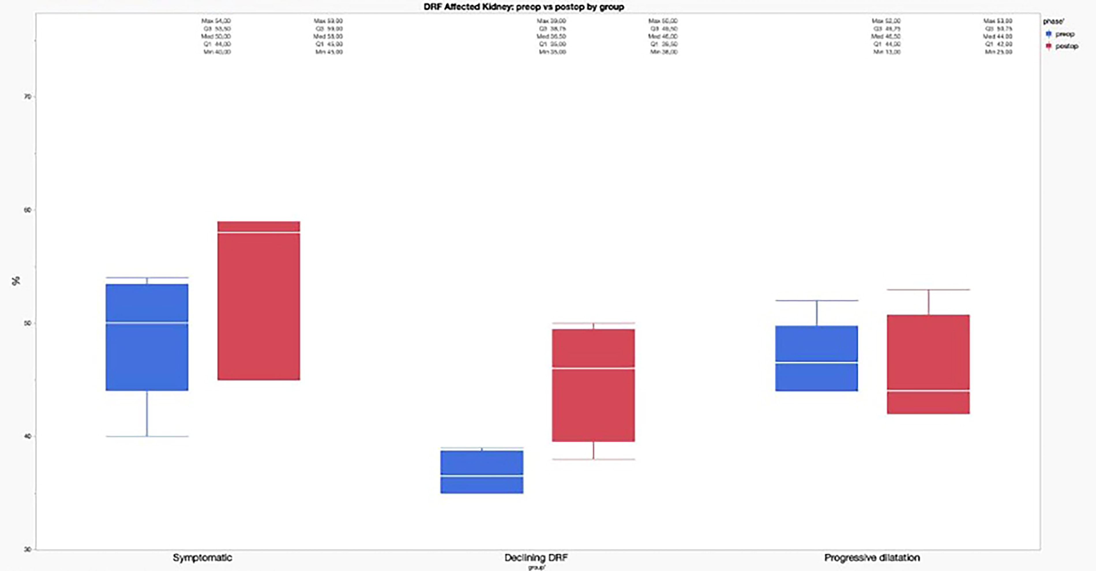
DRF Affected Kidney: preop vs postop by group. X-as: blue before EBD, red 6 months after EBD; Y-as: DRF in %. DRF = differential renal function; EBD = endoscopic balloon dilatation.
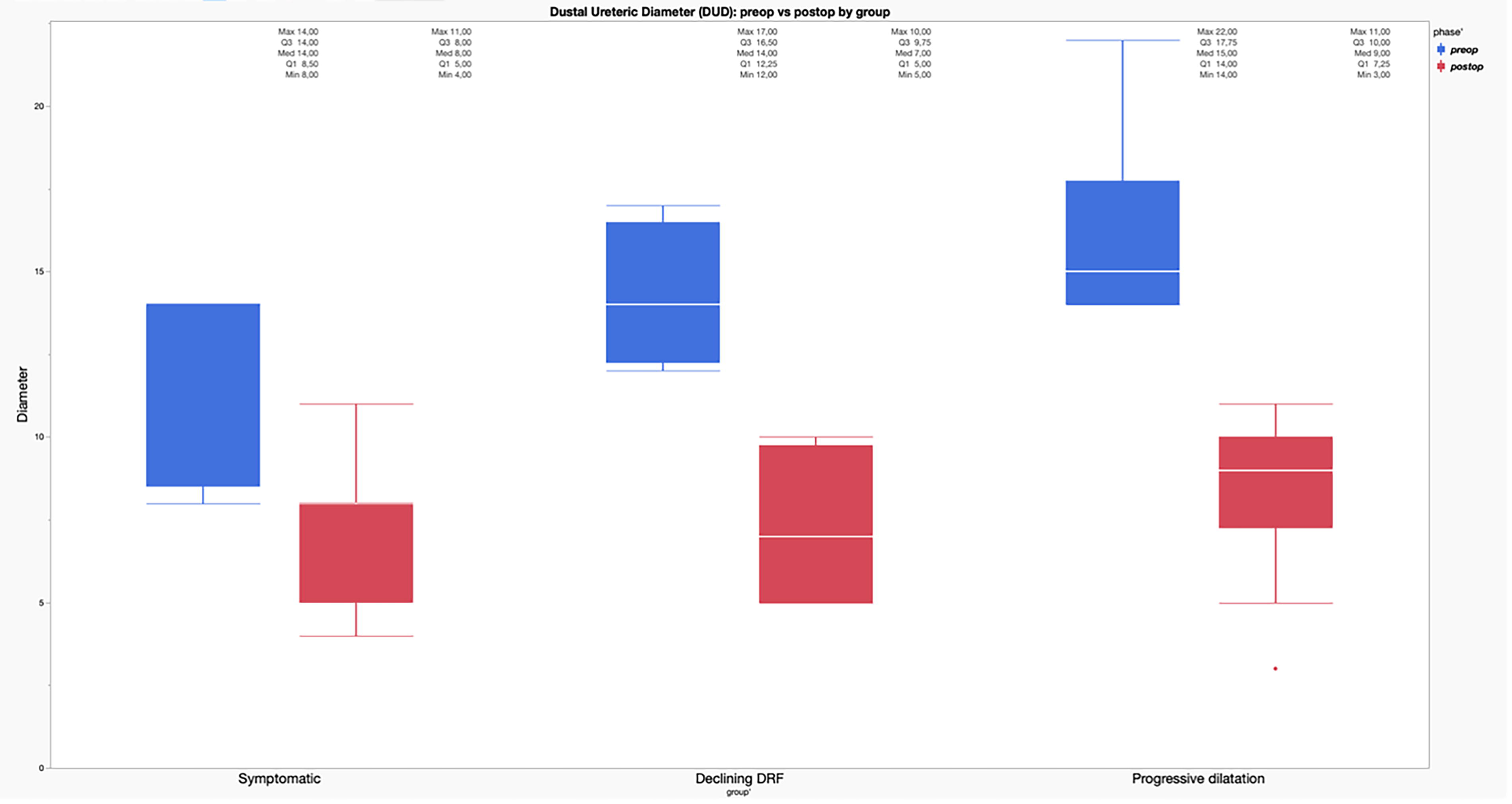
Distal Ureteral Diameter (DUD): preop vs postop by group. X-as: blue before EBD, red 6 months after EBD; Y-as: DUD in millimeters. EBD = endoscopic balloon dilatation.
There were 9 symptomatic cases, which included 11 ureters. In this category the mean DUD improved (p < 0.0001) from before EBD 11.4 (SD 3.1) mm to after EBD 5.6 (SD 3.5) mm, and the AP-diameter changed (p = 0.4702) from a mean of 9 (SD 3.4) mm to 7 (SD 4.4) mm. The DRF median had no significant change (p = 0.3750), from 50% (44%; 53%) to 58% (45%; 59%).
For the indication of declining DRF <40% (n = 5), a median improvement of DRF (p = 0.0313) from 36.5% (35%; 38.75%) to 46% (39.5%; 49.5%) was seen. Furthermore, other parameters showed a mean improvement as well: (p = 0.0121) DUD 14.4 mm (SD 1.9) to 8 mm (SD 2.8) and AP-diameter improved (p = 0.0256) from 23.5 mm (SD 7.1) to 12.3 mm (SD 5.9).
The indication of increasing dilatation >1.4 cm counted 11 cases with 12 ureters. The mean DUD improved (p < 0.0001) from 16.5 mm (SD 2.8) to 8.1 mm (SD 2.2) and the AP-diameter (p = 0.0168) from 22.1 mm (SD 5.1) to 14.3 mm (SD 4.4). There was no significant change (p = 0.7227) for the median DRF from 46.5% (44%; 49.75) to 44% (42%; 50.75%).
Independent of indication, we saw an overall improvement, as shown in Figure 6, of means as followed: DUD (p < 0.0001) 13.4 mm (SD 4.2) to 6.4 mm (SD 3.6), AP-diameter (p < 0.01) 18.8 mm (SD 7.9) to 11.5 mm (SD 5.5). The parameter DRF had no significant median improvement (p = 0.3750) 46% (39%; 50%) to 45% (42.5%; 52%).
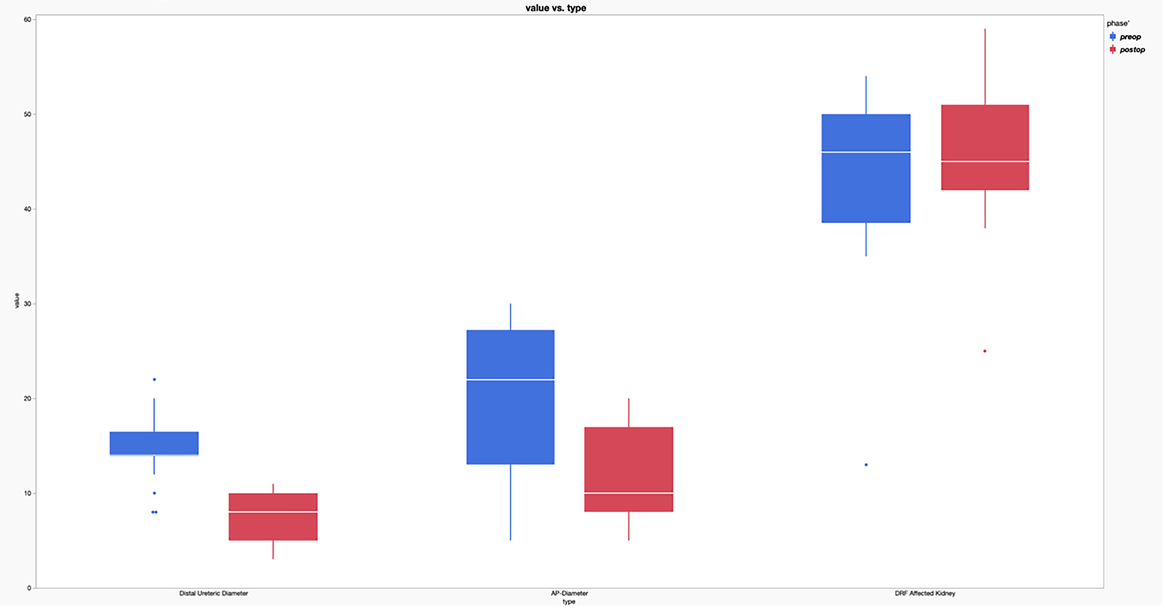
Evolution of each parameter preop vs postop. X-as: blue before EBD, red 6 months after EBD; Y-as: Value parameter (distal ureter diameter; anteroposterior-diameter; differential renal function). EBD = endoscopic balloon dilatation.
The indication of PUJ+VUJ disorder consisted of patients that initially were counseled for a pyeloplasty. During the procedure, there was a failure of the stent passing through the VUJ. In consequence, we changed intervention to EBD. No detailed separate analysis was performed for these cases.
Complications
Following the procedure, three patients (10%) developed n UTI with the DJ stent in situ. Two other patients (6%) required a uretero-renoscopic stent retrieval. One of these had a completely calcified DJ stent, which led to the need for a nephrostomy.
Beyond initial evaluation at 6 M, we further monitored children for the necessity of reintervention or appearance of possible complications.
Failures or need for further intervention
In total, 31 dilated ureters were treated with EBD. A second dilatation was performed in the first eight cases, but no difference was seen between those who received one or two dilatations. In consequence, we left the standard second dilatation. During further follow-up, two cases needed a more invasive intervention and are considered as failures. This is summarized in Figure 7.
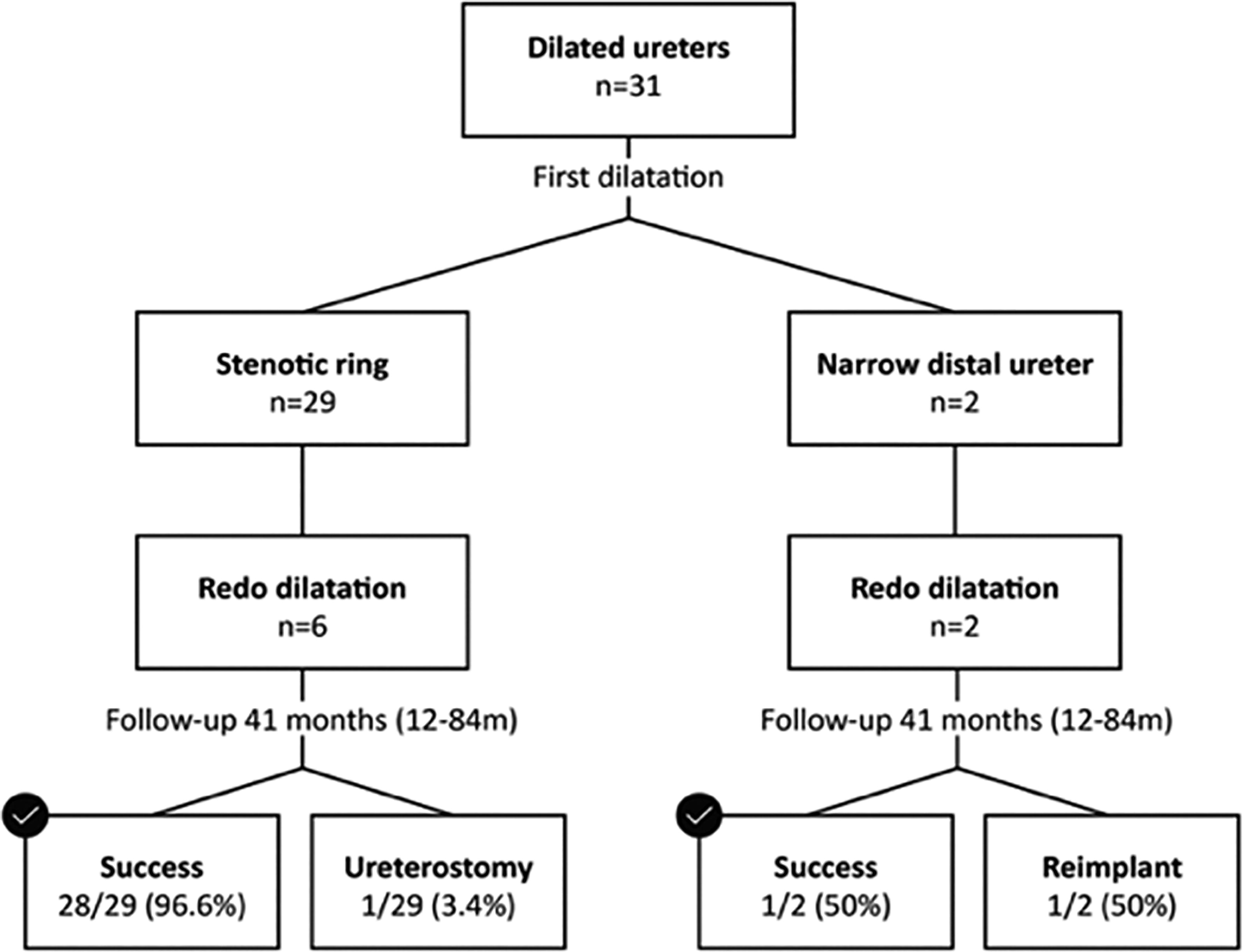
EBD intervention dependent of ureter obstruction. EBD = endoscopic balloon dilatation.
One failure originated from the symptomatic indication group. This case received an EBD of the right side because of recurrent infections and POM. However, this child also had a dysplastic contralateral kidney left kidney responsible of 14% of the total kidney function. Although after EBD the distal right ureteral diameter improved (from 10 mm to 4 mm) recurrent infections remained. He received a ureterostomy at the right side, after EBD, which was also insufficient to prevent his ongoing infections. A contralateral left nephrectomy (and reimplantation of the right ureter) was performed in the end.
The other failure was present in the increasing dilatation group. This boy was treated because of increasing dilatation above 14 mm and a DRF of 52%. A long, narrow distal ureter was identified during initial balloon dilatation. At 6 M an initial improvement of DUD was seen (14–9 mm) and DRF (increasing to 53%). In later, follow-up, 2 years after the initial EBD, hydroureteronephrosis increased, and a decrease of DRF to 40% was seen. This POM was considered a failure after EBD and needed reimplantation.
Radiation exposure
EBD therapy for each megaureter in groups (symptomatic, declining DRF <40%, and increasing dilatation >1.4 cm) had a mean radiation exposure of 0.058 mGym 2 (SD 0.031 mGym 2 ).
Discussion
This study used strict indications to include patients who required an intervention for a POM. We intervened by performing an EBD, which resulted in an improvement of the indication for treatment. Furthermore, the parameters DUD, AP-diameter and DRF showed an overall improvement 6 M after the EBD trial. The EBD intervention in 31 POMs had a success rate of 92.8%.
Therapeutic value
The success rate of EBD is reported to be dependent on the indication for therapy. Each indication for surgical procedure (symptomatic, increasing DUD, or decreasing DRF) clearly progressed in a positive way after EBD. This confirms the value of EBD in the treatment of POM in a structured way. Only for the indication of symptomatic patients, there was no significant change in the parameters DUD and DRF. Nevertheless, the values were pre-EBD close to normal. Nevertheless, the occurrence of symptoms disappeared after EBD.
The indication of increasing dilatation showed a good response on EBD for parameters DUD and AP-diameter, but no real change in DRF value was found. If the ureter is compliant and dilates without affecting renal function, then how long can we tolerate the situation without intervention. This requires a balance between a follow-up with mag three scans or an EBD trial. Further research through an RCT trial is warranted to make conclusions about this topic.
Skott et al. 10 published a systematic review that summarizes the outcomes of multiple studies9,11–14,16,17 describing the effect of EBD, concluding a 35%–97% success rate. This wide variety of a success rate can possibly be narrowed by adding a parameter, the length of the narrowing causing a POM. We concluded a clear difference of success rate depending on this length, 96.6% in narrow segments and 50% in longer narrow distal ureters.
Diagnostic value
Our study also stressed the diagnostic value of EBD. The use of a sufficiently long balloon inflated with contrast is a very important factor to appropriately assess the VUJ. Imaging can then show if there is an obstruction, as well as the kind of obstruction. According to results of the embryological study of Tanagho et al., 18 a narrow ring is typically present in the distal part of the ureter. A balloon that spans most of this distal part is essential for a correct evaluation so that no obstructive sites are missed. The study results implicate a decreasing success rate of EBD with an increasing length of stenosis. As there was a success rate of 96.6% with narrow rings, compared to a success rate of 50% in longer narrow distal ureters.
This finding was supported by a study of Chiarenza, S. et al., 13 where the patients were divided based on the length of the obstruction (<5 mm, 5–10 mm, >10 mm). The increasing length of obstruction also resulted in a decreasing success rate. The groups success rate after (single or multiple) EBD trials were: <5 mm was 100%, 5–10 mm was 92%, and >10 mm was where they had no success. Another important article by Mele, E. et al., 19 found a similar conclusion for EBD therapy. They describe an absence of a ring during inflation of the balloon as a significant risk factor for failure, with an odds ratio of 117.86% (6.27–2215.84), p = 0.0014. These findings support our suspicion that the length of stenosis/narrowing is an essential diagnostic parameter in POM.
Failures
The first failure, part of the narrow ring group, had an initial improvement after EBD. During follow up, the situation worsened, and further intervention was required. Recurrent infections only disappeared after a nephrectomy of the contralateral dysplastic kidney. As these ongoing symptoms where because of the dysplastic contralateral kidney rather than the POM, we can argue if this was a true failure of EBD. Excluding this patient results in a 100% success rate of EBD in the narrow ring group.
The second failure, a case with a longer narrow distal ureter, was a true failing of EBD with worsening parameters after 2 years of follow-up. Therefore, concluding a 50% failure rate of EBD in narrow distal ureters.
The evidence shows that narrow rings respond well to EBD therapy. Therefore, we recommend using EBD as a diagnostic tool to assess the underlying cause of obstruction. If a narrow ring is detected, dilation can be performed accordingly. However, if a longer distal ureteric narrowing is seen while dilatating, structured regular follow-up or immediate more invasive therapy can be suggested.
Conclusion
As most POM resolve spontaneously, strict indications are needed before treatment is offered.
Strong indications are symptomatic patients (e.g., infections) and declining DRF with dilated ureters. In those cases, EBD with a coronary dilatation balloon showed high success rates and provided diagnostic information about the length of the obstruction. Longer narrow ureteral segments are rare but result in a higher failure rate. Therefore, EBD can also predict the success rate and be used as a therapeutic and decision-making tool for whether to go for a more invasive corrective procedure, even during the same anesthesia.
Authors’ Contributions
J.M.: Writing. QB.: Data curation (equal). K.V.H.: Data curation (equal). D.T.: Data curation (equal). S.H.: Software and formal analysis. K.D.B.: Investigation and data curation (equal). G.D.W.: Conceptualization, supervision, and project administration. The undersigned declare that this article is original, has not been published before and is not currently being considered for publication elsewhere. They confirm that the article has been read and approved by all named authors. Signed by all authors as follows: J.M., Q.B., K.V.H., D.T., S.H., K.D.B., and G.D.W.
Footnotes
Author Disclosure Statement
No competing financial interests exist.
Funding Information
No funding was received for this article.