



Abstract
Objective:
To compare treatment plans created using the Eclipse and Prowess treatment planning systems for patients diagnosed with high-grade brain tumors in the left temporal lobe and to evaluate target volume coverage and doses delivered to critical organs using intensity-modulated radiotherapy (IMRT) technique.
Materials and Methods:
This retrospective comparative planning study included 15 patients. Plans were created on CT images using step-and-shoot IMRT with 6 MV photons. Eclipse used the Pencil Beam Convolution (PBC) algorithm, while Prowess used the Collapsed Cone Convolution (CCCS) algorithm. A total dose of 60 Gy in 30 fractions was prescribed. Dosimetric parameters were analyzed statistically.
Results:
Both TPSs achieved comparable target coverage, with no significant differences in conformity and homogeneity indices (p = 0.075 and p = 0.590, respectively) or dose–volume parameters (D95, D98, D2; p > 0.05). Eclipse provided significantly lower doses to the ipsilateral lens, brainstem, left optic nerve, cochlea, and whole brain V40 (p < 0.05).
Conclusion:
Both TPSs demonstrated adequate target coverage; however, Eclipse achieved statistically significant dose reductions in several critical structures, which may have clinical relevance in reducing toxicity risk.
Introduction
Glioblastoma (GBM) is the most common brain tumor in adults. 1 GBMs account for 12–15% of intracranial tumors and 60–75% of astrocytic tumors. GBMs represent approximately 13.9% of all brain tumors and 51.5% of malignant brain tumors. Although cases can occur in all age groups, they predominantly arise in adults over 40 years of age, with a median age at diagnosis around 66 years. 2 Despite technological advances, the prognosis remains poor; however, multimodal treatment including surgical resection, radiotherapy, and temozolomide has extended median survival to approximately 15–18 months.2–4
These limited survival outcomes necessitate the development of treatment planning systems (TPS) and their more effective use in clinical practice. In radiotherapy, TPS are software programs used to display the planned dose distribution on the patient or phantom in order to control the target volume and better protect normal tissues. The dose algorithms in treatment planning system software aim to ensure tumor control by calculating the energy of the radiation or electrons used in treatment, the width and depth of the target area, and the dose calculations. As a result of these calculations, the dose distribution for the target volume and normal tissues and the doses these tissues will receive are simulated with TPS.5,6 One of these TPSs, the Eclipse treatment planning system, can calculate dose distribution for different types of radiation, such as photons, electrons, and protons. Eclipse is a treatment planning system with many different algorithms used in radiotherapy planning. These algorithms in Eclipse are most commonly used for dose calculations. Algorithms such as Acuros, Anisotropic Analytic Algorithm (AAA), Cone Dose Calculation Algorithm (CDCA), and Pencil Beam Convolution (PBC) are used to calculate dose distribution in photon therapy.7–9 Another TPS, the Prowess treatment planning system, uses two different algorithms for conformal and intensity-modulated radiotherapy (IMRT) treatment planning. The dose calculation algorithms are “fast photon with or without effective path” for conformal therapy and “collapsed cone convolution superposition with or without heterogeneity (CCCS)”, which calculates the dose based on full heterogeneity correction and is a full three-dimensional dose calculation, for IMRT. 10 The extent to which these different algorithms produce clinically meaningful results can be determined through comparative evaluations. Comparing different TPSs is important in determining whether the differences in dose calculation between algorithms are clinically significant. Such comparisons help clinicians determine which system provides more reliable organ protection.6,11
Technological advances in radiotherapy, along with the development of new treatment devices and treatment planning systems, necessitate studies on the use of these advances in treatment. Previous studies have compared different treatment planning systems such as Eclipse, Prowess, Tomotherapy, KonRad, XiO, and Brainscan, mainly focusing on dose distribution, target coverage, and organ-at-risk protection. These studies generally reported comparable target coverage among systems, with some differences in conformity and organ dose sparing. However, the number of studies that directly compare Eclipse and Prowess systems for brain tumors remains quite limited.In the study, plans created with different treatment planning systems belonging to the same linear accelerator were examined; the clinical significance of the differences between the systems was revealed by comparing target volume and critical organ doses.
Materials and methods
Fifteen patients were included in the study. Inclusion criteria for the study were: patients over 18 years of age, who had undergone surgery for tumors located in the left hemisphere and had WHO grade 3 or 4 brain tumor pathology, who had pre- and post-operative brain MRIs, and who had received curative radiotherapy and concomitant temazolamide chemotherapy were included in this study. Patients diagnosed only by biopsy, those who did not receive their full radiotherapy and/or chemotherapy, those without an MRI, and those who had previously received radiotherapy to the same area for a different reason were excluded from the study. Brain tomography images of patients taken at 3 mm slice intervals using a computed tomography (Somatom Emotion model computed tomography) simulator device were transferred to treatment planning systems via the digital imaging and communications in medicine (DICOM) system. Subsequently, the target volume and critical organs were delineated and defined by the radiation oncologist. The clinical target volume (CTV) was generated by applying a 2 cm margin to the gross tumor volume (GTV) to account for microscopic tumor extension. The planned target volume (PTV) was then created by adding an additional 0.5 cm margin to the CTV to compensate for setup uncertainties and patient motion.
In each patient, the right and left lens, eye, optic nerve, cochlea, brain stem, chiasm, and entire brain were defined as critical organs. Taking into account the defined areas and critical organs, two separate treatment plans were created for each patient using the Eclipse Treatment Planning System (Eclipse V8.9.08 TPS, Varian Medical Systems Inc., Palo Alto, CA, USA) and the Prowess Panther Treatment Planning System (Prowess Panther V5.01 TPS, Prowess Inc., USA). In the Eclipse TPS, dose calculations were performed using the PBC algorithm, whereas in the Prowess Panther TPS, the Collapsed Cone Convolution/Superposition (CCCS) algorithm was utilized. The PBC algorithm was selected because it was routinely used in clinical practice at our institution during the study period. Treatment plans were created using the IMRT technique with 6 MV photon energy. The treatment was calculated to deliver a total dose of 60 Gy in 30 fractions, with 2 Gy per daily fraction. Treatment plans were created using the ‘Step and Shoot’ IMRT technique. In the planning process, the geometric and dosimetric parameters of a linear accelerator (Linac) equipped with an 82 leaf MLC system and 1 cm thick leaves were used.The treatment plan was designed so that 95% of the dose would cover at least 95% of the PTV.
All treatment plans were generated using the nine-field IMRT technique with fixed gantry angles of 18°, 45°, 80°, 99°, 137°, 188°, 239°, 269°, and 332°. To ensure a consistent and unbiased dosimetric comparison, the same beam geometry was applied in both treatment planning systems. The treatment plans were optimized independently in each system while maintaining the same planning targets and dose constraints. Dose calculations were performed using a 0.25 cm grid size in both systems.
Organ-at-risk dose constraints were determined according to QUANTEC guidelines and the RTOG 0225 protocol. For the brain, the V40 < 30% and V20 < 40% limits were evaluated based on the healthy brain volume (brain minus PTV). The maximum dose for the brainstem and optic chiasm was set at 54 Gy; additionally, the criterion that the volume receiving 60 Gy should not exceed 1% of the total volume was applied in accordance with the RTOG 0225 protocol. For the optic nerves, a maximum dose of 54–55 Gy; for the lens, 10 Gy; for the eyeball, an average dose of 35 Gy; and for the cochlea, an average dose of 45 Gy were considered.12–14 Although the QUANTEC guidelines do not specify a specific dose limit for the pituitary gland, the maximum tolerance value of 45 Gy proposed by Emami et al. in the literature was used as a reference. 15
The quality of treatment plans is assessed by analysing the homogeneity of the dose distribution and its conformity to the target volume using various indices. The Homogeneity Index (HI) and Conformity Index (CI) are among the most commonly used parameters for evaluating the suitability of radiotherapy plans in terms of geometry and dose distribution.
According to RTOG, HI is defined as the ratio of the maximum dose in the target volume to the prescribed dose value:

Imax: maximum isodose,
RI: reference isodose
This formula is used to evaluate the homogeneity of dose distribution in conventional fractionated radiotherapy plans. An HI value between 1.0 and 1.2 is considered ideal for acceptable dose homogeneity. The closer the HI value is to 1, the more homogeneous the dose distribution is considered to be. 16
According to RTOG, CI is defined as the ratio of the volume covered by the reference isodose line to the target volume (PTV):

VRI: Volume of the reference isodose
TV: Target volume (PTV)
According to RTOG, a CI value between 1 and 2 is acceptable. This formula indicates how well the applied dose conforms to the target volume. The closer the CI value is to 1, the more conformally the dose distribution is considered to have been applied to the target volume. 17
In this study, the CI and HI were calculated as part of the quality assessment of radiotherapy plans to examine the conformity and homogeneity of the dose distribution. The range of 1–2 for CI and 1.0–1.2 for HI were considered acceptable limits in the literature, and the obtained values were statistically analysed.
Based on the obtained data, the PTV doses were comparatively evaluated with the doses to the right and left lens, brainstem, chiasm, pituitary gland, whole brain, right and left eye, right and left optic nerve, and right and left cochlea. The minimum, average, and maximum dose values were calculated for each structure, and the levels of statistical significance were determined.
The statistical software IBM SPSS Statistics for Windows, Version 25.0 (IBM Corp., Armonk, NY, USA) was used to analyze the data. Descriptive statistics (mean, standard deviation, median, first and third quartiles) were calculated for categorical and continuous variables. Homogeneity of variance, a prerequisite for parametric tests, was assessed using the Levene test, while the assumption of normality was assessed using the Shapiro-Wilk test. To evaluate differences between two dependent measures, the paired t-test was used when parametric test conditions were met, and the Wilcoxon signed-rank test was used when they were not. The level of statistical significance was set at p < 0.05.
Findings
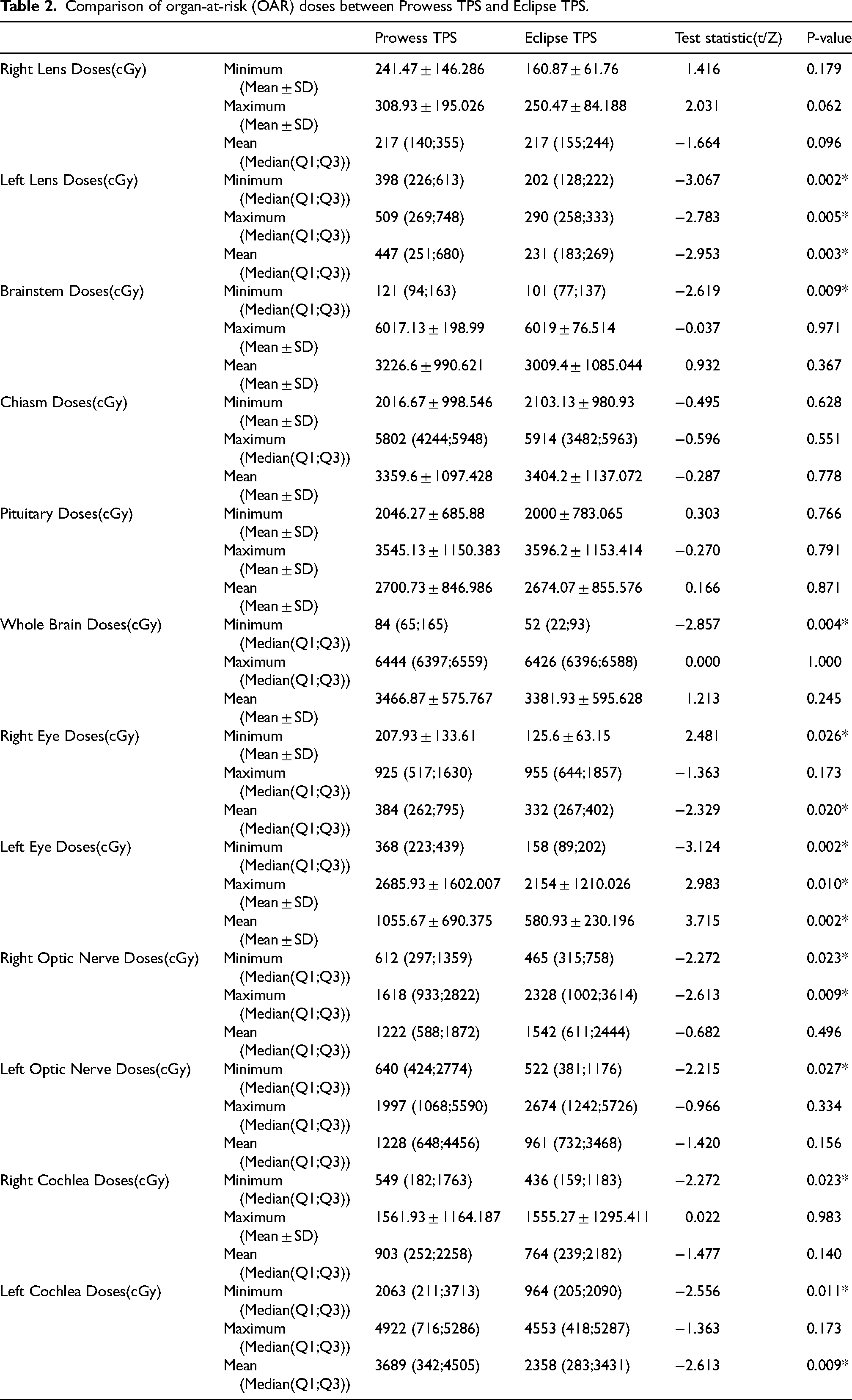
Radiotherapy plans were generated and dosimetrically evaluated for 15 patients diagnosed with high-grade brain tumors using the PBC algorithm in Eclipse TPS and the CCCS algorithm in Prowess TPS. The resulting dose-volume histograms (DVHs) for the PTV and all critical organs are presented in Figure 1. Additionally, DVH measurements are shown in Table 1 for the PTV, in Table 2 for critical organs, and in Table 3 for healthy brain tissue.
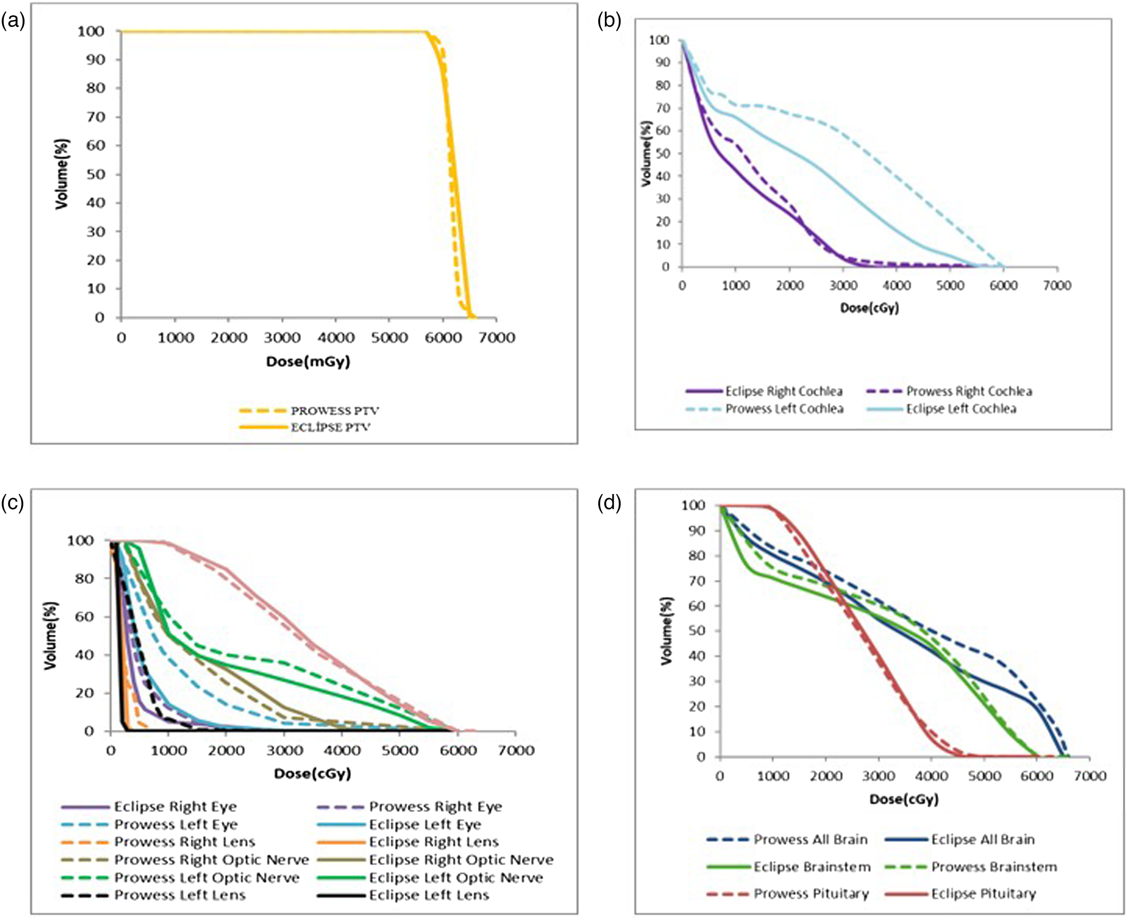
Dose–Volume Histogram (DVH) comparisons between Eclipse and Prowess treatment planning systems. (a) Target volume coverage (PTV); (b) cochlea dose distributions; (c) ocular structures including eyes, lenses, and optic nerves; (d) whole brain, brainstem, and pituitary gland.
Dosimetric parameters of the planning target volume (PTV) in Prowess TPS and Eclipse TPS.

Comparison of organ-at-risk (OAR) doses between Prowess TPS and Eclipse TPS.
DVH parameters for healthy brain tissue in Prowess TPS and Eclipse TPS.
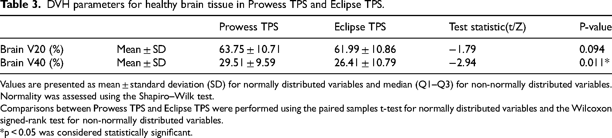
Values are presented as mean ± standard deviation (SD) for normally distributed variables and median (Q1–Q3) for non-normally distributed variables.
Normality was assessed using the Shapiro–Wilk test.
Comparisons between Prowess TPS and Eclipse TPS were performed using the paired samples t-test for normally distributed variables and the Wilcoxon signed-rank test for non-normally distributed variables.
*p < 0.05 was considered statistically significant.
The reference dose used in this study was 60 Gy, and the obtained HI values (Prowess HI: 1.13, Eclipse HI: 1.13) were evaluated as protocol-compliant according to RTOG criteria. The obtained CI values (Prowess CI: 1.25, Eclipse CI: 1.22) indicate that the dose distribution was applied appropriately to the target volume and is acceptable according to RTOG criteria. CI and HI index values are shown in Table 1. Upon examination of the homogeneity index and conformity index values, no significant difference was observed between the two planning systems (HI: p = 0.590, CI: p = 0.075).
Statistical analysis of the data obtained from the study revealed that the minimum and average doses applied to the target volume were lower in the Eclipse TPS compared to the Prowess TPS, but no significant difference was observed in maximum doses (minimum: p = 0.041, average: p = 0.027, maximum: p = 0.320). Upon comprehensive examination of the target volume dosimetric parameters, no statistically significant difference was detected between the Eclipse TPS and Prowess TPS groups in terms of D95, D98, and D2 values (D95: p = 0.321, D98: p = 0.332, D2: p = 0.590). These findings indicate that both planning systems produced plans with similar efficacy in terms of target volume coverage and dose distribution.
When critical organ doses were examined, no statistically significant difference was found between the groups regarding the minimum, average, and maximum doses of the right lens located on the opposite side of the target volume (minimum: p = 0.179, average: p = 0.062, maximum: p = 0.096). In contrast, for the left lens located on the same side, the minimum, average, and maximum doses were found to be higher in the results obtained with the Prowess TPS compared to those obtained with the Eclipse TPS (minimum: p = 0.002, average: p = 0.005, maximum: p = 0.003).
When examining brainstem, right cochlea, left optic nerve, and whole-brain doses, it was found that minimum dose values were lower in plans calculated with the PBC algorithm in Eclipse compared to those calculated with the CCCS algorithm in Prowess (brainstem: p = 0.009, right cochlea: p = 0.023, left optic nerve: p = 0.027, whole brain: p = 0.004). In contrast, no statistically significant difference was found between the groups in terms of maximum and average doses (brainstem: maximum p = 0.971, average p = 0.367; right cochlea: maximum p = 0.983, average p = 0.140; left optic nerve: maximum p = 0.334, average p = 0.156; whole brain: maximum p = 1.000, average p = 0.245). Although the V20 parameter, which indicates low-dose exposure of healthy brain tissue, was found to be lower on average in plans calculated with the PBC algorithm in Eclipse, no statistically significant difference was detected between the two planning systems (p = 0.094). The V40 parameter, indicating moderate dose exposure to healthy brain tissue, was found to be significantly lower in Eclipse plans (p = 0.011).
When pituitary doses were examined, no statistically significant difference was found between the minimum, average, and maximum doses obtained with Eclipse TPS and Prowess TPS (minimum p = 0.303, average p = 0.871, maximum p = 0.791).
When examining organ doses to the right optic nerve, plans calculated with the PBC algorithm in Eclipse yielded significantly lower values than Prowess TPS in terms of minimum dose; however, no significant difference was observed between the two systems in terms of average dose. Regarding maximum dose values, it was found that the Prowess TPS provided significantly lower doses compared to the Eclipse TPS (minimum p = 0.023, mean p = 0.496, maximum p = 0.009).
No difference was observed between the two treatment planning systems regarding the maximum doses applied to the left cochlea and the right eye, however, the values obtained from plans calculated with the CCCS algorithm in Prowess were higher than those calculated with the PBC algorithm in Eclipse in terms of minimum and average doses (left cochlea: minimum p = 0.011, maximum p = 0.173, average p = 0.009; right eye: minimum p = 0.026, maximum p = 0.173, average p = 0.020).
It was found that the minimum, maximum, and average doses for the left eye were lower in Eclipse compared to Prowess, and that the plans calculated with the PBC algorithm in Eclipse were closer to the ideal plan (minimum p = 0.002, maximum p = 0.010, average p = 0.002).
Discussion
The treatment planning system is an important part of radiotherapy. 18 The optimal dose distribution for the target defined in treatment should be achieved through accurate planning. 19 Treatment planning systems have various and complex dose calculation algorithms to accurately calculate the dose with the IMRT treatment technique. The clinical functionality of the treatment plan and the accuracy of dose calculations can be evaluated by comparing different TPSs. 20
Onay and colleagues compared craniospinal IMRT treatment plans created in Prowess TPS, Eclipse TPS, and Tomotherapy planning systems. In this study, dose distributions within the PTV were examined, and no significant difference was observed in homogeneity index values; however, it was observed that the PTV dose distribution was more homogeneous in plans created with Prowess TPS. In the same study, when the conformity index values were compared, it was noted that treatment plans made with the Tomotherapy planning system were better. On the other hand, since the maximum doses to critical organs were lower in Tomotherapy TPS, Tomotherapy was seen to be more advantageous compared to the other two TPS. 21
Eldesoky and colleagues evaluated the treatment plans of three different IMRT, TPS, namely KonRad TPS, XiO TPS, and Prowess treatment planning systems, for 11 different types of pediatric brain and head and neck cancer cases. In this study, it was observed that the target volume coverage was similar for all three TPS, but the critical organ doses, as seen in the dose volume histograms for the three TPS, were different from each other.This study found that all three treatment planning systems were technically successful in managing clinical target conditions according to internationally approved standards and were valid in terms of application principles in pediatric practice. However, it was noted that KonRad and Prowess TPS were more suitable for treatment planning than XiO TPS due to certain disadvantages of the latter. 20
In the study conducted by Petric and colleagues, treatment plans were created in the Brainscan and Eclipse treatment planning systems using the IMRT technique with 6 MV energy photons.
This study aimed to compare the clinical functionality of the treatment planning systems and to evaluate the clinical adequacy of the dose calculation accuracy of both TPSs. As a result, it was stated that both TPSs were largely equivalent in terms of target coverage and healthy tissue protection, but that the Eclipse TPS was inadequate in accurately calculating the dose for highly modulated fields. 22
Although various studies in the literature have compared different treatment planning systems for brain tumors, studies directly comparing the Eclipse and Prowess treatment planning systems are limited. Therefore, the present study aims to contribute to the existing literature by evaluating the dosimetric differences between these two systems. Previous studies have generally reported that target volume coverage is similar across different treatment planning systems; however, differences in dose calculation algorithms and optimization strategies may lead to variations in the doses delivered to organs at risk. These findings are also consistent with the results obtained in our study.
In our study, the homogeneity index and conformity index values were found to be similar for both planning systems. Additionally, no statistically significant difference was detected between the two systems regarding PTV D95, D98, and D2 parameters. These results indicate that both TPSs demonstrate clinically adequate performance in terms of target volume coverage and dose homogeneity.
When critical organ doses were evaluated, statistically significant differences were observed between the two planning systems in some structures. Upon examining lens doses, it was observed that the minimum, maximum, and average dose values for the left lens were significantly lower in the plans calculated with the PBC algorithm inEclipse. Similarly, the fact that the minimum, maximum, and average dose values for the left eye were found to be lower in the PBC algorithm in Eclipse suggests that this system may offer a potential advantage in terms of protecting ocular structures. When brain stem doses were evaluated, the minimum dose value was found to be significantly lower in plans calculated with the PBC algorithm, while no significant difference was detected between the two systems regarding maximum and average doses. When all brain doses were examined, it was determined that the minimum dose value was significantly lower in plans calculated with the PBC algorithm in Eclipse.
A different trend was observed when the optic nerves were evaluated. It was determined that the maximum dose to the right optic nerve was significantly lower in plans calculated with CCCS in Prowess. However, no significant difference was found between the two systems in terms of average doses. When cochlear doses were examined, it was observed that the minimum and average dose values, particularly for the left cochlea, were significantly lower in plans calculated with the PBC algorithm in Eclipse.
When examining the V20 and V40 parameters, which indicate low and moderate dose exposure in healthy brain tissue, it was observed that V20 values did not show a statistically significant difference between the two planning systems. In contrast, the V40 parameter was found to be significantly lower in plans calculated with the PBC algorithm in Eclipse. The QUANTEC guidelines state that an increase in the high-dose volume of normal brain tissue may increase the risk of late-stage neurological toxicity. Therefore, minimizing the moderate and high-dose volumes as much as possible is clinically important. The fact that V40 values were found to be lower in plans calculated with the PBC algorithm in our study suggests that this algorithm may offer a potential advantage in reducing moderate dose exposure to healthy brain tissue. However, the absence of a statistically significant difference in the V20 parameter indicates that both planning systems demonstrate similar performance in terms of low-dose distribution. Based on our findings, both planning approaches provide clinically acceptable target coverage and dose homogeneity. However, in clinical scenarios where tumors are located adjacent to critical ocular structures or the brainstem, plans calculated with the PBC in Eclipse may offer advantages due to its lower dose delivery to these organs. Conversely, plans calculated with the CCCS in Prowess demonstrated lower maximum doses in certain structures such as the right optic nerve, which may be advantageous in specific anatomical situations. Therefore, the selection of treatment planning approach should consider algorithm characteristics, tumor location, and organ-at-risk priorities. Comparative evaluation of TPS performance under different clinical conditions can support individualized treatment planning and optimize patient outcomes. Overall, while plans calculated with the PBC algorithm were associated with lower dose values in some critical structures, plans calculated with the CCCS algorithm yielded more advantageous results in others. This situation may stem from differences in the optimization strategies and dose calculation algorithms used in the various treatment planning systems. The PBC algorithm used in the Eclipse system calculates photon scattering based on specific mathematical assumptions. The CCCS algorithm used in the Prowess system, however, employs a more advanced dose calculation approach based on a three-dimensional energy distribution model. Dose calculation algorithms may differ in terms of heterogeneity correction, scattering modeling, and methods for calculating dose distribution in modulated fields. Indeed, various studies have demonstrated that different photon dose calculation algorithms can yield different dose calculation results, particularly in heterogeneous environments and regions with high dose gradients. Fogliata and colleagues reported that different dose calculation algorithms can produce significant dose differences in heterogeneous environments. Similarly, a study by Knoos and colleagues demonstrated that different commercial TPS algorithms can generate varying dose distributions in highly modulated fields and regions near critical structures. Therefore, the emergence of algorithm-dependent dose differences is an expected occurrence, particularly in regions with high dose gradients near critical structures such as the optic nerves, optic chiasm, and brainstem.23,24 Therefore, in clinical situations where the protection of critical organs is a priority, it is important to carefully evaluate the dosimetric characteristics of planning systems.
From a clinical perspective, it cannot be definitively stated whether certain dose differences found to be statistically significant will result in a noticeable difference in toxicity regarding clinical outcomes. However, a lower maximum dose in serial organs could theoretically contribute to reducing the risk of late-term radiation toxicity.
In our study, the V60 < 1% criterion defined in the RTOG 0225 protocol for the brainstem and optic chiasm was met in all cases. However, it was observed that the criterion of keeping the maximum dose below 54 Gy could not be met in some cases. For brain tissue, the V20 values recommended in the QUANTEC guidelines were evaluated, and values exceeding the recommended limits were detected in some cases. This situation demonstrates that, as reported in the literature for high-grade glioma cases, exceeding volumetric dose constraints for normal brain tissue may be unavoidable due to the prioritization of wide safety margins and tumor coverage.12–14 In contrast, it was observed that the dose constraints defined in the QUANTEC guidelines for the lenses, optic nerves, eyes, and cochleae were met in all cases. Although the QUANTEC guidelines do not specify a specific dose limit for the pituitary gland, the maximum tolerance value of 45 Gy proposed by Emami and colleagues in the literature was used as a reference. In our study, the planned maximum dose for the pituitary remained below this limit; this indicates that pituitary protection was ensured during the planning process. 15
The most significant limitation of this study is the small sample size (n = 15). A small sample size may limit statistical power and reduce the generalizability of the results. Additionally, the study is limited to high-grade brain tumors located in the left hemisphere. Results may vary across different tumor locations and clinical scenarios. Furthermore, only specific algorithm versions and a single energy level were used. Studies conducted on larger patient cohorts and under different clinical conditions are important for validating the findings.
Moreover, although the PBC algorithm in Eclipse demonstrated lower doses in some critical structures, the CCCS algorithm used in Prowess may provide more accurate dose calculations in heterogeneous regions, which can offer advantages in certain clinical scenarios. Therefore, both systems have complementary strengths, and TPS selection should be tailored to the tumor location and patient-specific priorities. Both treatment planning systems achieved comparable target coverage. The observed differences in organ-at-risk doses are more likely related to the dose calculation algorithms rather than any inherent superiority of the systems themselves. Accordingly, the choice of treatment planning system should be based on clinical requirements, algorithm characteristics, and anatomical factors.
In conclusion, the dosimetric findings of this study are specific to the comparison between the PBC algorithm implemented in Eclipse TPS and the CCCS algorithm used in Prowess TPS. The observed differences should be interpreted as a consequence of variations in dose calculation approaches rather than inherent superiority of one system over another, and are not generalizable to all versions or configurations of these systems. Therefore, treatment planning system selection should be based on algorithm characteristics, tumor location, and clinical priorities related to organ-at-risk sparing.
Footnotes
Ethics approval
This retrospective study was approved by the Ethics Committee for Non-Interventional Research of Necmettin Erbakan University, Meram Faculty of Medicine (Approval No: 2020/2717, Date: 17 July 2020). Due to the retrospective design of the study,the requirement for informed consent was waived by the ethics committee.
Funding
The authors received no financial support for the research, authorship, and/or publication of this article.
Declaration of conflicting interests
The authors declared no potential conflicts of interest with respect to the research, authorship, and/or publication of this article.