



Abstract
Traumatic brain injury (TBI) leads to significant public health concerns due to cognitive decline and increased risks of neurological conditions like Alzheimer’s disease and chronic traumatic encephalopathy. Preclinical models are essential for exploring how mild TBI leads to neuronal dysfunction and neurodegeneration. Using a mouse model, we applied repetitive, mild, side-alternating impacts to induce rapid head rotational acceleration–deceleration. A novel odor-based learning and memory task was developed to address TBI-related vision impairments. Our findings revealed that this side-impact model specifically affects the hippocampus, evidenced by activated CD68+ microglia appearing in the dentate gyrus, stratum lacunosum-moleculare, and corpus callosum. Importantly, no olfactory dysfunction was observed. However, injured mice exhibited learning and memory deficits in an olfaction-based task. These results suggest that repetitive mild TBI damages hippocampal regions, leading to cognitive dysfunction characterized by impaired learning and memory, as demonstrated by this novel behavioral method.
Keywords
Introduction
Mild traumatic brain injury (mTBI) is estimated to comprise 75% of all traumatic brain injuries (TBIs), with a reported one in four adults and nearly 4% of children under the age of 17 having experienced a concussion.1–4 While the symptoms in patients with mTBI are typically transient, repetitive mild TBI (rmTBI) may be associated with long-term neurological impairment, including learning and memory deficits. 5 RmTBI also results in neuronal and glial morphological changes, imbalance in excitatory and inhibitory processes, white matter dysregulation, and altered expression of genes involved in apoptosis, stress response, metabolism, and synaptic plasticity.6–10 Clinical studies further show that rmTBI occasionally produces persistent cognitive and psychiatric impairments and may be associated with diseases such as Alzheimer’s and chronic traumatic encephalopathy (CTE). 11 Given the significant clinical prevalence of cognitive deficits following rmTBI, preclinical studies should employ appropriate behavioral tasks to better assess TBI-related behavioral outcomes and treatment. Selecting an appropriate behavioral task involves many considerations, including not only which regions are directly impacted by TBI but also their function and connectivity, such that connected regions may also be indirectly affected by mTBI through diffuse axonal injury.12,13
Visual impairment after TBI is common in clinical settings and various preclinical animal models of closed-head TBI, 14 with rotational acceleration–deceleration forces15,16 and without.17–19 However, most tasks used to assess learning and memory in mice after TBI are vision-dependent, such as the Morris water maze. Considering the visual system deficits observed in many rodent models of closed-head rmTBI 14 and the reliance of established animal behavioral models on vision, a behavioral test that uses alternative sensory systems will assist in accurate interpretation. 20
Unlike humans, who rely heavily on vision, mice use smell as their dominant sense. 21 Therefore, using an olfaction-based task for mice is potentially favorable to test learning and memory.22,23 Olfaction is a preferable sensory modality for memory-based behavior tests due to its pivotal role in memory storage and retrieval, distinct anatomical organization, and broad applicability across species and clinical applications, such as Alzheimer’s disease.21,24 Existing olfaction-based learning tests have been shown to be reliable and effective, with some olfaction-based tasks using scents such as a predator odor (PO) as a powerful fear stimulus to test an animal’s innate response to danger, detecting certain cognitive abnormalities 20 and functions of specific brain regions. 25
In this study, we identified specific brain regions impacted by rmTBI—mainly in the hippocampal molecular layer, lateral geniculate nucleus, optic tract, dorsal lateral geniculate nucleus, and superior colliculus—through an analysis of microglial activation. The selective vulnerability of these regions underscores the potential effects of rmTBI on visual pathways and cognitive function,14,26 questioning the validity of vision-based learning and memory tasks and necessitating a vision-independent behavioral test to examine memory and learning outcomes. We first evaluated the olfactory system in mice post-rmTBI using the olfactory habituation and discrimination task,27,28 confirming it remained intact. With this confirmation, we developed the olfaction-based learning and memory (OBLM) test, a novel assay that assesses learning and memory independently of vision.
Materials and Methods
The described studies were carried out according to the protocols outlined by the Boston Children’s Hospital Institutional Animal Care and Use Committee. Each study complied with the National Institutes of Health (NIH) Guide for the Care and Use of Laboratory Animals.
For all experiments, adult C57BL/6J male mice (Jackson Laboratories, Bar Harbor, ME), aged 8–9 weeks and weighing 23–26 g, were housed in a reverse-light room (inverse 12-h light/dark cycle) with ad libitum access to food and water. Animals were allowed to acclimate for 3–4 days before experimentation. Supplies used for this study are listed in Table 1.
List of Materials

Repetitive mild closed-head TBI
The closed-head weight drop method combined with rotational acceleration–deceleration forces was used as described in previous studies16,29 (Fig. 1A). Animals were anesthetized using 4% isoflurane in 100% oxygen for 45 sec and positioned on a delicate tissue (Kimwipe, Irving, TX). A 54 g metal bolt was dropped from a height of 127 cm (50 in.), targeting the area between the coronal and lambdoid sutures, resulting in a lateral impact (Fig. 1B). Animal recovery was monitored until they could self-right. The time between removal from anesthesia to the animal’s self-right, loss of consciousness, was recorded (Fig. 1C). We tested a range of injury frequencies and found that administering side-alternating impacts more than four times from a 50-in. height (using a weight-drop device) produced a consistent level of injury, as indicated by comparable numbers of astrocytes and microglia counts in the hippocampus. Specifically, there was no statistically significant difference in injury amount. Based on these findings, we decided to combine both injury models. Notably, even in the 10-hit model, olfactory function was not significantly different from sham controls, suggesting that olfaction is unlikely to be affected in the six-hit model.
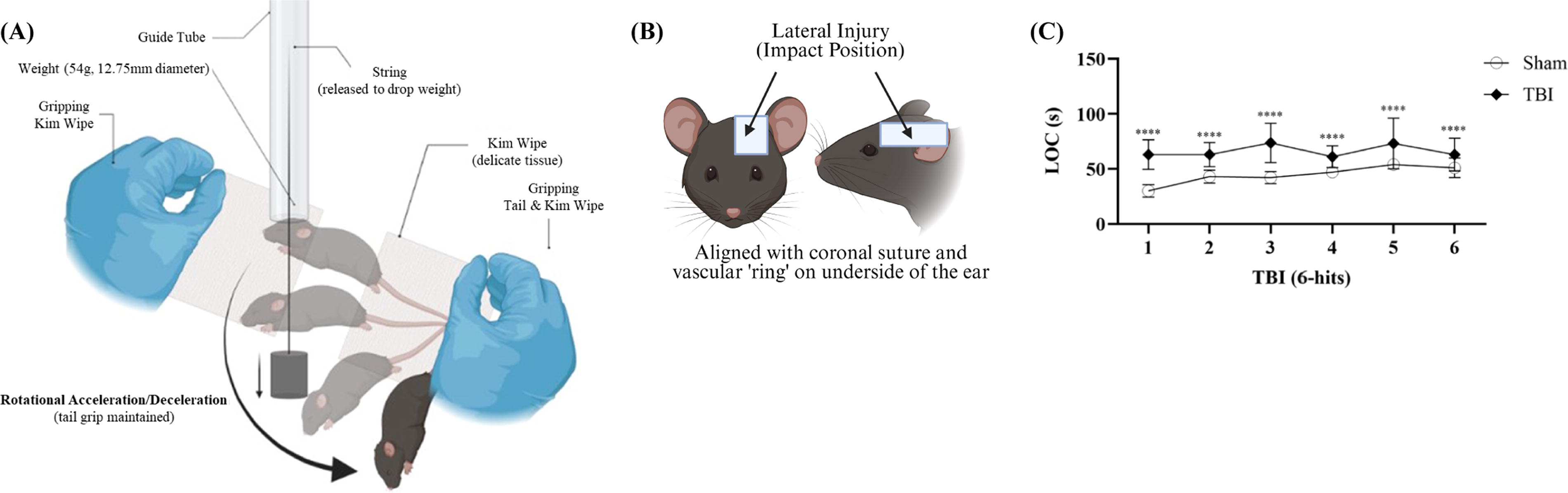
The closed-head weight drop.
Study timeline
All behavioral tests were conducted during the animal’s active phase. Mice were randomly assigned to one of three groups. Group 1: Mice underwent 10 side-alternating rmTBI (10-hits) spaced 24 h apart, alternating sides with five impacts per brain hemisphere. Subgroups included sham (n = 5), naïve (n = 5), and 10-hits (n = 8). The olfactory habituation and discrimination task, conducted 1 week after their last injury, confirmed that the 10 impacts did not impair the olfactory system in the 10-hit group. Group 2: Mice completed the OBLM test 1 week after their last injury. Subgroups included sham (n = 13) and six side-alternating rmTBI (six-hits, n = 13). Brain tissue was collected from each group for histological analysis at two time points: 10 days after the last injury, following the completion of the OBLM test (n = 8/group), and 1 month post-injury, after the open field test (OFT; n = 5/group). Additionally, a separate six-hits cohort (n = 3) was analyzed for histology 3 days post-injury without undergoing behavioral tests (Fig. 2). Group 3: This group, subdivided into sham and optic nerve crush (ONC)-treated sham (both without TBI), included sham (n = 8) and ONC-sham (n = 8). It served as a control in the novel OBLM test to evaluate optic nerve damage effects observed post-TBI.15,16

Study timeline. For the injury, animals received either 10 side-alternating rmTBI impacts (10-hits), spaced 24 h apart, with 5 impacts per brain hemisphere, or 6 side-alternating rmTBI impacts (6-hits), spaced 24 h apart, with 3 impacts per brain hemisphere. Group 1 performed the olfactory habituation and discrimination task with sham (n = 5), naïve (n = 5), and 10-hits (n = 8). Group 2 performed the OBLM test 1 week after their last injury with sham (n = 13) and 6-hits (n = 13). Brain tissue was collected at two time points: 10 days post-injury (at the end of the OBLM test, n = 8/group) and immediately after the OFT conducted 1 month post-injury (n = 5/group). Additionally, a separate 6-hits cohort (n = 3) underwent histological analysis 3 days post-injury without behavioral tests. Group 3 performed the OBLM test to assess the effects of optic nerve damage observed post-TBI with sham (n = 8) and ONC-treated sham (n = 8). OBLM, olfaction-based learning and memory; OFT, open field test; ONC, optic nerve crush; rmTBI, repetitive mild traumatic brain injury.
Immunohistochemistry
Animals were anesthetized and perfused with 0.9% saline followed by 4% paraformaldehyde (pH 7.5) at 3 days, 10 days, and 1 month post-rmTBI. Forty-micrometer brain sections were obtained using a cryostat (Leica Biosystems, Buffalo Grove, IL; CM1950). Coronal sections from frontal to caudal (starting at ∼bregma −1.94 and finishing at ∼bregma −2.92) aspects of the hippocampus were chosen per animal for immunohistochemistry analysis (Table 1). Diaminobenzidine staining: Brain sections were incubated with a primary antibody against ionized calcium-binding adapter molecule 1 (IBA-1; 1:200), further incubated with biotinylated secondary antibody (1:300), followed by treatment with avidin-biotin complex peroxidase and diaminobenzidine solution. Slides were imaged at 40× magnification using a MoticEasyScan slide scanner (Motic Microscopes, San Antonio, TX). For the IBA-1 immunohistochemistry analysis, images were processed using QuPath, 30 an open-source software for automated quantification. 31 A cell detection algorithm was designed to identify and count positively stained microglia within the region of interest (ROI) around the molecular layer of the dentate gyrus and stratum lacunosum-moleculare of the hippocampus. Thresholds (super pixel = 0.5000) were established to capture microglia morphology reflecting activation-related changes (relatively larger size of IBA-1+ cells). Data were acquired from the average of the replicate images, two coronal sections per animal with four images per ROI. Immunofluorescent staining: For Cluster of Differentiation 68 (CD68) staining, the brain sections were incubated with an anti-CD68 antibody (1:100) overnight at 4°C. The secondary antibody was conjugated with Alexa Fluor™ 488 (1:1000), and 4′,6-diamidino-2-phenylindole (1:2000) was used for counterstaining. The brain sections were further analyzed using a Zeiss LSM 980 confocal microscope. CD68 analysis was performed using Fiji (ImageJ, USA) software. The ROIs were defined around the dentate gyrus’s molecular layer and the hippocampus’s stratum lacunosum-moleculare. First, background fluorescence was subtracted using the software’s built-in function. A threshold value was then determined and applied uniformly to all images to isolate CD68 expression from background fluorescence. Subsequently, noise (outliers smaller than 3 pixels) was removed, and the mean fluorescence intensity was calculated.
Optic nerve crush
The ONC procedure has been described in previous studies.32,33 Briefly, all animals were anesthetized with an intraperitoneal injection of ketamine. A single consistent person performed the procedure. A conjunctival incision was made over the dorsal aspect of one eye. The orbital muscles were separated to expose the optic nerve at its exit from the globe. The crush was always carried out 0.5 mm behind the eyes using 45° angled jeweler’s forceps (Dumont #5). The crush was performed with maximum pressure to close the forceps for 5 sec. The sham group underwent the same procedures without ONC. The same methods were conducted for bilateral ONC in both optic nerves.
Behavior testing
Animals were habituated in the behavior testing room for ≥15 min before starting behavioral tests.
Olfactory habituation and discrimination task
The olfactory habituation and discrimination task consists of repeated presentations of a control (distilled water), two distinct nonsocial odors, and two distinct social odors27,28 (Fig. 3A). Time spent near the odor stimulus indicated interest in the stimulus. Animals habituated in a test static cage with clean bedding and a dry stimulus delivery swab for 30 min before beginning the task. The dry swab was replaced for three consecutive 2-min presentations of five different odors, for a total of 15 two-min trials. Swabs were dipped in distilled water, followed by almond extract (1:100) and banana extract (1:100). Two different neutral social odors were obtained by sweeping dry swabs for 15 sec through the bedding of two different conspecific cages that had been soiled for at least 3 days, each containing four adult male C57BL/6J mice. After completing all odor stimulus trials, animals returned to their home cages. Testing was conducted under dim white lighting (30 lux). Data were obtained from the animal tracking software EthoVision, Version 17 (Noldus, Leesburg, VA), using nose- and center-point detection with dynamic subtraction. Metrics collected include the cumulative and percent time the animal’s nose was within a 3 cm diameter around the tip of the odor stimulus swab (“sniff zone”; 0.0 cm zone exit threshold). Total entrances into the area where the olfactory stimulus was presented, denoted as “sniff zone,” latency to entrance into sniff zone, and average distance to the sniff zone were also collected.

An olfactory habituation and discrimination task was conducted in sham, naïve, and injured groups to evaluate olfactory function.
Olfaction-based learning and memory test
The OBLM behavioral chamber consists of two segments: one main rectangular arm (77 × 7 cm) and a square-shaped area (7 × 7 cm), denoted the “start zone,” positioned adjacent to the center of the main arm with a manual retractable door that blocks the mouse’s access to the main arm. Both segments are enclosed with walls of 18 cm in height. Out of the 77-cm-long arena floor, 35 cm features a cross-haired pattern, denoted as the “danger zone.” The remaining 42 cm is smooth acrylic, denoted as the “safe zone” (Fig. 4A). The odor stimulus was administered using a 1.5-mL capless tube and surgical gauze. During habituation, the odor stimulus consisted solely of clean and dry gauze. During the learning trials, however, 1 mL of 100% bobcat urine, considered a PO to rodents, 34 was pipetted onto the gauze and allowed to absorb for at least 15 min. The behavior was observed and recorded live using a web camera positioned 1.8 m above the behavioral chamber under white light conditions (30 lux). Prior to the behavioral test, animals were acclimated to handling by the same investigator for 2 days to mitigate any fear response.

The OBLM test.
The OBLM test occurred over 3 days (Fig. 4B). On day 1, a 10-min trial began with a neutral conical tube (gauze without odor) positioned against the back wall within the danger zone. The animal was placed in the start zone, then allowed to enter the main arena and freely explore. This first trial controlled any response to odor stimuli or environment novelty (acclimation). On day 2, each animal participated in three 3-min learning trials with the PO-containing conical tube in the danger zone to establish an association between the textured danger zone and the mouse’s innate fear response. Following these learning trials, there was a 10-min extinction trial to test short-term memory (STM)/working memory (WM) by exploring the arena without the conical tube. Avoidance of the danger zone and freezing behavior were measured to assess conditioned fear response (learned behavior). After the STM test, three additional learning trials were conducted. The chamber was thoroughly cleaned before and after each learning trial to prevent cross-contamination between trials. The intertrial interval was maintained at 2–3 min. On day 3, approximately 24 h after the initial learning trial, another 10-min extinction trial was conducted to assess long-term memory (LTM) of PO and the textured arena floor association. The data were obtained from EthoVision (Noldus, Leesburg, VA) using activity analysis, the deep learning algorithm, center-point detection, and dynamic subtraction. The zone exit threshold was set at 0.0 cm. Metrics collected included distance traveled, mean distance from the odor stimulus and the point, percent cumulative duration in each zone, and cumulative duration of freezing in each zone. Freezing was defined as time spent below 0.8% mobility, and the outlier filter averaging interval was one sample. Immobility parameters were defined by titrating the EthoVision settings to produce the same total immobility as the hand calculations of two experimenters blinded to an animal group for the same trial. Those settings then defined the scoring for all the other trials.
Open field test
The OFT evaluated general emotionality and has been used to identify various behavioral phenotypes. 35 Briefly, this study utilized OFT to evaluate locomotor activity, exploratory activity, and an anxiety-like phenotype via thigmotaxis. The open field measures 40 × 40 cm—a 20 × 20 cm square defines the center, and the surrounding 10-cm-wide zone forms the periphery (Med Associates, Fairfax, VT). Mice were placed in the corner of a square chamber and allowed to explore freely for 15 min. The chamber was cleaned between animals to reduce confounding olfactory trails. All testing was conducted under dim white lighting (35 lux). Data were obtained via ANY-maze software (Wood Dale, IL). Movement was tracked via three 16-beam IR arrays on the X, Y, and Z axes for positional and rearing tracking. Metrics collected included the total distance traveled, percent cumulative time, percent cumulative time excluding rest time, and time spent rearing, all within the center and periphery. Data were collected in three 5-min time bins.
Statistical analysis
Statistical comparisons for behavioral data and histology were performed using SPSS (Version 28.01; IBM, Armonk, NY) and Stata (Version 17, StataCorp, College Station, TX). Data visualization was done in GraphPad Prism 10 (GraphPad Software Inc., San Diego, CA). The OBLM behavioral test was conducted independently in two cohorts, and the data were assessed for homogeneity of variances before being combined. Sample size calculations were based on previous studies, accounting for expected performance differences between sham and injured groups. As determined by the Shapiro–Wilk test, data with normal distributions and homogeneity of variances were analyzed using repeated measures of one-way, two-way, or three-way analysis of variance (ANOVA), followed by Bonferroni’s test for multiple comparisons. For comparisons that did not meet assumptions of normality, homogeneity of variance, or outlier criteria, nonparametric testing (independent-samples Kruskal–Wallis test) or linear regression clustered by animal was used. Multiple comparison adjustments were limited to Dunn’s multiple-comparison test with Bonferroni correction. Grubbs’ test was used to identify statistical outliers (all outliers were removed from data analysis). Correlation between IBA-1 expression and behavioral outcomes was assessed using Pearson’s correlation coefficient. The receiver operating characteristic (ROC) analyses were conducted to evaluate the discriminatory power of IBA-1 expression and behavioral outcomes between sham and six side-alternating rmTBI groups, with the area under the curve (AUC) used as a measure of accuracy. Significance was set at a p value of less than 0.05. Summarized data are presented as means ± standard error of the mean (SEM), unless otherwise specified.
Results
Histological study of microglial activation
Mild concussive TBI with rapid rotational acceleration of the head leads to axonal injury, with affected brain regions extending beyond the impact site to include axon-projected areas. 12 In this side-impact model, we first wanted to examine which brain regions were injured to determine what behavioral tasks we could apply to validate functional changes. Since the injury induces acute inflammation, 36 microglial activation was used as a marker of tissue damage in this study to determine the impacted brain regions. First, we conducted a comprehensive brain mapping analysis from the frontal to caudal regions in the six-hit, side-alternating mTBI group. Using IBA-1 as a microglial marker, we identified key regions with significant morphological changes of IBA-1+ cells. Specifically, regions such as the hippocampal molecular layer, lateral geniculate nucleus, optic tract, and superior colliculus exhibited higher densities of IBA-1+ cells, which persisted at 3 days, 10 days, and 1 month post-rmTBI (Fig. 5A), suggesting a heightened neuroinflammatory response following rmTBI. Moreover, we analyzed microglial marker IBA-1 morphology at the same time points. The results showed significant changes in microglial morphology as early as 3 days (Fig. 5C; p = 0.0001) and 10 days (Fig. 5C; p = 0.0392) post-injury compared with sham, with these changes being more pronounced than in the sham group. The activated microglia displayed an amoeboid shape, with enlarged somas and shorter, less ramified processes (Fig. 5B). One month after the last injury, the number of activated microglia was significantly reduced compared with day 3 (Fig. 5C; p = 0.0128). These changes were confined to both sides of the molecular layer of the dentate gyrus and the stratum lacunosum-moleculare of the hippocampus, consistent with the alternating side impact of the TBI. Additionally, CD68, a marker of activated phagocytic microglia, showed a dramatic increase in the same regions where microglial morphology changed, particularly 3 days after the last injury (Fig. 5D,E). By 10 days and 1 month post-rmTBI, microglial morphology had returned to near-sham levels (Fig. 5C), and CD68 expression was still significantly increased at 10 days (Fig. 5E; p = 0.0063) and diminished at 1 month post-injury (Fig. 5E; p = 0.2123). Furthermore, activated microglia with altered morphology and visually increased CD68 marker were found in the optic tract (Fig. 5F) at all time points, indicating potential damage to the visual pathway.

Increased activated microglia in the hippocampus and optic tract after rmTBI.
Olfactory habituation and discrimination task: Olfactory function
Given the olfactory deficits linked to both TBI and age-related neurodegeneration and the visual deficits in our mouse model of rmTBI,15,16 we performed the olfactory habituation and discrimination task to verify that injured mice can detect and respond to smells similarly to sham mice (Fig. 3A). During this task, intact olfaction was indicated by reduced interaction with repeated odors (habituation), increased interaction with novel odors, and a preference for social over nonsocial odors. We assessed whether isoflurane, the anesthetic used during TBI, affects olfaction by comparing sham (isoflurane-exposed) and naïve (nonexposed) mice. Trial number was a significant predictor, while stimulus type and group condition were not (Fig. 3B; p = 0.001, p = 0.548, p = 0.689). This suggests that both naïve and sham mice similarly interacted with odors, influenced mainly by the number of odor presentations. We then assessed odor interaction behavior between sham animals and those subjected to 10 instances of alternating lateral impact. Findings showed no significant difference in interaction time between the sham and injured groups (Fig. 3C; p = 0.915). These findings suggest that neither isoflurane exposure nor rmTBI significantly affects the olfactory system. Thus, olfaction remains a reliable sensory measure in our novel OBLM test.
Olfaction-based learning and memory test
The OBLM test was conducted on sham and six-hits groups. We measured avoidance of PO by assessing time spent in the “safe zone” during behavioral criteria assessments (Fig. 6A–C). Sham animals significantly avoided the PO during learning and WM tests but not during the LTM test (Fig. 6A; WM p < 0.001). Injured animals did not significantly avoid the PO and spent less time in the “safe zone” than sham animals. We also analyzed freezing behavior in the “safe” and “danger zones” during all assessments. Sham animals showed significantly more freezing in the “safe zone” with the PO and during the WM test but not the LTM test, suggesting they could re-learn without the PO (Fig. 6B; WM p < 0.001). Injured animals showed no significant freezing changes across assessments. This indicates sham animals can learn and remember the STM association with the PO but fail to retain the LTM, while injured animals show deficits in learning and memory. Additionally, injured animals exhibited significantly higher locomotor activity (Fig. 6C; p = 0.002), suggesting reduced freezing, increased danger zone time, and greater overall activity compared with sham animals. These behavioral differences highlight the impact of injury on learning, memory, and activity.
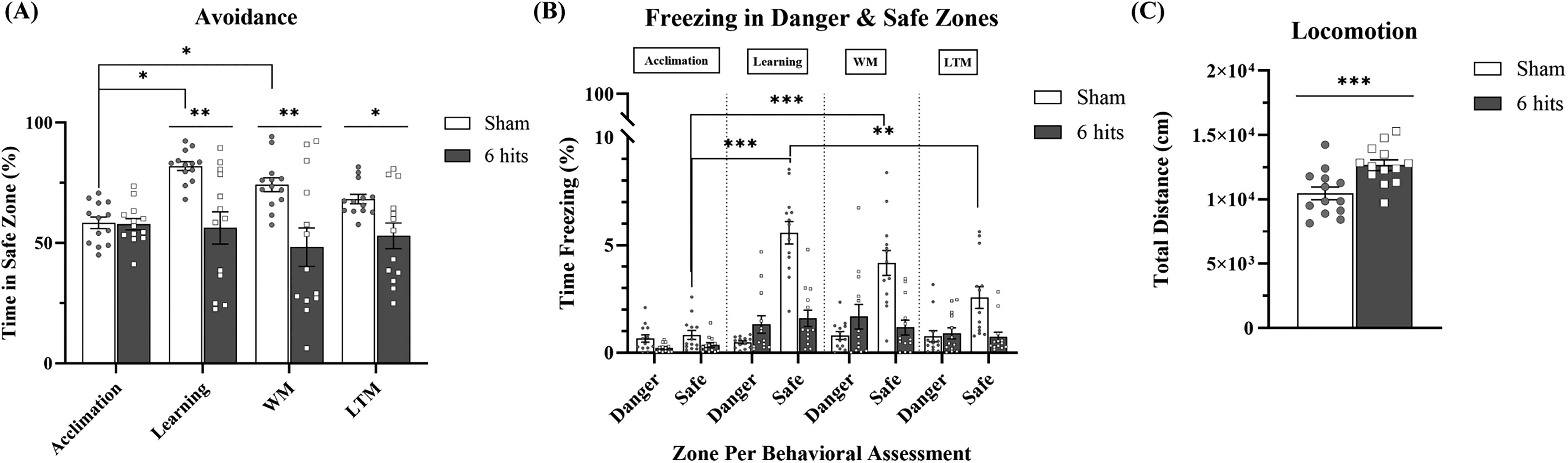
Six alternating lateral impacts presented potential impairments in learning and memory during the OBLM test. All data were obtained from EthoVision software.
Open field test: Locomotion
During the olfaction-based study, we found that the distance the injured mice traveled was longer than that of the sham group. Here, we further assessed locomotor activity in sham and TBI (six-hits) groups. Although there was no significant difference in overall locomotor activity between the groups during the 15-min trial (Fig. 7A), when locomotion data were broken down into three 5-min intervals, the injured animals exhibited hyperactivity during the first interval compared with the sham group (Fig. 7B; 0–5 min, p = 0.04). As the trial progressed, the locomotor activity of the injured animals decreased to levels comparable with the sham group (Fig. 7B). These findings highlight distinct differences in behavioral responses to the anxiety-inducing environment between sham and injured animals.

An increase in locomotion was observed in the six alternating lateral impacts group.
Microglial activation correlates with early memory impairment in TBI animals
To examine whether the injury quantified by the number of microglia correlated with memory deficits in six-hits rmTBI, we assessed the expression of IBA-1 protein levels and memory performance using the OBLM test. At 10 days post-last injury, no correlation was observed between IBA-1+ cells and STM performance (Fig. 8A; R2 = 0.2215, p = 0.2215); however, a moderate correlation was observed between increased IBA-1+ expression and impaired LTM performance (Fig. 8B; R2 = 0.6031, *p = 0.0234). This suggests that increased number of activated microglia is correlated with worsened memory retention at this acute stage post-TBI. Furthermore, the ROC curve analysis for IBA-1 expression demonstrated a strong ability to distinguish between sham and TBI groups (Fig. 8C; AUC = 0.9063, p = 0.0063), indicating high predictive accuracy. This result indicates IBA-1 positivity is a strong discriminator of neuroinflammation between sham and TBI groups. Similarly, the ROC curve for the OBLM test behavioral outcomes (Fig. 8D; AUC = 0.8281, p = 0.0274) suggests a robust discriminatory between the two groups. Together, these findings underscore the importance of both IBA-1 expression and behavioral metrics in characterizing TBI-related changes.

Acute microglial activation correlates with memory impairment in TBI animals.
Olfaction-based learning and memory
Here, we tested whether blind mice without TBI performed similarly to shams. The mice were subjected to ONC, which resulted in a significant vision deficit. 37 Both sham and sham with ONC mice performed OBLM tests; no significant differences were detected between the two groups (Fig. 9), indicating that the OBLM test is not vision-dependent.

The ONC group presented a learning and memory phenotype similar to sham during the OBLM test. Damage to the optic nerve, as occurs during injury, leads to impaired vision. This study investigated whether blind mice without TBI exhibited performance similar to sham mice, since blindness can potentially influence movement, cognitive abilities, and memory. The mice underwent ONC, which caused a significant loss of vision. The sham and ONC-treated sham groups were tested on OBLM tasks; quantitative data were collected, presenting no significant differences between groups. This suggests that blindness alone does not impair OBLM abilities. Data: each individual point represents a single animal; mean ± SEM; no significant difference was determined by p > 0.05; n = 8/group. LTM, long-term memory; OBLM, olfaction-based learning and memory; ONC, optic nerve crush; SEM, standard error of the mean; TBI, traumatic brain injury; WM, working memory.
Discussion
Our comprehensive brain mapping analysis from the frontal to caudal brain regions at 3 days, 10 days, and 1 month revealed that alternating lateral rmTBI resulted in bilateral inflammation, as evidenced by an increase in IBA-1+ cells, specifically in the hippocampal molecular layer, lateral geniculate nucleus, optic tract, and superior colliculus. This inflammation pattern, particularly prominent in regions associated with memory, learning, and visual processing, suggests that rmTBI disrupts neural circuits essential for cognitive and sensory functions. Despite the significant inflammatory response in these areas, olfactory function remained unaffected, as demonstrated in the olfactory habituation and discrimination task.
To further explore cognitive impairment related to rmTBI, we developed a novel PO-based, vision-independent learning and memory task. This task effectively identified deficits in learning and memory, providing an assessment method to target hippocampal-dependent memory independent of visual function. 26 Our findings suggest rmTBI from alternating lateral impacts may increase locomotor activity, implying potential changes in motor control or behavioral responses due to repeated injury. We offset the impact to the side of the head to mimic the rotational and angular forces commonly experienced by athletes who sustain sport-related concussions.38,39 Biomechanical investigations into athletic injuries have indicated that angular head accelerations are more likely to cause injury. Schneider and colleagues conducted a study involving a computer simulation of head impacts on soccer players, revealing that the risk of injury from angular head accelerations surpassed that from linear head accelerations. 40 Moreover, lateral impacts resulted in higher angular and lower linear head accelerations than frontal impacts.40,41 Rapid rotational acceleration may cause injury in different brain regions, depending on head impact location. 42 This model improves on our previous studies, which used an impact on the front middle of the head and created more diffuse brain injuries in both the hippocampus and cortex. 43 The current side-impact model resulted in more focal injury, while the vision pathway was also damaged (Fig. 4). The increased active microglia in the optic tract indicates wallerian degeneration due to axonal injury. 44 Since injury induces acute inflammation,45,46 we used microglia markers, IBA-1, a pan-microglial marker, and CD68, an activated phagocytic form of microglia,46,47 to show which brain regions are injured. Immunostaining revealed increased microglia number and activation concentrated in the regions of the molecular layer of the dentate gyrus and stratum lacunosum-moleculare of the hippocampus, 48 where the axons from the entorhinal cortex communicate with the dendrites from the hippocampus.26,49,50
Notably, these findings are consistent with previous reports from other laboratories, which have also observed elevated microglial activation within these hippocampal subregions following various TBI models.17,19,51,52 Exact sites of axonal injury are unknown, but our results are consistent with the expected consequences of these axonal injuries: rapid downstream axonal degeneration (wallerian degeneration) and inflammation.49,53–58 Among other entities, TBI is a risk factor for Alzheimer’s disease and CTE, and the entorhinal cortex is one of the key brain regions where characteristic pathological changes occur.59,60
Olfaction plays a crucial role in daily life.21,61 The olfactory receptor neurons originate in the olfactory bulbs and project to the primary olfactory cortex, which includes regions such as the amygdala, piriform cortex, entorhinal cortex, and anterior olfactory nucleus. 62 The entorhinal cortex is a crucial network hub for learning, memory, and navigation. 49 Disruptions such as neurodegeneration in connections between the entorhinal cortex and hippocampus can impair these functions, which is one of the earliest pathological features of Alzheimer’s disease.26,53,55 Our rmTBI model demonstrated increased activated microglia within the hippocampal molecular layer and stratum lacunosum-moleculare, suggesting damage in these connections. However, the extent of this pathway disruption is yet to be quantitatively assessed.
To investigate whether traumatic hippocampal injury may result in learning and memory decline, we developed a novel olfaction-based, vision-independent behavioral test to assess learning and memory in the mice. Supplementing vision-dependent rodent behavior tests with those dependent on other sensory systems is important in rodent models of rmTBI to increase accuracy and repeatability within experimental paradigms. Doing so will help identify the neurostructural and neurochemical changes correlated with rmTBI behavioral symptoms, which will then translate to the development of effective rmTBI therapies. The olfactory habituation and discrimination task, a pivotal aspect of our research, was initially formulated by Arakawa and colleagues. 41 Arbuckle and colleagues 28 subsequently modified it to establish the normal functioning of the olfactory system and related behaviors in mice. Widely adopted in the field, this task offers a dependable way of assessing mice’s olfaction within different research fields. Adapting from the test above, we developed a novel vision-independent behavior test to assess spatial learning and memory. The OBLM test uses repeated presentations of a PO paired with a uniquely textured portion of the behavioral chamber floor. It tests the recollection of that association in both STM and LTM.
No olfactory dysfunction was observed even after 10 instances of alternating lateral impact. In the social olfaction test, the rmTBI mice exhibited behavior comparable with the sham controls at the designated time point, indicating intact olfactory function. We then applied a model with fewer injuries, six alternating lateral impacts, to assess potential impairments in learning and memory. The data revealed that sham and rmTBI mice spent approximately 50% of their time in the safe zone during the acclimation phase (day 1). However, on the testing days (days 2 and 3), the TBI mice demonstrated a reduced ability to learn compared with the sham group. Specifically, the TBI mice failed to remember the PO and spent more time in the danger zone during the subsequent 2 days, indicating STM and LTM deficits. In contrast, the sham mice retained STM of the PO and demonstrated the ability to re-learn when the PO was removed from the arena. The study suggests that learning likely depends on intact STM (evident within 10 min), while memory consolidation occurs over more than 24 h, involving both short- and long-term recall. These findings indicate that this behavioral task effectively differentiates learning and memory deficits in TBI mice, including those with vision impairments, from sham controls. Our observations show that the sham group’s avoidance of PO, as a measure of fear, during both the learning trials and the STM test (OBLM test, day 1) confirms that our novel OBLM test accurately evaluates risk aversion, behavioral inhibition, associative learning, and WM or STM. Notably, the sham group demonstrated increased avoidance with each subsequent PO presentation during the learning trials, further validating our test’s ability to assess learning capacity. In contrast, the injured group did not show significant changes in avoidance of PO or the PO-associated environment after PO removal. Because the Olfactory Discrimination and Habituation Task confirmed the absence of olfactory deficits, it is expected that these changes in avoidance behavior result from injury alone. We also investigated whether vision impairments might influence learning and memory performance, as blindness can alter animal behavior. As shown in Figure 7, vision impairment did not affect performance in the olfaction-based tasks, confirming that blindness did not cause the observed deficits in learning and memory for the TBI group. The OBLM task’s ability to assess complex, higher-order cognitive functions, typically mediated by the frontal lobe, in a vision-independent manner distinguishes it among other cognitive assessments.63–66
Our findings highlight a strong relationship between hippocampal injury and memory performance following six-hit rmTBI. At 10 days post-last injury, elevated microglial activation in the hippocampus, as indicated by the increased number of IBA-1+ cells, correlates with impaired LTM but not STM. The STM in this task was defined immediately after learning. The acute activation of microglia reflects the extent of injury. This association underscores the critical role of the hippocampus in cognitive deficits post-rmTBI and suggests that this olfaction-based memory test can be used for evaluation of hippocampal injury-related LTM deficits but not STM. Furthermore, the ROC curve analyses highlight the potential of IBA-1 expression and behavioral metrics as tools for distinguishing between sham and TBI groups. Whereas the IBA-1 could be a strong biomarker for neuroinflammatory changes associated with TBI and that the right functional assessments can effectively capture TBI-related deficits, further supporting their diagnostic relevance. It is worth mentioning that certain neuronal injury markers, particularly neurofilament light chain, are increased rapidly within 24 h and go down quickly as well after injury (our unpublished data). This makes these biomarkers unable to be used for evaluating injury levels at subacute and long-term timepoints. Meanwhile the activated microglia morphology lasts fairly longer. It becomes a relatively reliable biomarker for validating injury in the subacute phase. Together, these findings suggest that combining molecular and behavioral measures could enhance the accuracy of TBI diagnostics. Future studies should aim to validate these findings across diverse experimental and clinical contexts to establish their translational potential.
Despite its strengths, there are some limitations in this study. First, there is no direct evidence pinpointing the exact location of axonal injury. While microglial activation in the hippocampal regions is linked to the entorhinal cortex, particularly in layers II and III, we hypothesize that the axons connecting the hippocampus and entorhinal cortex are compromised. However, no acute neuronal death or loss was observed in the hippocampus or other brain regions, including the entorhinal cortex. To address this, we plan to use the CLARITY technique 67 and 3D imaging with axonal tracing dyes to identify the specific regions of axonal injury. Second, this study includes only a single time point, as the primary aim was to develop an OBLM test. In future research, additional time points will be incorporated to investigate whether the injury leads to LTM deficits. Third, we did not examine a range of injury severities. In future studies, we will apply milder and more severe injury models of TBI to determine whether the OBLM test can accurately differentiate the magnitude of deficits based on injury severity. Fourth, the small sample size and single time point for the OBLM test represent a limitation in calculating the correlation between IBA-1-positive cell numbers and behavioral outcomes, as well as in conducting the ROC analysis for both parameters. Further studies are warranted to address this by incorporating larger sample sizes and looking at acute, subacute, and chronic time points to more accurately assess the relationship between IBA-1 expression and behavioral outcomes post-TBI. Lastly, a limitation of this study is the exclusive use of male subjects. Given the known sex differences in olfactory processing, 68 neuroinflammatory responses, and behavioral outcomes following brain injury, our findings may not be fully generalizable to females. Future studies should include both sexes to better understand potential sex-specific effects and enhance the translational relevance of the findings.
Conclusions
This study demonstrated that rmTBI induced by alternating lateral impacts can lead to hippocampal dysfunction and memory deficits. Given that indirect optic nerve injury15–17,19,69 is common in preclinical rodent models of TBI, traditional approaches such as the Morris Water Maze (MWM) may not be suitable for assessing memory in this context.14–16,70,71 Our findings suggest that a novel olfaction-based, vision-independent learning and memory test offers a more appropriate method for evaluating neuronal function following rmTBI and may be a valuable tool for future studies. Ultimately, no single behavioral assay should be used in isolation. We advocate for integrating MWM with visually independent tests such as our novel OBLM test and psychophysical tests of vision to provide a more comprehensive understanding of cognitive outcomes in TBI models.
Authors’ Contributions
F.G.-C.: Idea, conceptualization, methodology, supervision, writing—original draft, review and editing. E.J.M.: Idea, conceptualization, methodology, supervision, writing—original draft, review and editing. Y.Y.: Methodology, writing—review and editing. G.O.: Methodology, writing—original draft, review and editing. K.W.: Methodology, writing—original draft, review and editing. N.N.: Methodology, writing—original draft, review and editing. A.T.-S.: Methodology, writing—original draft, review and editing. C.L.: Methodology, writing—original draft, review and editing. E.A.K.: Methodology, writing—original draft, review and editing. J.L.: Methodology, writing—original draft, review and editing. R.L.S.: Methodology, writing—original draft, review and editing. R.M.: Idea, conceptualization, methodology, supervision, writing—original draft, review and editing. W.P.M.: Idea, conceptualization, methodology, supervision, writing—original draft, review and editing. J.Q.: Idea, conceptualization, methodology, supervision, writing—original draft, review and editing.
Footnotes
Acknowledgments
We would like to thank Dr. Lindsey Vansandt, Director of the Imperiled Cat Signature Conservation Project for the Center for Conservation and Research of Endangered Wildlife (CREW) at the Cincinnati Zoo & Botanical Garden, who supplied valuable materials for this study. Dr. Vansandt also provided valuable cat behavior and biology expertise, including predator–prey relations and urinary analysis. We would also like to thank the urine donor, Citrine the Cat, for contributing to this study. We sincerely thank Dr. Nathaniel Hodgson of the Animal Behavior and Physiology Core at Boston Children's Hospital for his invaluable contributions to the design and construction of the apparatus used in this study. His expertise and support were instrumental in enabling the experimental work.
Author Disclosure Statement
The authors have no competing interest to disclose.
Funding Information
(1) This work is part of the NFL-LONG study, funded by a grant from the National Football League. Part of the work was performed in the Animal Behavioral and Physiology Core at Boston Children’s Hospital (CHB IDDRC, 1U54HD090255). (2) This work was supported by T32HD040128 from the National Institute of Childhood Diseases and Human Development (NIH).
Transparency,Rigor,and Reproducibility Summary
The study design and statistical analysis plan were determined based on prior work conducted in our lab using behavioral and histological data. Mice were randomly assigned to one of three groups, with subgroups designed to assess behavioral and histological outcomes following rmTBI and ONC. The olfactory habituation and discrimination task confirmed that 10 impacts did not impair olfactory function in the 10-hits group. Sample sizes for behavioral assessments and histological analyses were based on previous findings from our lab. They were designed to detect significant group differences with sufficient power, which indicated >80% power to detect both an overall significant effect of the repeated-measures ANOVA (the primary statistical analysis), and a post hoc difference between the injured group versus sham with p < 0.05 after correction for multiple comparisons. A total of 76 mice were included in the study. Exclusions due to technical reasons, mortality, or noninterpretable data were zero. Mice were randomly assigned to groups using a random number generator. Investigators conducting behavioral tests and data analyses were blinded to group assignments. Histological analyses were performed at multiple time points: 10 days post-injury, immediately after behavioral testing at 1 month, and 3 days post-injury in a separate cohort without behavioral testing. Standardized tissue collection and staining protocols ensured consistency of histological assessments.