




Abstract
This research aimed to explore the effects of varying porosities and pore sizes in porous titanium on osteointegration following mandibular defect reconstruction in goats. Porous titanium products were fabricated using spark plasma sintering technology combined with powder metallurgy techniques. Based on experimental requirements, standard porous titanium samples were categorized into three groups: Group A with 50%–70% porosity and 100–300 μm pore size; Group B with 70%–85% porosity and 300–500 μm pore size; and Group C with no porosity. These samples were implanted into the mandibular defects of goats, which were euthanized three months after implantation for analysis. The assessment of osteointegration involved general observations, X-ray, micro-CT, biomechanical testing, and histological examinations. Results indicated that Group B's porous titanium exhibited superior osteointegration, making it the most suitable material for enhancing cellular and tissue growth.
Introduction
Numerous hard tissue-repair materials, notably titanium, and its alloys, have been widely adopted due to their exceptional corrosion resistance, biocompatibility, low density, and high specific strength. 1 However, the mismatch between their elastic modulus and that of natural bone often impedes proper load transmission to bone tissue, resulting in what is known as stress shielding at the bone-implant interface. This mismatch leads to reduced osteointegration of the implants.2,3 The introduction of porous structures within these titanium implants enhances stress distribution across the interface, which significantly reduces the intensity and Young's modulus, thereby effectively mitigating stress shielding and promoting better integration with the natural bone.4,5
Currently, assessments of bone tissue implants incorporating various porosities and pore sizes have been conducted; however, identifying the most effective porous structure parameters remains a challenge.6–8 The intricate relationship between porosity, mechanical strength, and the elastic modulus requires meticulous consideration to ensure that the selected porosity and pore size of porous titanium achieves an elastic modulus closely aligned with that of natural bone. This alignment is critical for enhancing osteointegration and facilitating osteoblast cell growth, thereby improving the clinical outcomes of bone tissue repairs.9–11
Recent advances in the rational design of bioactive materials have shown significant promise in bone hemostasis and defect repair. For instance, Gai et al. have demonstrated innovative strategies for designing bioactive materials that enhance bone healing processes. 12 Additionally, studies on mandibular remodeling, such as the work by Li et al., offer valuable insights into how different bite-altering devices impact bone structure, highlighting the relevance of animal models in testing the efficacy of new materials. 13 Furthermore, Yu et al. explored the targeting of specific molecular pathways to promote bone formation while preserving cardiovascular health, underscoring the multifunctional potential of bioactive materials in clinical applications. 14 These studies provide a broader context for understanding the challenges and opportunities in developing materials for hard tissue repair, supporting the rationale behind the use of porous titanium in the present study.
The application of porous titanium requires meeting key criteria for successful clinical use. Biocompatibility is critical, as the implant must not induce adverse reactions and should integrate well with bone tissue. Mechanical strength must be retained despite porosity, allowing the implant to withstand physiological loads, while the elastic modulus should match that of the surrounding bone to avoid stress shielding. Osteoconductivity is also essential, with the porous structure facilitating bone growth and effective osseointegration. This study focuses on optimizing pore size and porosity to achieve a balance between mechanical performance and biological compatibility for clinical applications. This research addresses the gap in optimizing porous titanium implants by refining pore size and porosity to enhance both osteointegration and mechanical stability, areas where previous studies have been insufficient.
In this comprehensive study, we explored the impacts of different porosities and pore sizes in titanium on osteointegration after their implantation into the mandibles of goats. Our research aims to provide a clearer understanding of how these variables influence the healing and integration processes, thereby guiding the development of next-generation biomaterials that synergize optimal mechanical properties with superior biocompatibility. Such advancements hold significant potential to revolutionize the field of medical implants, making them more effective and suitable for a variety of clinical applications.
Materials and methods
Animals
Thirty goats, each aged 6 months and weighing between 20–25 kg, were obtained from the Animal Laboratory Center at Sichuan University for this study. Throughout the treatment period, comprehensive monitoring of behavior, posture, reactivity, and general appearance was conducted to assess the well-being of the animals. Rigorous protocols were implemented to minimize any pain or discomfort experienced by the goats during the experiment. All procedures and interventions were closely supervised and periodically reviewed to ensure adherence to ethical standards. The study received approval from the Animal Experiment Ethics Committee of the State Key Laboratory of Oral Diseases at Sichuan University (SKLOD), affirming compliance with all relevant ethical guidelines for animal care and use.
Porous titanium
Porous titanium implants were collaboratively designed and developed by the Institute of Rare Earth and Nanomaterials at Sichuan University and our research team. These materials were fabricated utilizing an innovative method that combines spark plasma sintering technology with traditional powder metallurgy techniques. Three distinct types of porous titanium materials were produced, each differing in porosity and pore size to facilitate targeted research analysis. Each implant is a circular specimen measuring 10 mm in diameter and 5 mm in thickness. Specifically, Group A features a porosity range of 50% to 70% and pore sizes between 100 and 300 μm; Group B exhibits a porosity of 70% to 85% with pore sizes ranging from 300 to 500 μm; Group C is designed with no porosity.
The sintering process for these implants involved using a SPS machine. The sintering temperature was set at 1100°C, and the pressure was maintained at 50 MPa, with the process lasting for 10 min. This ensured adequate bonding between the titanium particles and the formation of a stable, porous structure.15–16
Before use, all porous titanium implants were meticulously cleaned, dried, and subsequently sterilized under high temperature and pressure to ensure their suitability for surgical application and to maintain the highest standards of research integrity. The post-processing of the implants included ultrasonic cleaning in ethanol to remove any surface contaminants, followed by drying at 100°C for 2 h. Finally, the implants were sterilized in an autoclave at 121°C and 15 psi for 30 min to ensure sterility before surgery. 17
Surgical methods
From each group, ten goats were randomly selected for the study. Animals were anesthetized using 3% pentobarbital sodium administered at a dosage of 1ml/kg through intraperitoneal injection. To ensure a sterile environment for surgery, the surgical field was disinfected with a 10% povidone-iodine solution, and pain management was addressed by injecting 0.5% lidocaine along the planned incision line. A 5-cm horizontal incision was then made at the lower edge of each side of the mandible. After the incision, the periosteum was carefully separated to fully expose the mandible. Cylindrical bone defects, each with a diameter of 10 mm, were precisely created between the anterior teeth and the first premolar using a low-speed drill, operating at speeds between 900 and 1200 rpm under continuous saline irrigation to prevent overheating.
Each bone defect was then filled with one of the three groups of porous titanium implants, each differing in porosity and pore size. To stabilize the mandible and prevent potential fractures or displacement resulting from the bone defects, four-hole titanium plates from Double Sheep Medical Instrument Co., Ltd, Suzhou, were secured along the lower margin of the mandible on both sides of each defect. Closure of the surgical sites involved suturing the periosteal membrane with 6-0 nylon and the skin with 4-0 nylon to ensure optimal healing.
Postoperatively, the animals were administered antibiotics intramuscularly, specifically penicillin at a dosage of 40U/day—for one week to prevent infection. Additionally, the skin wounds were meticulously cleaned daily with iodine tincture to further mitigate the risk of infection. To facilitate recovery, the goats were provided with soft food for one week following the surgery.
Sample preparation
Three months post-procedure, all animals were humanely euthanized using an overdose of 3% pentobarbital sodium and administered intravenously while under systemic anesthesia induced by ketamine. Following euthanasia, the mandibles were carefully harvested and promptly immersed in 4% paraformaldehyde to ensure rapid and effective fixation, preserving the tissue integrity for subsequent histological examination.
Plain radiographic examinations
After carefully removing the soft tissues, each dissected mandible was accurately positioned on an occlusal film, ensuring that the lingual side was in direct contact with the film. Lateral plain radiography was subsequently performed using a Philips X-ray unit. The unit operated under standardized conditions, set at 70 kV and 8 mA, with a brief exposure time of only 0.06 milliseconds to capture detailed radiographic images.
Specimen preparation and Micro-CT scanning
Specimens were carefully prepared and scanned using a Micro-CT 80 scanner (Scanco Medical, Bassersdorf, Switzerland). For optimal imaging, each specimen was positioned in a mid-sized sample holder with the mandible body plane oriented vertically to the X-ray tube. Micro-CT scans were conducted at settings of 60 kV and 180 mA.
Image analysis and data extraction
Serial images obtained from each scan were analyzed using the recommended software, VGStudio MAX 2.1. Critical parameters were quantified using the same software to ensure consistency in data analysis. The parameters of interest that were calculated included the bone volume to total volume ratio (BV/TV), trabecular number (Tb.N), trabecular thickness (Tb.Th), trabecular spacing (Tb.Sp), and structural model indexes (SMI). These parameters are essential for assessing the micro-architectural properties of the bone and the effectiveness of the implant material
Histological analysis
Removed specimens were initially fixed in 4% paraformaldehyde. Subsequently, half of these samples underwent a dehydration process using a graded series of ethanol and were then embedded in resin. Thin sections of 10 μm were prepared using a hard tissue slicer and grinder (SP 1600, Leica, Germany) and stained with toluidine blue for detailed microscopic examination. Histological evaluations were conducted with a high-resolution biological microscope (DXM 1200, Nikon, Japan), focusing on cellular morphology and tissue integration.
Biomechanical examinations
The biomechanical properties of the remaining samples were assessed using an INSTRON4034 universal material testing machine (model 4034, INSTRON, USA). Tests were performed under controlled room temperature conditions where unidirectional pressure was systematically applied at a loading rate of 1 mm/min. The resilience and compressive strength of the samples were measured and compared with their pre-implantation properties. Experimental data were meticulously recorded and analyzed to evaluate changes in mechanical integrity over time.
Statistical analysis
All collected data were statistically processed using the SPSS software package, version 21.0 (SPSS, Chicago, IL). Results are presented as mean ± standard deviation (SD). To determine the statistical significance of the findings, a one-way analysis of variance (ANOVA) was employed. A p-value of less than 0.05 was considered to indicate statistical significance, ensuring that the results are robust and reliable.
Results
Superior osteointegration and stability of group b implants in mandibular reconstructions
The postoperative recovery process was uniformly uneventful across all animal subjects, indicating a high level of tolerability for the surgical procedure and the biomaterial used. Radiographic evaluations conducted immediately after surgery and periodically during the recovery phase consistently confirmed the stability of the implants within the mandible. These X-rays revealed no signs of implant loosening, shedding, or displacement, suggesting robust mechanical stability.
Most notably, the implants from Group B not only demonstrated stability but also exhibited the highest degree of osteointegration. This was observed as a significant new bone formation that seamlessly integrated with the surrounding bone tissue at the material-bone interface. This integration was quantitatively superior when compared to the other groups, as detailed in subsequent quantitative analyses (Figures 1 and 2). The superior performance of Group B implants suggests an optimal balance of porosity and mechanical properties conducive to bone ingrowth and stabilization within the challenging environment of the mandible.

Creation of cylindrical bone defects.
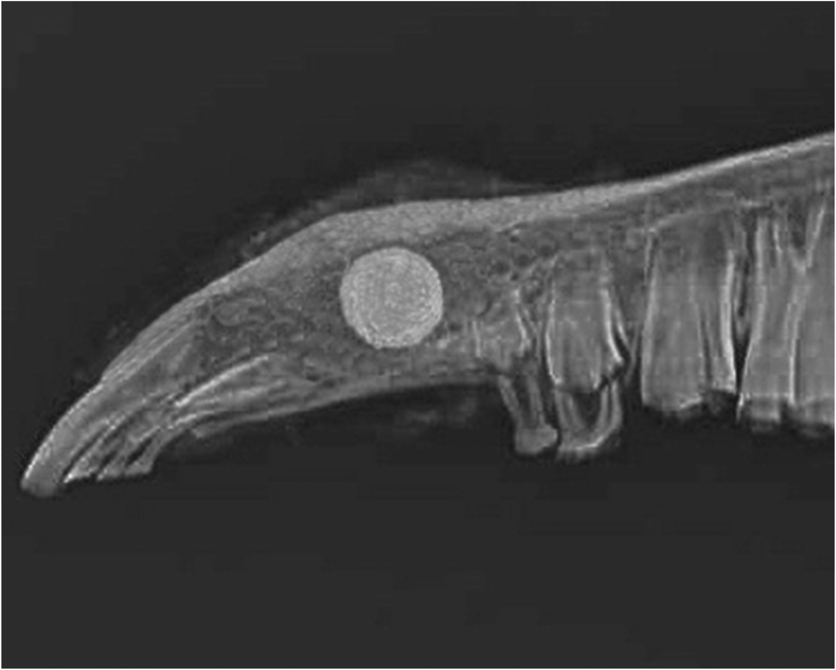
Stability of implants in postoperative X-rays.
The consistent stability and effective osteointegration observed in Group B underscore the potential of tailored porous titanium implants in clinical applications, particularly for mandibular reconstructions where mechanical stability and rapid bone integration are crucial for successful treatment outcomes.
Twelve weeks post-surgery, micro-CT analyses demonstrated that Group B exhibited the most substantial 3D-porous bone formation among all groups, indicating a direct and favorable response to the implant's structural design. This group's distinct porosity and pore size facilitated enhanced osteoconduction, leading to superior bone ingrowth within the porous matrix. Statistical evaluations further substantiated these observations, revealing a robust positive correlation between increased porosity levels and significant improvements in key bone structural metrics. Specifically, the bone volume to total volume ratio (BV/TV), trabecular number (Tb.N), and trabecular thickness (Tb.Th) were markedly higher in areas with greater porosity. Conversely, trabecular spacing (Tb.Sp) and structural model index (SMI) values showed a significant decrease with increasing porosity, underscoring the role of optimized porous structures in promoting more dense and structurally coherent bone tissue integration (Table 1).
The parameters of material-bone interface of three groups at 12 week x ± SD.

The parameters of material-bone interface of three groups at 12 week x ± SD.
These findings underscore the pivotal role of pore architecture in bone remodeling and emphasize the need for meticulously engineered porosity parameters to optimize the efficacy of bone implants, especially in load-bearing scenarios like mandibular reconstructions.
Histological evaluations provided deeper insights into the differential patterns of bone growth among the experimental groups. In Group A, new bone primarily formed in a direction parallel to the material surface, suggesting limited but direct engagement with the implant structure. This type of growth indicates some degree of osteoconduction but possibly insufficient pore size or distribution to encourage more extensive bone ingrowth.
In contrast, Group B displayed remarkable bone formation with extensive inward growth and high connectivity within the pores. This group's histological samples showed that new bone not only filled the available pore spaces but also interconnected across the pores, creating a robust structural network. This suggests that the porosity specifications for Group B are well-optimized for promoting not just bone ingrowth but also integration, crucial for the structural stability and functionality of the implant.
Group C, however, demonstrated minimal bone interaction with the implant, with scarce new bone formation visible at the titanium-bone tissue interface. This lack of significant bone formation could be attributed to the absence of porosity, which is essential for bone tissue to anchor and grow into the implant structure effectively.
These histological findings emphasize the critical influence of pore characteristics on bone integration and regeneration. The results clearly demonstrate that appropriate porosity is vital for facilitating not only the initial bone ingrowth but also its subsequent interconnection and maturation, which are crucial for the long-term success of bone implants in clinical settings (Figures 2, 3 and 4).

Micro-CT analysis of bone formation.

Histological evaluation of new bone formation.
Biomechanical testing conducted after the implantation process demonstrated significant increases in both elastic modulus and compressive strength for the implanted groups, underscoring the mechanical efficacy of the implant materials used. Specifically, the changes in these mechanical properties are meticulously documented in Tables 2 and 3. Twelve weeks post-implantation, both the compressive strength and elastic modulus of the implants were considerably higher compared to their pre-implantation values, with statistical significance (P < 0.05). This indicates a substantial improvement in the mechanical integrity of the bone-implant interface over the recovery period.
Elastic modulus of two groups before and after material implantation Gpa.

Elastic modulus of two groups before and after material implantation Gpa.
Compressive strength of two groups before and after material implantation Gpa.

Furthermore, the growth rates for these mechanical properties in Group B were significantly greater than those observed in Group A. This disparity suggests that the higher porosity and specific pore architecture in Group B not only supports bone ingrowth but also contributes to the enhanced mechanical properties of the implants.
These biomechanical findings highlight the crucial role of implant design, specifically porosity, in influencing the mechanical outcomes post-implantation. The enhanced mechanical properties observed in Group B align with the superior bone integration and growth patterns identified in histological and micro-CT analyses, confirming the holistic success of the optimized porous structure. These results contribute significantly to our understanding of the interplay between material science and biological integration, paving the way for the development of more effective and robust implants in orthopedic and dental reconstructive surgeries. This comprehensive approach ensures that future implant designs can achieve both biological compatibility and necessary mechanical durability.
As of now, titanium and its alloys are primarily employed as compact implants in clinical practices. These materials are recognized for their excellent biocompatibility; however, their elastic modulus, which is often greater than 100 GPa, significantly surpasses that of natural bone.18–20 Such mechanical discrepancies frequently result in implant loosening. The development of porous titanium has addressed this problem by allowing adjustments in porosity and pore size to better align the implant's elastic modulus with that of bone tissue.21–23 This adjustment promotes new bone growth into the implant's pores and enhances the formation of a robust bonding interface. 24
Research by Cyster and colleagues has shown that an increase in pore size significantly enhances the contact area between the implant and the bone interface, which is conducive to the adhesion and proliferation of osteoblasts within the pores, thereby facilitating mechanical integration between the material and bone. 25 Furthermore, Gotz and his team demonstrated that pore sizes exceeding 300 μm can enhance bone and capillary formation under certain conditions. 26 In vivo experiments have corroborated that greater porosity and increased surface area are beneficial for the exchange of nutrients and osteogenic factors, thereby promoting osteogenesis. 27 However, the optimal pore size and porosity for bone formation still require precise determination.
Various methods are employed to prepare porous titanium, including powder metallurgy methods, loose sintering methods, pore-forming agent methods, pulp foaming methods, organic foam impregnation methods, and self-propagating high-temperature sintering methods.28–32 Each of these techniques demands stringent control over factors such as pore size, porosity, corrosion resistance, and biological activity, highlighting the need for further technological advancements.
Our study demonstrated that the porosity and pore size of porous titanium specimens prepared through spark plasma sintering and powder metallurgy could be meticulously controlled, validating the suitability of this method for producing porous titanium implants. Our assessments, including X-ray, Micro-CT, histomorphological analysis, and biomechanical testing, indicated that porous titanium with a pore size larger than the diameter of osteoblasts and an interconnected 3D structure considerably supports the ingrowth of new bone tissue and the transport of bodily fluids. Titanium with a porosity of 70% to 85% and a pore size of 300 to 500 μm showed enhanced osteoconductivity and bone-binding capabilities. These groups exhibited the highest levels of new internal bone formation, characterized by mature lamellar bone structures, suggesting a seamless integration of material and bone. The internal 3D structure of the pores facilitates the formation of artificial bone-material complexes with mechanical properties comparable to those of natural bone. The adequate compressive strength can support the mechanical loads of mandibular segmental defects while reducing the elastic modulus of titanium, offering novel approaches for the biomimetic repair of mandibular defects. The development of high-porosity titanium material holds considerable promise as an advanced material for bone regeneration.
These results of our study provide a robust basis for the future clinical application of porous titanium. Nevertheless, the molecular mechanisms underlying how porosity and pore size affect osteogenesis are still unclear.31–33 Future research should investigate these mechanisms at the molecular and genetic levels.34,35 By incorporating specific bioactive molecules into the surface modification of porous titanium, it may be possible to induce the proliferation and differentiation of osteoblasts at the bone-material interface.36–38 Understanding the molecular mechanisms of cytokines and signaling pathways that influence osteoblast proliferation and differentiation during material-bone integration is essential. These investigations are critical not only for advancing our understanding but also for applying these insights to develop next-generation implant materials that combine superior mechanical properties with enhanced biological functionality.
Conclusion
Our study has investigated the effects of varying porosities and pore sizes of titanium implants on osteointegration in a goat model. Our results indicate that titanium specimens with 70–85% porosity and pore sizes between 300 and 500 μm significantly outperformed other configurations in promoting bone ingrowth and integration. The optimized porous structure not only enhanced mechanical stability but also improved biological compatibility, making it a promising option for mandibular defect reconstruction.
The application of SPS enabled precise control over the material properties, resulting in implants that closely mimic the mechanical characteristics of natural bone. Additionally, our thorough post-processing methods ensured the sterility and integrity of the implants before surgical application. These findings highlight the potential of tailored porous titanium implants in clinical settings, particularly in improving outcomes for bone repair and regeneration. Future research should aim to further elucidate the underlying biological mechanisms and explore additional modifications to enhance the performance of these implants across various clinical applications.
Footnotes
Funding
The authors received no financial support for the research, authorship, and/or publication of this article.
Declaration of conflicting interests
The authors declared no potential conflicts of interest with respect to the research, authorship, and/or publication of this article.