



Abstract
Summary
Here we report two cases of infection caused by Mycobacterium arupense in HIV-infected patients who had received Mycobacterium avium complex medication after primary treatment with antituberculous drugs. The causative agents were isolated from the respiratory and blood specimens of the patients. The identification was based on conventional and molecular tests. Our study provides further evidence on the role of this microorganism in clinical cases.
INTRODUCTION
In the developed countries, presentations of HIV-related pulmonary disease are changing from opportunistic infections to chronic obstructive lung disease and pulmonary neoplasms due to combination antiretroviral therapy (cART). However, opportunistic infections remain frequent in developing countries as a result of limited access to cART. 1
We describe here in the first report of Mycobacterium arupense (M. arupense) infection in two cases of HIV-infected patients who have received M. avium complex (MAC) medication after primary treatment for tuberculosis.
CASE REPORTS
Case one
Strain AFP-0007 was isolated in pure culture from three different sputum samples and one broncho-alveolar lavage (BAL) specimen of a 55-year-old Iranian HIV-positive man suffering from chronic pulmonary disease in 2009.
The patient was hospitalized in 2004 due to fever, weight loss, chronic chest pain and productive cough. The patient's tuberculin skin test was 6 mm in diameter and the chest X-ray was normal. He had a past history of oral candidiasis with HIV seropositivity and received antiretroviral therapy in 2002. The initial laboratory examination of sputum samples by Ziehl–Neelsen (ZN) staining revealed the presence of a few coccobacillary acid-fast organisms which yielded pure culture of M. tuberculosis, confirmed by IS6110 based-PCR. He was successfully treated for pulmonary tuberculosis (TB) in 2004 with standard TB treatment including isoniazid, rifampicin, streptomycin and ethambutol for duration of nine months. In spring 2009, the patient was hospitalized again in another centre due to mild fever and chronic cough. Sputum samples were evaluated for mycobacteria; all of the specimens were culture positive for slowly growing Mycobacterium sp. Chest X-ray was normal and tuberculin skin test was negative. The patient was put on anti-TB treatment but, after a month, direct examination of sputum was still positive. HIV status of the patient showed a viral load of 800,000 copies/mL with 80 CD4 lymphocytes/mm3. Other laboratory indexes were: platelet count, 3 × 10⁹/L; alanine aminotransferase level, 130 U/L; alkaline phosphatase level, 400 U/L; albumin concentration, 20 g/L; globulin concentration, 15 g/L C-reactive protein (CRP) up to 50 mg/dL and erythrocyte sedimentation rate (ESR) up to 100 mm/hour.
Following primary identification of the isolated mycobacteria as non-tuberculous mycobacteria (NTM) by phenotypic tests, to rule out any contamination a BAL specimen from patient was examined, yielding a positive smear and culture for the same isolate. The patient treatment regimen was shifted from anti-TB drugs to clarithromycin (500 mg twice daily), ethambutol (15 mg/kg daily) and rifabutin (150 mg/day) due to a suspicion of M. avium complex (MAC) infection. In addition, cART was started simultaneously with tenofovir/emtricitabine and efavirenz for HIV treatment. The patient had received 4 months of anti-TB treatment before switching to MAC infection treatment.
Direct examination of sputum remained positive in follow-up examination after a month, but the culture was found to be negative. A subsequent follow-up examination after one year revealed that both culture and direct smear were negative and the patient was declared clinically and microbiologically cured. The isolated Mycobacterium sp. (AFP-0007) was subsequently identified as M. arupense using sequence analysis. The HIV status at the end of the M. arupense history was: viral load 300,000 copies/mL with 400 CD4 lymphocytes/mm3.
Case two
A 40-year-old male AIDS patient was hospitalized in January 2010 due to fever, weight loss, and cough for approximately four weeks with simultaneous cytomegalovirus infection. His past medical history included oesophageal candidiasis. The patient had been receiving zidovudine/lamivudine therapy since August 2009, when his CD4 count had dropped (800 CD4 lymphocytes/mm3) and because of viral suppression (HIV RNA 125,000 copies/mL), indinavir was also added to patient's regimen in October 2009.
On admission, he was severely immunocompromised (18 CD4 lymphocytes/mm3 and HIV RNA 1,000,000 copies/mL). His laboratory tests showed an elevated CRP up to 43 mg/dL and ESR up to 75 mm/hour. Other tests results were: platelet count, 10 × 109/L; alanine aminotransferase level, 130 U/L; alkaline phosphatase level, 220 U/L; albumin concentration, 20 g/L; globulin concentration, 11 g/L; CRP up to 43 mg/dL and ESR up to 75 mm/hour. The patient's tuberculin skin test was negative and the chest X-ray was normal.
Slowly growing mycobacteria were recovered from two independent blood cultures from the patient in 2010. Combination treatment of clarithromycin (500 mg twice daily), ethambutol (15 mg/kg daily) and rifabutin (150 mg/day) were initially prescribed for treatment of disseminated MAC, while waiting for later laboratory confirmation. A few weeks later, the patient died due to liver failure. The isolated Mycobacterium sp. was subsequently identified as M. arupense.
It is necessary to mention that the current work was approved by the committees of medical ethics of Ahvaz Jundishapour University of Medical Sciences (Ahvaz, Iran), Imam Khomeini hospital (Tehran, Iran) and Masoud Laboratory (Tehran, Iran).
MICROBIOLOGY
Acid-fast organisms were detected in clinical specimens of case one by ZN staining. The isolates from both cases grew rapidly (5–7 days) at 30℃ and slowly (10–12 days) at 37℃ on Lowenstein (LJ) medium. They had negative results for niacin production, nitrate reduction, arylsulfatase activity (3-day), urease production, iron uptake, tolerance to 5% NaCl and growth on MacConkey agar without crystal violet. The isolates also shown positive results for heat stable catalase, arylsulfatase activity (14-day) and tween 80 hydrolysis.
Susceptibility testing was performed using in-house microdilution plates for slowly growing NTM. The results of susceptibility testing were as follows: Isolates were susceptible to ethambutol (minimum inhibitory concentration [MIC] 0.25–0.5 µg/mL), clarithromycin (MIC 0.5–1 µg/mL) and rifabutin (MIC ≥0.25 µg/mL), but were resistant to rifampicin (MIC >32 µg/mL), linezolid (MIC >128 µg/mL), streptomycin (AFP-0007; MIC >32 µg/mL and AFP-0008; MIC >128 µg/mL), ciprofloxacin (MIC >32 µg/mL) and moxifloxacin (AFP-0007; MIC >32 µg/mL and AFP-0008; MIC >64 µg/mL).
Following DNA extraction, amplification and sequencing of the nearly full length of 16S rRNA gene, 2 partial segment of hsp65 gene 3 and rpoB gene, 4 was carried out for each isolate. The obtained sequences of 16S rRNA, hsp65 and rpoB genes were aligned and compared with all existing sequences of slowly growing mycobacteria retrieved from GenBank database using the jPhydit program. 5
The identical 16S rRNA gene (1463 bp) sequences of the strains AFP-0007 and AFP-0008 gave an identical sequence to that of M. arupense ATCC BAA-1242T (GenBank accession No. X93032). Figure 1 represents the neighbour-joining tree,
6
based on 16S rRNA sequences of the strains AFP-0007 and AFP-0008 matched sequences to that of M. arupense. The identical hsp65 gene (597 bp) and rpoB gene (710 bp) sequences of the isolates also showed the identical sequence to that of M. arupense ATCC DSM 44942T.
Phylogenetic tree of nearly complete sequences of the 16S rRNA gene using the neighbour-joining method with Kimura's two-parameter distance correction model. The tree shows the genetic relationship between strains AFP-0007 and AFP-0008 and other Mycobacterium species. Nocardia asteroides ATCC 19247T was included as an outgroup. The percentages indicated at nodes represent bootstrap levels supported by 1000 re-sampled data sets. The bars indicate sequence differences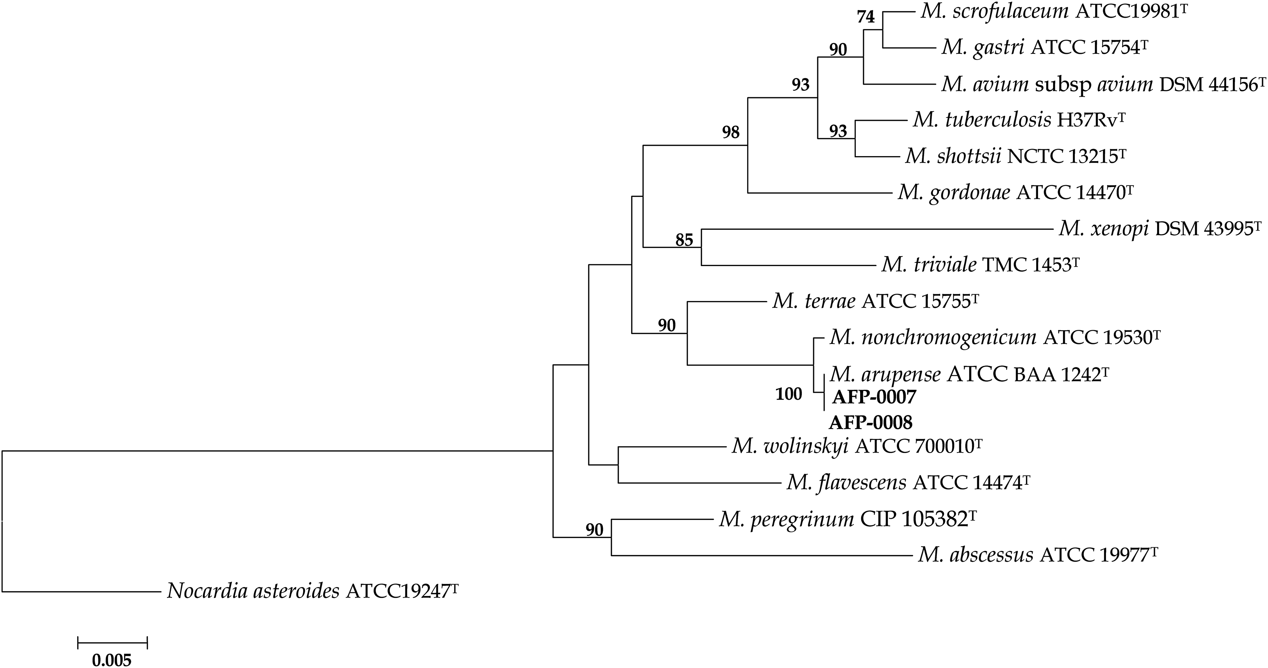
Nucleotide sequence accession numbers
The GenBank accession numbers of the 16S rRNA, hsp65 and rpoB gene for AFP-0007 as representative isolate are as follows: JQ617910, JQ617911 and JQ617912.
DISCUSSION
In TB-endemic countries, particularly Iran, mycobacterial specimens are presumed to be TB by presence of acid fast bacilli in smear microscopy and are not routinely cultured 7 or screened for NTM strains mainly due to lack of expertise methodology. Consequently, the patients are started on treatment with anti-TB drugs that have low activity or are inactive against infections caused by other species of mycobacteria. 8
M. arupense was introduced as novel species in 2006 as a result of five-year isolation and identification of strains which genotypically match to Mycobacterium sp. MCRO-6 from 65 human clinical samples. 9 Tendovaginitis, 10 tenosynovitis in a patient with diabetes mellitus, 11 flexor tenosynovitis in a patient, 12 pulmonary infection in malignant patient, 13 osteomyelitis of the wrist, 14 were reported due to infection with M. arupense. To our knowledge, the present study is the first report of pulmonary infections in two independent HIV-infected patients in a developing country. Our patients illustrated both the potential pathogenicity of M. arupense and its occurrence in Iran. Repeated isolation of strains in pure culture from a BAL specimen in Case one, as well as the severely immunocompromised condition and isolation of organism from blood samples in Case two, revealed that the isolates were clinically significant and met American Thoracic Society criteria. 8
Due to the importance of isolation of M. arupense among HIV patients in our study, conventional culture and identification tests were performed and the antimycobacterial therapy was undertaken based on susceptibility testing results for better management of the infection. Conventional methods failed to identify the clinical isolates at the species level, so, sequence analysis of 16S rRNA gene, rpoB gene and hsp65 region was carried out and definitive identity of the isolates was determined as M. arupense. The present report has highlighted the problems in detection and identification of NTM in clinical practice in developing countries like Iran, and misdiagnosis of such infections as TB. Application of molecular methods such as sequencing of the 16S rRNA, hsp65 and rpoB genes is crucial for the accurate identification of such opportunistic pathogens with ability to cause serious infections in immune compromised individuals such as AIDS patients.
In conclusion, M. arupense can cause pulmonary infection in immunocompromised patients such as those with HIV infection. Consequently, rapid molecular identification tests are required for accurate identification of mycobacterial species and control of related infections by proper antimicrobial treatment strategies.
Footnotes
ACKNOWLEDGEMENTS
The authors are grateful to research affairs, Ahvaz Jundishapur University of Medical Sciences, Ahvaz, Iran, for financial support (Grant No. 87101). Our appreciation goes to Director General of the Masoud Laboratory (Tehran, Iran) and Department of Infectious Diseases of Imam Khomeini Hospital, Tehran, Iran, for providing the patients’ medical records.