
Abstract
Background
The pathogenesis of male genital lichen sclerosus (MGLSc) is controversial. Incriminated factors include infection with human papillomavirus (HPV) and autoimmunity (e.g. Human Leukocyte Antigen [HLA]). To address the roles of HLA and HPV in MGLSc we studied adult Caucasian males with a clinical and histological diagnosis of MGLSc. The men in the study attended two specialised Male Genital Dermatoses Clinics between July 2011 and September 2012 and were selected and phenotyped from the clinical records. DNA was extracted from blood and paraffin-embedded biopsy sections, for HLA and HPV typing, respectively. HLA allele frequencies were compared with those derived from the UK-based Caucasian population. Eighty-eight cases of MGLSc were identified. HPV DNA was detected in 33/88 (37.5%) cases of MGLSc. HPV16 was the most prevalent type found: 11/88 (12.5%) MGLSc. No statistically significant HLA associations were established but HLA-B*35, -B*51, -C*15, -DRB1*04, -DRB1*10 (predisposition) and -DQA1*01 (protection) were revealed as alleles of interest. HPV16-associated MGLSc cases showed no statistically significant association with HLA genotype. The relationship between HPV and MGLSc suggests a passenger effect rather than a pathogenic role. HLA is not associated with MGLSc nor co-existent HPV16.
Introduction
Male genital lichen sclerosus (MGLSc) is an acquired cutaneous disease of uncertain aetiology. Lichen sclerosus (LSc) affects mainly the genitalia of men and women. MGLSc causes acute and chronic inflammation (balanoposthitis) with scarring that can result in sexual and urological dysfunction and is associated with an increased risk of penile squamous cell carcinoma (SCC). 1
Postulated aetiopathogenic factors in GLSc include genetic influences such as Human Leukocyte Antigen (HLA), autoimmunity, infection with human papillomavirus (HPV) (and other agents e.g. hepatitis C and Borrelia), environmental factors and chronic occluded exposure to urine. 1 A genetic influence for disease susceptibility is suggested by case reports of familial LSc.2,3 The HLA system contributes centrally to the immunocompetence of the human organism. HLA antigens are cell surface glycoproteins present on the surface of almost all cells that are responsible for lymphocyte recognition and “antigen presentation.” 4 HLA associations have been published for female genital LSc and MGLSc.5–8 HLA typing might be a useful surrogate for immunophenotypic susceptibility and prognostication. Moreover, HLA (protective and associative) relationships have been identified in patients with HPV-related diseases such as recurrent respiratory papillomatosis (HPV6/11), cervical cancer, head and neck cancers, cutaneous warts and non-melanoma skin cancer. 9 HLA type may determine the expression of HPV-associated diseases and the predisposition to certain HPV types. Also, HLA (especially HLA-C) may be a proxy for modulation of viral infections such as HPV. 10 Viral infections such as HPV have been certainly associated with penile SCC. This work was undertaken to explore further the roles and relationships of HLA and HPV in MGLSc.
Materials and methods
Ethical considerations
Ethical approval was obtained through the NHS Research Ethics Service (NRES); NRES Committee London – Riverside, REC references 07/H0706/58, amendment number 2.2. Research governance was secured from and assured by the Research and Development Department at the Chelsea & Westminster and University College Hospitals London NHS Foundation Trusts.
Patients
Adult males with a clinical and histological diagnosis of MGLSc were selected using simple, non-random (sequential) sampling between July 2011 and September 2012 from attendances at the specialised Male Genital Dermatoses Clinics, Chelsea & Westminster and University College Hospitals, London. Only patients with clinical and histological diagnosis of MGLSc were recruited for this study.
Human papillomavirus typing
DNA was extracted from archival formalin fixed and paraffin embedded (FFPE) tissue by using standard Qiagen protocols, with the addition of the following stringent anti-contamination procedures: extended tissue lysis, a longer elution time, and a smaller elution volume. DNA from cutaneous wart-associated HPV types was detected using a Luminex-based platform (HSL-PCR/MPG), using broad-spectrum primers and probes to detect both the types specified above and the additional HPV types 7, 4, 40, 43, 48, 50, 60, 65, 88, 91, 94, and 95. 11 Thus, 23 cutaneous wart-associated HPV types were detectable. Genital and Betapapillomavirus types were distinguished using 2 separate commercial reverse hybridization line probe assays. The genital assay identified 25 HPV types: high-risk HPV types 16, 18, 31, 33, 35, 39, 45, 51, 52, 56, 58, 59, 66, 68, and 70 and low-risk HPV types 6, 11, 34, 40, 42–44, 53, 54, and 74 (HPV SPF10- DEIA-LiPA25 system, version 1 [based on licensed Innogenetics technology]; Labo Biomedical Products, Rijswijk, the Netherlands). 12 , 13 The Betapapillomavirus assay contained probes that detected 25 HPV types: HPV 5, 8, 9, 12, 14, 15, 17, 19, 20, 21, 22, 23, 24, 25, 36, 37, 38, 47, 49, 75, 76, 80, 92, 93, and 96 (Diassay B.V., Rijswijk, The Netherlands). 14
Human leucocyte antigen tissue typing
DNA was extracted from fresh frozen whole-blood specimens, using standard Qiagen protocols (Qiagen, Crawley, United Kingdom) 15 and underwent HLA typing by a validated laboratory (Anthony Nolan Histocompatibility Laboratories, London), using sequence-specific oligonucleotide probes were used for HLA typing on a commercial Luminex-based platform (One Lambda LABtype SSO typing, VH Bio, Gateshead, United Kingdom). HLA allele frequencies for Caucasoid individuals derived from UK-based populations were also available as control subjects. 16
Statistics
Descriptive and analytical statistics for HPV and HLA data were computed with the use of software programs: Microsoft Excel and Epi Info (Centres for Disease Control and Prevention http://www.cdc.gov/epiinfo/[downloaded 1st October 2010]). For categorical data, the chi-squared test was utilized together with Yates’ correction for continuity. Advice regarding HLA allele counting and immunophenotypic analysis was provided by the Anthony Nolan Histocompatibility Laboratory. A substantial Bonferroni correction for multiple testing was applied to each HLA analysis, to account for all potential allele comparisons. Corrected p-values of less than 0.05 were regarded as significant.
Results
Eighty-eight cases of MGLSc were studied (supplementary material 1). The mean age at diagnosis of MGLSc was 47 years (range 13–81 years; median 45 years). Five of the 88 (5.7%) reported a previous history of genital warts but HPV-infected cells (koilocytosis) were not seen in the penile biopsies. Three of the 88 (3.4%) reported no apparent history of genital warts but koilocytosis was seen in the penile biopsies. Twelve of 88 (13.6%) were ex-smokers and fifteen of 88 (17%) were current smokers. Eight of 88 (9.1%) had associated organ-specific autoimmune conditions including vitiligo and thyroid disease. None had extragenital LSc on complete cutaneous examination. Eight men had eczema and six men had psoriasis.
Human leucocyte tissue typing data
The HLA profiles of 88 cases of MGLSc are shown in supplementary material 2. Only the alleles of relevant associations that have been investigated are shown (Table 1).
HLA alleles and MGLSc – only alleles of potential interest are listed.

CI, confidence interval; MGLSc, male genital lichen sclerosus.
Overall, there were no significant differences in frequency among the HLA-A and HLA-DQB1. There was a significantly increased frequency of -B*35 (p = 0.0033), -B*51 (p = 0.015), -C*15 (p = 0.00083), -DRB1*04 (p = 0.0064) and -DRB1*10 (p = 0.02). There was a reduced frequency of -DQA1*01 in the patient group and this was statistically significant following Yates’ correction (p = 0.05). After Bonferroni correction, the above differences no longer reached statistical significance.
Human papillomavirus typing data
HPV was detected in 33/88 (37.5%) cases of MGLSc. A wide spectrum of HPV types was detected. Genital types were the most prevalent at 30/88 (34.1%) followed by 6/88 (6.8%) beta types and 4/88 (4.5%) cutaneous (wart) types (Table 2). HPV16 was the most prevalent HPV type: 11/88 (12.5%). Lesions have been defined as harbouring a mixed infection if more than one HPV type from any one HPV group (cutaneous, genital or beta) is identified. Fourteen cases of MGLSc harbouring multiple cutaneous or genital HPV types were identified.
HPV in MGLSc.
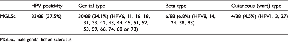
MGLSc, male genital lichen sclerosus.
HLA and HPV interaction data
Subgroup analysis has been performed according to HPV16 infection status because this oncogenic type was the most prevalent genotype seen in this work. Further analyses for other HPV types were not attempted as the numbers were too small.
When HPV16-infected MGLSc cases were compared with controls no significant difference in the allele frequencies was observed in any of the tested HLA classes.
Discussion
This is, to our knowledge, the largest and most comprehensive study of HPV and HLA profiles in MGLSc (and the first of their putative interaction) using contemporaneous, broad-spectrum, specific and sensitive molecular technology.
HPV has been linked with LSc.17–19 The prevalence of HPV infection in asymptomatic men ranges from 1.3 to 72.9%, with most studies reporting ≥20%. 20 Variation in prevalence is likely to be due to differences in sampling techniques, the populations studied, anatomic sites sampled (e.g. scrotum, shaft, glans penis/coronal sulcus) and the HPV DNA detection method employed. The use of a more sensitive sampling technique i.e. a pre-wetted Dacron swab rather than a cytobrush or collecting a urine sample can result in a higher HPV prevalence estimate. Similarly, HPV prevalence is higher when samples are collected from a greater number of anatomic sites.
Drut et al. 17 detected 16/23 (70%) HPV DNA in prepuces of boys treated surgically for phimosis with a histological diagnosis of MGLSc. Another study reported no evidence of HPV DNA in 82 boys who underwent circumcision for persistent phimosis, including six with histological evidence of MGLSc. 21 von Krogh et al. 22 found high-risk HPV DNA in 4/19 (21%) men with a clinical and histological diagnosis of MGLSc. Nasca et al. 19 reported the presence of HPV DNA in 8/46 (17.4%) cases of clinically and histologically proven MGLSc compared with 4/46 (8.7%) healthy controls. HPV DNA was positive in 6/18 (33%) histologically proven MGLSc cases described by Prowse et al. 18
In terms of HPV subtypes, Drut et al. 17 detected HPV6, 16 and 18, amongst which type 6 was the most prevalent at 8/16 (50%). von Krogh et al., 22 used low-risk (HPV types 6, 11, 34, 42, 43 and 44) and high-risk (HPV types 16, 18, 31, 33, 35, 39, 45, 51, 52, 54, 56 and 58) oligoprobe mixtures and hence the breakdown of each HPV subtype was not established. Nasca et al. 19 detected HPV16, 18 and 45, amongst which type 16 was the most prevalent at 6/8 (75%). In the work by Prowse et al., 18 HPV16 was present in all HPV positive MGLSc samples: 3 cases with HPV16, 1 case with HPV16 and 18, 1 case with HPV16 and 33, and another case with HPV16, 18, 33, 51. High-risk HPV was detected in six of 50 (12%) circumcised foreskin samples in asymptomatic boys before first sexual intercourse. All positive samples showed HPV16. 23 Regarding the differential prevalence of HPV types in men, an Americas study showed that 50.5% of men were positive for at least one known HPV type and 25.7% of men were positive for multiple HPV genotypes. 24 In our work, HPV16 was the most prevalent HPV type at 11/88 (12.5%) in keeping with the published literature. All HPV subtypes reported in the literature (HPV types 6, 16, 18 and 51) were seen in our work except HPV type 33. Multiple HPV infection was seen in 14/88 (15.9%). In addition, several previously unreported HPV subtypes: 1, 3, 11, 14, 24, 27, 31, 42, 43, 45, 52, 53, 59, 66, 74, 68 or 73, 93 have also been found in MGLSc in our work. All these findings raise the question whether some of these HPV were merely skin surface, background “passenger HPV” or “driver HPV.”25 Driver HPV is causally significant in a lesion, whereas passenger HPV refers to a bystander or opportunistic infection that is incidental to disease/tumour pathogenesis that may be present in only a minority of cells within pathological tissue/tumour mass. A descriptive study of a large number of MGLSc showed no clinical correlation with HPV infection. 26 Furthermore, no specific HPV-associated gene expression patterns were found. 27
Our work, showing HPV DNA to be present in 33/88 (37.5%) of men with GLSc, is not dissimilar to previous virological findings in adult MGLSc. The previous studies are smaller numbers and employed limited HPV typing techniques where only certain genital types HPV were routinely tested. Other HPV types such as cutaneous and beta types have not routinely been sought by other investigators. Our work describes the largest number of GLSc studied virologically and has deployed novel, validated molecular HPV typing techniques capable of detecting a broad spectrum of HPV types-73 in all – cutaneous (wart), genital and beta HPVs. The PCR-based detection system used in our study is especially efficient in amplifying smaller amplicons from formalin-fixed, paraffin-embedded tissue samples which often yield poorly amplifiable DNA. Amplicons are detected in an enzyme-linked immunosorbent assay using a mixture of HPV-specific probes that recognize a broad range of genotypes in comparison to type-specific PCR.
On balance, in the light of our findings and those of others, it is arguable that HPV is a passenger phenomenon in MGLSc and not pathogenic. This argument is further sustained by the following considerations.
Firstly, none of the MGLSc patients had clinical evidence of genital warts at presentation; although five of the 88 (5.7%) reported a previous history of genital warts, HPV-infected cells (koilocytes) were not seen in their penile biopsies; three of the 88 (3.4%) reported no apparent history of genital warts but koilocytosis was seen in the penile biopsies. Yet 33/88 (37.5%) of MGLSc in this work showed the presence of HPV DNA. Techniques for virological presence are clearly more sensitive than clinical assessment (patient history and clinician examination) and histology, but the time dynamic and field change propensities of HPV infection will impact on these discrepancies.
Secondly, the PCR techniques only detect the presence of HPV. It is unable to differentiate whether these are driver or passenger HPV. 28 Therefore, the possibility exists that HPV detected by PCR in MGLSc could be background rather than driver HPV.
Finally and independently, a recent review by Hald et al. has concluded similarly; 75% percent of sexually active people acquire HPV during their lifetime, and therefore HPV alone is not likely to be a cause of LSc. 29
This is the second study of putative HLA associations in adult MGLSc and is larger and more comprehensive than that of Azurdia et al. 8 Our initial results suggested that -B*35, -B*51, -C*15, -DRB1*04 and -DRB1*10 were associated with susceptibility to MGLSc and -DQA1*01 with protection against MGLSc but statistical significance was lost with multiple corrections. The majority of previous studies looking at the association between GLSc and HLA have been done in women: it has been reported that HLA-DQ7 and DRB1*12 confer susceptibility whereas DRB1*03 protects against vulvar LSc. 7 HLA-DQ3 and specifically -DQ7 are more frequent in vulvar LSc than controls, suggesting a role in susceptibility to GLSc. 5 , 6 HLA-DR17 was found to be less frequent with GLSc than controls. 5 Further analysis indicated that the -DR4/-DQ8 haplotype was less frequent in vulval LSc with structural changes compared with those without, implying a role in disease progression. 9 A marginally increased frequency of DR11 (p = 0.05), DR12 (p = 0.04) and DQ7 (p = 0.05) were previously claimed in a small study of MGLSc. 8 However, these associations were not seen in our work.
This is the first study to investigate the interactive relationship of HLA and HPV in MGLSc. As previously discussed, genital HPV infection is extremely common. However, only a small fraction of infections would appear to progress to persistent disease and cancer, suggesting that other determinants contribute to the pathogenesis. One potential cofactor may be the host cellular immune response to HPV infection, mediated by HLA-restricted T lymphocytes. The HLA haplotype of an individual may lead to clearance or predispose to persistence of HPV infection. The exact nature and mechanism whereby the host gene products regulate HPV oncogene expression and prevent HPV-induced cell immortalization remain elusive. While certain alleles have a documented detrimental effect on HPV infection, some HLA alleles have a protective role. 30
In conclusion, the role of HPV in MGLSc is likely to be a passenger phenomenon and not pathogenic. Immunogenotype seems not to play a role in the pathogenesis of MGLSc but larger patient numbers are needed to investigate the relationships, particularly any association with HPV16 and downstream risk of SCC; this work is in progress. The precise pathogenesis of MGLSc remains unknown but a compelling body of evidence points to the pivotal role of chronic intermittent occluded urinary exposure and there are here also translatable implications for clinical and investigative attitudes to GLSc in children and women.
Supplemental Material
sj-pdf-1-std-10.1177_0956462420949395 - Supplemental material for Immunogenetics and human papillomavirus (HPV) in male genital lichen sclerosus (MGLSc)
Supplemental material, sj-pdf-1-std-10.1177_0956462420949395 for Immunogenetics and human papillomavirus (HPV) in male genital lichen sclerosus (MGLSc) by Tang Ngee Shim, Catherine A Harwood, Steven GE Marsh, Frances M Gotch, Wim Quint, Maurits N de Koning, Nick Francis, Charles Jameson, Alex Freeman, Suks Minhas, Michael Dinneen, Asif Muneer and Christopher B Bunker in International Journal of STD & AIDS
Supplemental Material
sj-pdf-2-std-10.1177_0956462420949395 - Supplemental material for Immunogenetics and human papillomavirus (HPV) in male genital lichen sclerosus (MGLSc)
Supplemental material, sj-pdf-2-std-10.1177_0956462420949395 for Immunogenetics and human papillomavirus (HPV) in male genital lichen sclerosus (MGLSc) by Tang Ngee Shim, Catherine A Harwood, Steven GE Marsh, Frances M Gotch, Wim Quint, Maurits N de Koning, Nick Francis, Charles Jameson, Alex Freeman, Suks Minhas, Michael Dinneen, Asif Muneer and Christopher B Bunker in International Journal of STD & AIDS
Footnotes
Declaration of conflicting interests
The authors declared no potential conflicts of interest with respect to the research, authorship, and/or publication of this article.
Funding
The authors disclosed receipt of the following financial support for the research, authorship, and/or publication of this article: This work was supported by Skin Treatment and Research Trust (START) and Sir John Fisher Foundation.
References
Supplementary Material
Please find the following supplemental material available below.
For Open Access articles published under a Creative Commons License, all supplemental material carries the same license as the article it is associated with.
For non-Open Access articles published, all supplemental material carries a non-exclusive license, and permission requests for re-use of supplemental material or any part of supplemental material shall be sent directly to the copyright owner as specified in the copyright notice associated with the article.