



Abstract
Epididymitis is a common cause of scrotal pain presentation in sexual health clinics; however, it is unclear what fraction is attributable to transmissible infections. We, therefore, reviewed the aetiologies causing epididymitis. A retrospective data analysis of all cases of epididymitis diagnosed from January 2018 to December 2018 in three sexual health clinics was conducted, collecting demographics, results, management and symptom resolution at two weeks follow up. A total of 127 cases of epididymitis (mean age 32 years, heterosexual 97, MSM 30) were included. Among them 14 cases (11%) were caused by sexual transmitted infections (<35 years n = 9; >35 years n = 5): seven cases of chlamydia, six gonorrhoea, one syphilis and one trichomonas vaginalis. There were three cases of urinary tract infection diagnosed. All cases were treated with antibiotics recommended by the British Association for Sexual Health and HIV (BASHH). At two weeks follow up post-treatment 10 (7%) were symptomatic; 91% did not attend for follow up. Sexually transmitted infections were associated with acute epididymitis in 11% of this study cohort.
Introduction
Acute epididymitis is a clinical syndrome consisting of pain, swelling, and inflammation of the epididymis with or without testicular involvement. Pain is usually of gradual onset and localized behind the testes, followed by scrotal swelling in 24 to 48 hrs. 1 It is usually unilateral, but bilateral epididymitis can also occur. E Causes of epididymitis can be bacterial, viral, and inflammatory. Sexually transmitted infections (STI), most commonly Chlamydia trachomatis and Neisseria gonorrhoeae, are common causes in men below 35 years of age. Urinary tract infection (UTI) pathogens are common causatives for men above 35 years of age, 2 especially if they underwent recent urological procedures, and in men of any age practicing insertive anal sex. 3 Symptoms of urethritis, urethral discharge indicates an STI cause likely, while dysuria, frequency, urgency, and fever are thought to be indicative of a urinary tract infection. A clinical finding of unilateral firm swelling and tenderness of the epididymis is sufficient for clinical diagnosis of epididymitis and a recommendation to administer treatment. 4 A delay in treatment can lead to complications - scrotal abscess, reactive hydrocele, testicular infarction, orchitis, and chronic epididymitis. 4 Investigations in our sexual health clinics for epididymitis at the time of this study included urethral gram staining, urethral gonorrhea culture, first-pass urine for chlamydia and gonorrhoea nucleic acid amplification tests (NAAT), urine dipstick and mid-stream urine (MSU) culture, and seroloical tests for syphilis and HIV. 4
Empirical antibiotics for epididymitis as recommended by British Association for Sexual Health and HIV guidelines 4 are a combination of ceftriaxone 1 gram intramuscular one dose, followed by doxycycline 100 mg oral twice daily for 14 days. If gonorrheoa is ruled out by microscopy or NAAT, ceftriaxone may be omitted. Quinolones, preferably ofloxacin 200 mg oral twice daily for 14 days is recommended for UTI pathogens. If there are associated severe systemic symptoms and sepsis, hospital admission and intravenous antibiotics are considered.
We conducted a review of cases of epididymitis diagnosed in our sexual health clinics to identify common causes and outcomes of management.
Methods
Data on cases of epididymitis diagnosed from January 2018 to December 2018 were collected from the electronic patient record from our three sexual & reproductive health clinics including demographics, STI screening results, and treatment provided. Data were analyzed using Microsoft Excel software. Epididymitis was assumed to be resolved if the patient did not attend for follow up at two weeks post-treatment.
Results
A total of 127 cases of epididymitis were included. Mean age 32 years (Range: 15–67 years), heterosexual 97 (76%), men who have sex with men (MSM) 30 (24%), White ethnicity 75 (59%), Black African/Caribbean 24 (19%), other ethnicities 28 (22%). 14 individuals (11%) were diagnosed with sexually transmitted infection (<35 years n = 9; >35 years n = 5). There were seven cases of chlamydia, six gonorrhoea, 1 syphilis and 1 trichomonas vaginalis (Figure 1).

Incidence of sexually transmitted infections.
A urine dipstick was performed on 95 (75%) of our cohort and 20 (20%) had a positive urine dipstick for leukocyte esterase and/or nitrites. A mid-stream urine (MSU) sent for 54 (42%) patients. Positive growth was isolated from three MSU culture (Table 1).
Urine dipstick results and UTI confirmation.

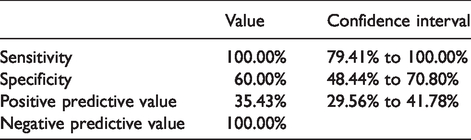
A total of 103 (81%) patients underwent urethral gram stain microscopy and in 38 (36%) non-specific urethritis was diagnosed. Among them, six had a confirmed STI by NAAT results and 2 had a confirmed urinary tract infection. Absence of pus cells (<5 polymorphonuclear leucocytes per high power field x1000) on urethral gram stain and negative leukocyte esterase on urine dipstick had a 100% correlation with the absence of STI in our cohort (Table 3). Among patients who did not have urethral gram stain microscopy (n = 24), five had an STI diagnosis.
All patients were given antibiotics as recommended by BASHH guidelines after clinical diagnosis at the first visit. At two weeks follow up 10 (7%) were symptomatic, among which 1 had an epidydimal cyst, 1 required a change in antibiotics, 1 developed chronic epididymitis, and seven did not attend further follow up and considered to have resolved symptoms. A major proportion (81%) did not attend for follow up after the first visit and assumed to have achieved symptoms resolution. No systemic or urological complications requiring in-patient admission were identified in our cohort.
Discussion
Sexually transmitted infections accounted for only 11% of epididymitis in our annual cohort. NAAT tests for chlamydia and gonorrhea in sexual health clinics of England have a sensitivity of 92% and specificity of 99% for male urine. 5 Urethral chlamydia and gonorrhoea infection were causative for approximately 5% of epididymitis each in our cohort. This is significantly low when compared to studies from developing countries where up to 78% of cases can be caused by chlamydia. 6 A previous study in the US found chlamydia causing two-third cases of idiopathic epididymitis. 7
The etiological pathogen should not be assumed based on the age cut off (<35 years & >35 years) for men and should be guided by sexual practices. Chlamydia and gonorrhoea caused epididymitis among both these age groups (Table 2). Among confirmed UTI, two men were from <35 years age group in our cohort (1 MSM, 1 Heterosexual). STIs were an identified as a cause of epididymitis in a higher proportion of MSM compared to heterosexual men (17% vs 9%). There was one case of syphilitic orchitis in MSM in our cohort. Syphilitic orchitis is a known but a rare manifestation of early infectious syphilis.
STI and UTI by age group.

Urinary tract pathogens were isolated in only 2% of cases in our study. Urine dipstick was reported to be useful to rule out a UTI if both leucocyte esterase and nitrite were negative. 9 However, a positive test should be confirmed by MSU culture. A negative urine leukocyte esterase on dipstick correlated 100% with no growth in MSU culture in our cohort. Not all patients in our cohort had urine dipstick and MSU culture performed which limits this conclusion. Non-STI uro-genital pathogens, mainly E.coli, are a common cause of epididymitis in developing countries – up to 28% in one study. 10 Multiplex PCR based tests for UTI can improve the detection rate of uropathogens when widely available. 11 Identification of UTI pathogens should trigger a referral pathway to urology services for further evaluation of prostate and urinary tract pathology.
Trichomonas vaginalis (TV) was identified in one patient by urine NAAT in our study. It is considered a rare cause of epididymitis. 12 Only symptomatic men of black African or Caribbean ethnicity were tested for TV NAAT in our clinics due to higher prevalence of TV than other ethnic groups. 13 TV was reported to be associated with 10% of infective epididymo-orchitis in another Ukrainian study. 14 Further studies in the UK are required to explore its association with epididymitis.
Mycoplasma genitalium NAAT was not a part of our routine testing protocol during the time of our study. STI negative non-specific urethritis in our cohort (n = 28; 73%) could have been caused by any untested pathogens - M. genitalium, Herpes virus, Ureaplasma urealyticum, adenovirus, and anaerobic bacteria. The resolution of symptoms in our study was assumed if the patient did not attend for follow up after 2 weeks post-treatment. It is unknown if they attended other sexual health clinics or urology services post-treatment.
There are a lack of data on the incidence of trivial trauma during coitus causing testicular pain. In one middle eastern setting, trauma was reported to be causative for 9% scrotal pain presentation in the emergency department. 15 Referred pain from lumbar or sacral nerve roots impingement, urolithiasis, varicocele, abdominal aortic aneurysm, retrocecal appendicitis, and retroperitoneal tumor can cause scrotal pain.
Viruses like mumps, rubella, coxsackie, echovirus, lymphocytic choriomeningitis virus, parvovirus are common etiologies of orchitis among adolescents. 16 Sexual health clinics do not routinely test for viral pathogens for epididymitis. The findings from this study are setting specific in a well resourced clinical setting. It may not be generalized to other developing health care settings where tropical infections like tuberculosis, brucellosis, schistosomiasis, and hydatidosis should be explored. 17
BASHH recommends commencing antibiotics at the first visit after clinical diagnosis. This has resulted in the potential overuse of antibiotics with no pathogens identified on urine NAAT and MSU culture in our cohort. Treatment with broad-spectrum antibiotics for epididymitis in the absence of pathogen is a concern in the era of increasing multidrug resistance. 18 A negative urine dipstick for leukocyte esterase and absence of pus cells (<5/hpf) on urethral smear gram stain microscopy had 100% negative predictive value for STI in our cohort (Table 3). Clinicians should consider this observation to study a protocol variation for conservative treatment and follow up for such cases to promote antibiotics stewardship in the community.
Combined value of urethal smear gram staining and urine dipstick for leucocyte esterase to diagnose STI [N = 111].
Footnotes
Acknowledgements
The authors thank Andrea Svinciakova for her support in data collection.
Declaration of conflicting interests
The authors declared no potential conflicts of interest with respect to the research, authorship, and/or publication of this article.
Funding
The authors received no financial support for the research, authorship, and/or publication of this article.
