



Abstract
Background
Syphilis is known as the “great imitator” because of its polymorphic clinical manifestations. Condyloma lata are an uncommon mucocutaneous manifestation of secondary syphilis, generally localized in intertriginous areas, such as the genitals and anus. Extragenital localization of condyloma lata is considered unusual.
Methods
A case study of extra-genital condylomata is presented. To contextualise the case, a literature review of extra-genital condylomata lata was subsequently undertaken. The authors searched MEDLINE/PubMed, Scopus and Excerpta Medica/EMBASE English-language sources using the following keywords: “condyloma lata”, “condylomata lata”, and “condyloma latum”.
Results
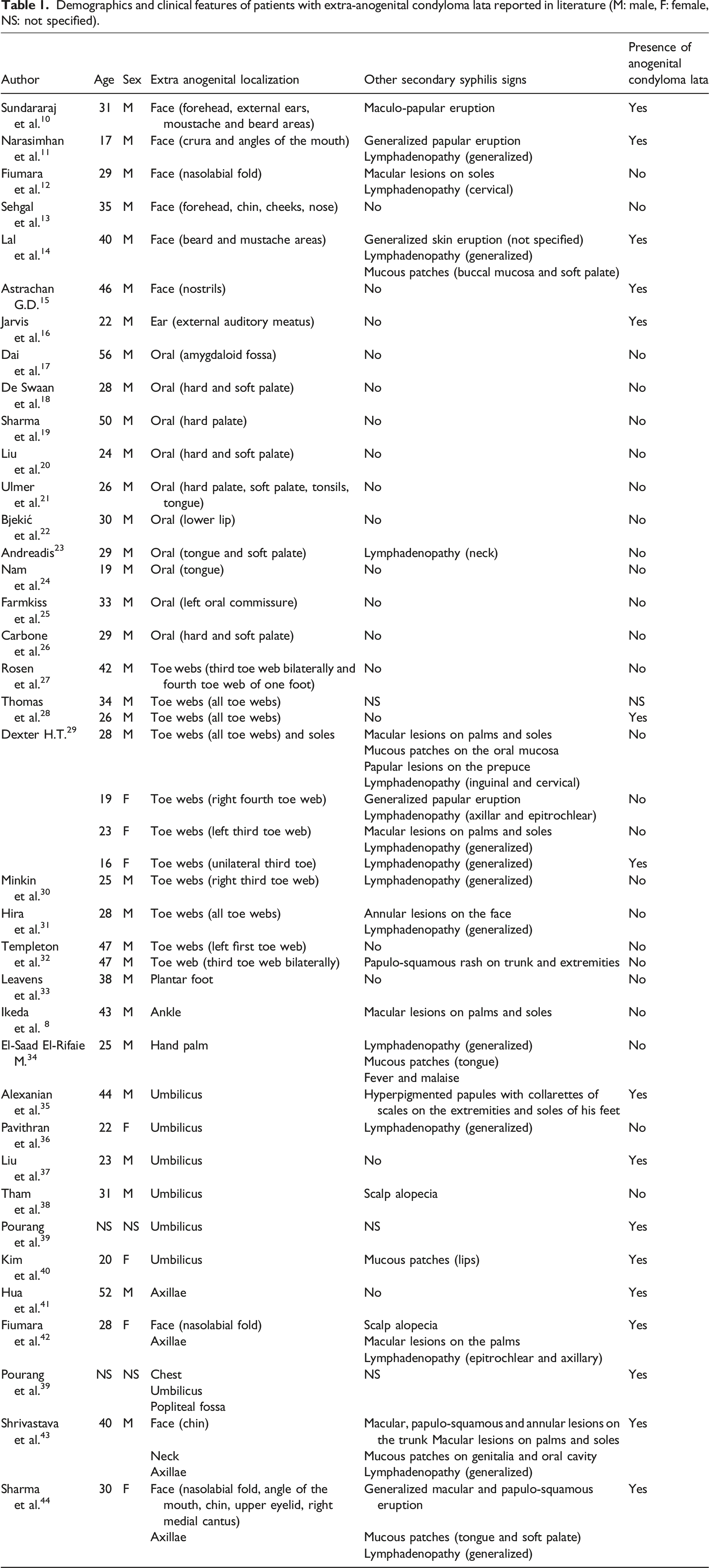
Thirty-five papers (from 1940 to 2021) describing an extra-anogenital localization of condyloma lata were found and are summarized in Table 1. Patients were mainly males (82.1%), with a mean age of 31.9 years. Most of them showed other manifestations of secondary syphilis (53.9%). In a minority of cases (39.0%), concomitant anogenital condyloma lata were present, thus making the diagnosis easier. The toe web localization was the most documented (26.2%) followed by the oral cavity (23.8%).
Conclusion
In the presence of extra-genital condyloma lata, the differential diagnosis is not always clear, especially when no other muco-cutaneous lesions are observed. In the case of eroded or wet lesions involving any cutaneous fold, associated or not with other cutaneous manifestations, a sexual history should be obtained, and syphilis must be considered.
Introduction
Syphilis is a bacterial sexually transmitted infection (STI) with worldwide diffusion caused by the spirochetae Treponema pallidum subspecies pallidum (TP).1–3 Its course is conventionally divided into three stages interspersed by latency periods.1–3 The clinical manifestation of primary syphilis is generally univocal, with a chancre appearing in the site of the infection, associated with regional lymphadenopathy. Conversely, secondary syphilis can produce a wide variety of signs and symptoms,1–3 enough to give syphilis the pseudonym of “great imitator”. 4 The authors report a case of secondary syphilis presenting with multiple condyloma lata with both genital and extragenital involvement.
Case report
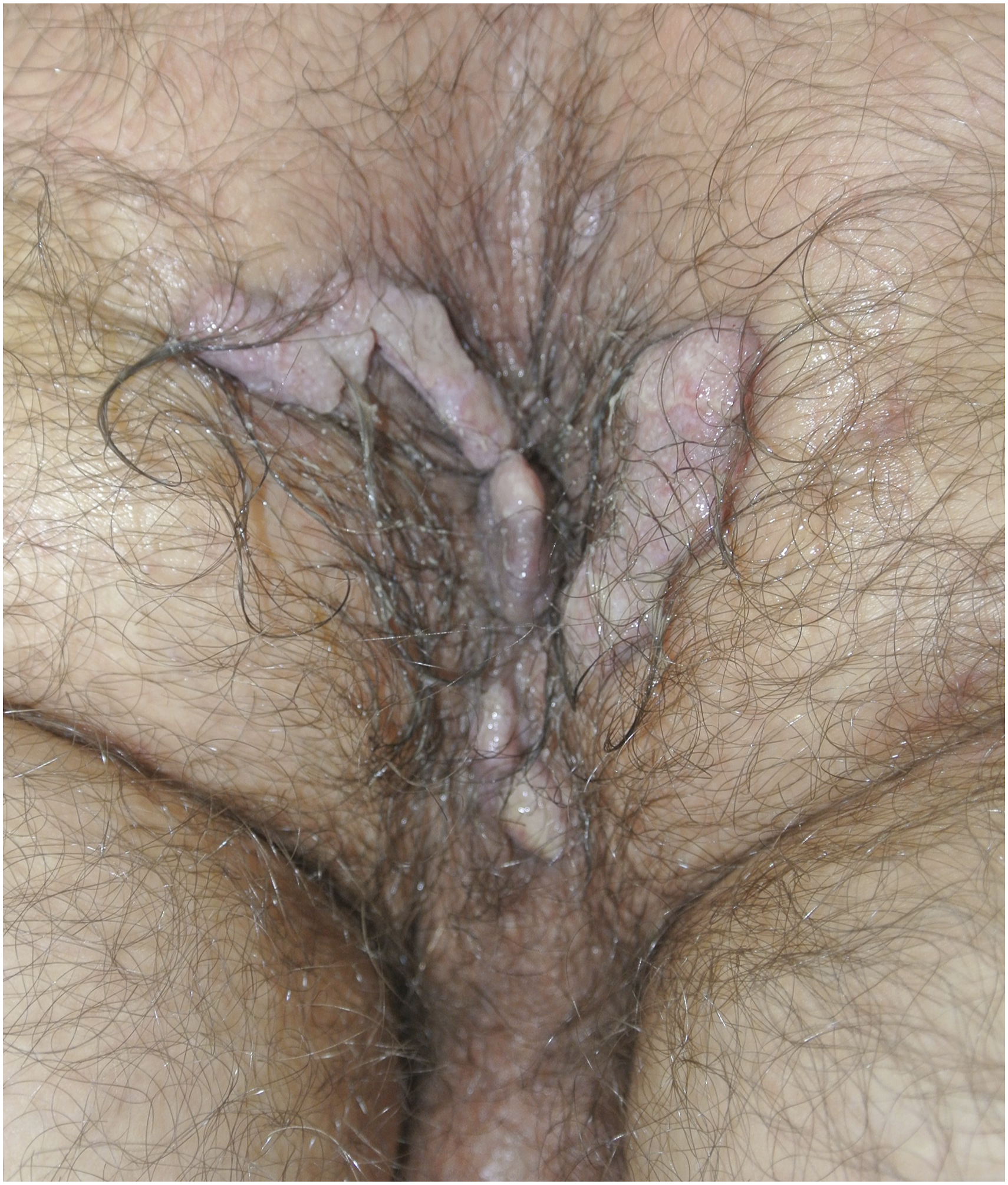
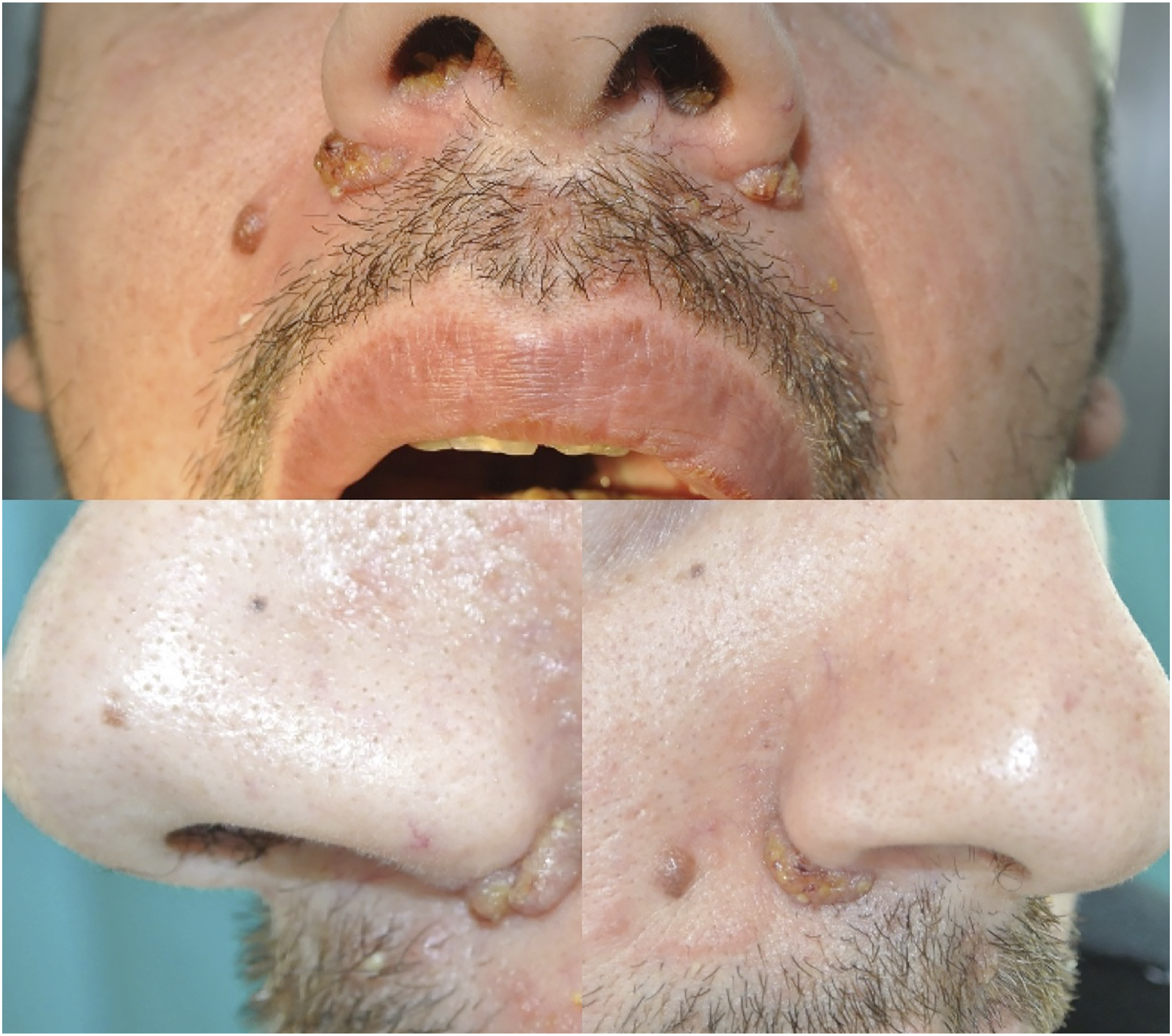
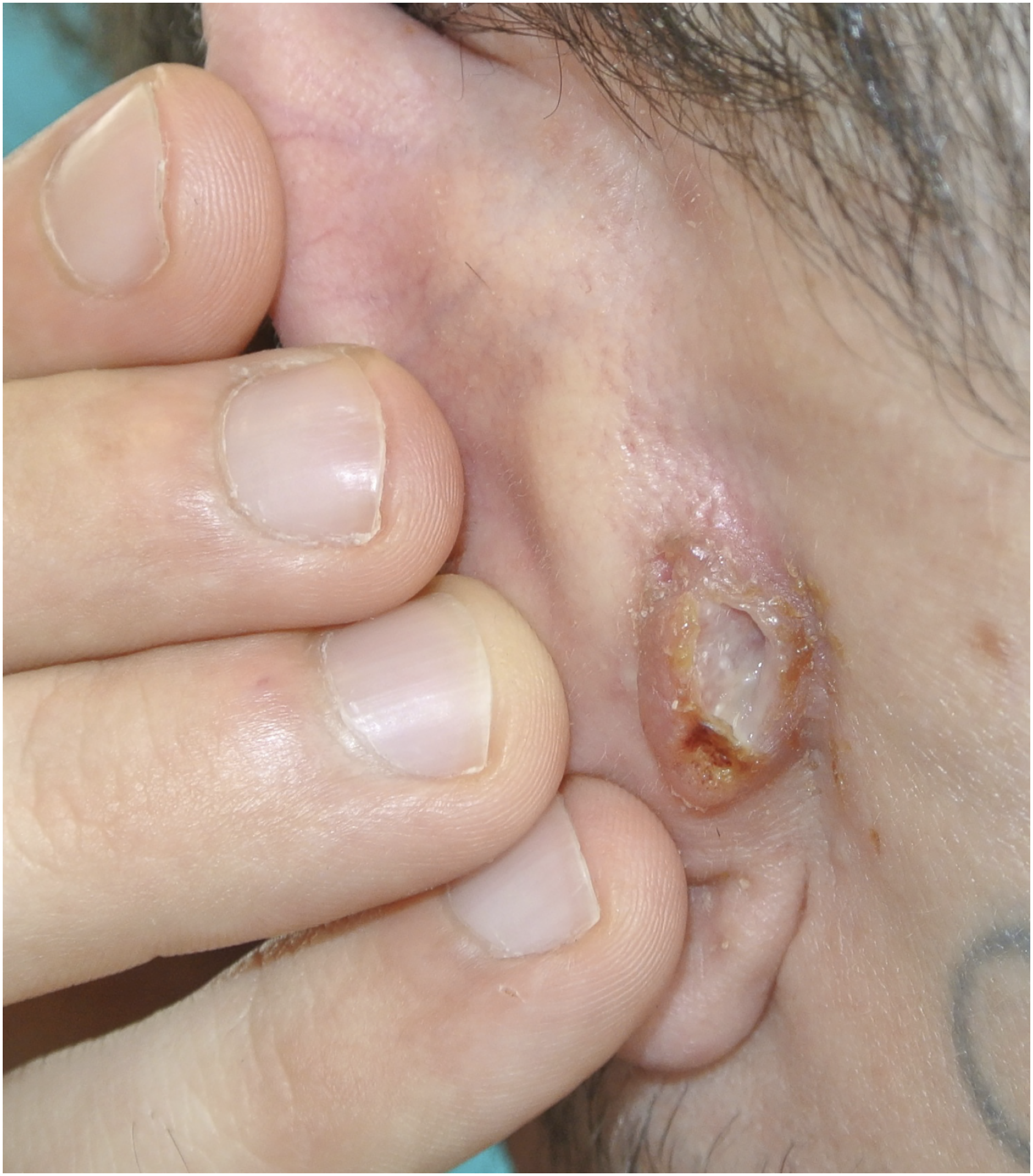
A 30-year-old male came to our attention complaining of multiple cutaneous asymptomatic lesions which had appeared during the previous month. He was in good general health, without a history of drug assumption, but he reported numerous episodes of heterosexual unprotected intercourse with multiple partners. Physical examination revealed a few erythematous indurated annular lesions of the penis (Figure 1) and numerous papules coalescing into multilobulated moist cauliflower-like masses in the perineal region (Figure 2); masses similar to these, but of a smaller size, involved the nostrils as well as the nasal and the retroauricular folds (Figures 3 and 4). Moreover, small, rounded alopecic patches, with a “moth-eaten” appearance, were visible on the scalp. No other cutaneous or mucosal manifestations were observed, but a diffuse lymphadenopathy was detected. The results of routine blood tests (complete blood count, liver and renal function) were normal, while enzyme-linked immunosorbent assay (ELISA) for anti-TP IgM and IgG was positive. Treponema Pallidum Particle Agglutination (TPPA), positive at a titer of 1:80, and Rapid Plasma Reagin (RPR), positive at a titer of 1:64, confirmed the diagnosis of syphilis. The diagnosis of secondary syphilis was supported by the demonstration of the presence of TP in the perineal and retroauricular lesions through a nucleic acid amplification test (NAAT), performed using a commercial, CE marked, Real Time PCR kit targeting a 47 kDA portion of the gene encoding the membrane lipoprotein of TP. Given their clinical aspect and the presence of TP within the lesion, the flexural masses were diagnosed as condyloma lata. According to the International Union Against Sexually Transmitted Infections (IUSTI) guidelines,
5
a single dose of penicillin G benzathine (2.4 million units intramuscularly) was administered. A complete resolution of all the lesions and the negativization of RPR was recorded after 3 months. Annular syphiloderm of the penis. Condyloma lata of the perineum characterized by moist multilobulated cauliflower-like masses with superficial erosion. Condyloma lata of the nostrils and nasal folds, mimicking viral warts. Retroauricular condyloma latum presenting as an ulcerated nodular lesion.
Discussion of the case
In its secondary stage, syphilis becomes a systemic disease due to hematic diffusion of TP, which typically occurs 3–12 weeks after the resolution of the chancre but can be concurrent as well. As opposed to the primary stage, secondary syphilis can produce a wide variety of signs and symptoms,1–3 enough to give syphilis the pseudonym of “the great imitator”. 4 Secondary syphilis most commonly consists of adnexal and mucocutaneous lesions but can also show generalized lymphadenopathy, malaise, sore throat, body aches and low-grade fever. Organic involvement is rare, but possible. Although syphilitic roseola and papulous lenticular syphiloderm are the most typical cutaneous presentations, numerous other types of syphiloderms have been described.1–4 In case of annular manifestations, different dermatoses - such as dermatophytosis, annular granuloma and subcutaneous lupus erythematosus - should be considered. In such cases, a positive serology is not sufficient for diagnosis and a histopathological examination is generally required. 6 In our case, the demonstration of TP presence within the lesions supported the diagnosis of secondary syphilis making skin biopsy unnecessary. Although no NAAT for TP has been approved by the U.S. Food and Drug Administration, the method that specifically amplifies the tpp47 gene is well known for its high sensitivity and high specificity. 7 In our report, the flexural vegetating masses were consistent with condyloma lata, which are muco-cutaneous manifestation occurring in up to a third of patients with secondary syphilis. Condyloma lata (also named condylomata lata) are characterized by papules or nodules, very rich in treponemas, that coalesce in warm, moist areas; the surface can be smooth, verrucous, hypertrophic, or covered with exudate. Their typical localizations are the anogenital region and the medial thighs, given their association with heat, moisture and friction.1–3 The main differential diagnosis is condyloma acuminatum due to Human Papilloma Virus infection. Extragenital localization of condyloma lata is considered to be unusual. 3 To the best of our knowledge, we report here the first case of condyloma lata behind the ears; literature reports only the involvement of the external auditory meatus. In the presence of laboratory-confirmed syphilis, the diagnosis of condyloma lata is mainly clinical, even though the TP-detection by DFM or TP-NAAT can be useful, avoiding invasive procedures such as skin biopsy. Dermoscopy can be helpful too: red to milky-red globules, glomerular vessels and a whitish pink network can be observed on the raised border, while a yellowish structureless area and multiple, white, small, round structures can be seen in the center of the lesion.8–9
Literature review - Methods
Extragenital localization of condyloma lata is considered to be unusual. The literature contains only case reports and a few case series. We searched MEDLINE/PubMed, Scopus and Excerpta Medica/EMBASE english-language sources using the following keywords: “condyloma lata”, “condylomata lata”, and “condyloma latum”. Only papers reporting a case or a case series of extra-genital condyloma lata were selected. Most of the authors validated the diagnosis of condyloma lata through histological examination or dark-field microscopy. These are the main limitations: first, most of the data come from single case reports (only four case-series were found), consequently the laboratory diagnosis is not standardized. Second, in two cases the diagnosis of condyloma lata is not specified, in seven cases the diagnosis is only clinical and in two of them the iconography is not qualitatively valid. Lastly, some clinical data are missing in three different cases.
Literature review - Results
Demographics and clinical features of patients with extra-anogenital condyloma lata reported in literature (M: male, F: female, NS: not specified).
Conclusion
In conclusion, although the anogenital area is the typical localization, condyloma lata have been described in many other sites, making differential diagnosis not always straightforward, especially when no other muco-cutaneous lesions are observed. Therefore, in case of eroded or wet lesions involving any cutaneous fold, with or without other cutaneous manifestations, a sexual history should always be obtained, and syphilis must be considered. In addition to specific serology, at least one direct test for TP-detection (DFM or TP-NAAT) should be performed in order to confirm the diagnosis. Syphilis remains “the great imitator” and a high index of suspicion is required.
Footnotes
Acknowledgements
The patient in this manuscript have given written informed consent to publication of her case details.
Declaration of Conflicting Interests
The author(s) declared no potential conflicts of interest with respect to the research, authorship, and/or publication of this article.
Funding
The author(s) received no financial support for the research, authorship, and/or publication of this article.
Contributorship
F.B. and G.M. equally contributed (data collection and writing) to this work and both qualify as first authors; S.R. contributed to data collection and first revision of the manuscript; M.C. and A.V.M. reviewed the manuscript. All authors have contributed significantly, and all authors are in agreement with the content of the manuscript.
