



Abstract
Primary colonic lymphoma is an infrequent malignancy among other large bowel malignancies, and the risk of the spread of tumor cells through a spleno-colic fistula is a unique finding and hence noteworthy. We report a case of a 55-year-old man living with HIV on anti-retroviral treatment for 12 years, who presented to the emergency room with complaints of generalized weakness and left-sided abdominal discomfort. Further examination and evaluation revealed massive splenomegaly with a thickened splenic flexure of the colon and spleno-colic fistula. The diagnosis of lymphoma with spread was made following laparotomy and histopathological examination of the colon and spleen.
Keywords
Introduction
Almost 25%–40% of people living with HIV (PLWHA) develop malignancy and around 10% will have non-Hodgkin lymphoma (NHL). 1 High viral load with low CD4 cell count is one of the major risk factors associated with increased risk of malignancy. Lymphomas may rarely present as intrabdominal fistulas 2,3 Colon, especially, tends to connect abnormally to nearby organs and in extremely rare cases to the spleen. Most reports have mentioned Crohn’s disease as the cause; one case was attributed to colorectal adenocarcinoma whereas only three have been documented in association with B cell lymphomas.2–4 The median survival period in HIV-related lymphoma patients is 8–20 months, which is significantly reduced when compared with HIV-negative counterparts. 4 We report the case of a 55-year-old man living with HIV who was brought to the emergency department with left-sided abdominal discomfort and progressive asthenia who was diagnosed to have primary colonic NHL spread to the spleen through an unusual spleno-colic fistula.
Case summary
A 55-year-old man living with HIV on antiretroviral therapy (ART) for 12 years with tenofovir, lamivudine, and dolutegravir, presented to the emergency room with chief complaints of worsening asthenia and abdominal discomfort on the left side of the abdomen with a dragging sensation. The symptoms had been worsening over the past few months while the dragging sensation in the abdomen had gradually increased over the last 4 years. He had completed anti-tubercular therapy for cervical lymph node tuberculosis in the last 2 years.
On arrival, he was pale, ill-appearing, and hemodynamically stable with a BP of 120/80 mmHg, heart rate of 88/min, and SpO2 of 98% on room air. Upon examination, grade III splenomegaly was evident on abdominal examination without any peripheral lymph node enlargement. He was admitted to the emergency medical ward, analgesics were given and workup was sent. The differential diagnosis under consideration was myeloproliferative disorder, disseminated tuberculosis, splenic abscess, and portal vein/splenic vein obstruction.
Laboratory investigations revealed severe anemia (Hemoglobin (6 g/L), leucopenia TLC (3.02 x10⁹/L) and thrombocytopenia (platelets 82x109/L) for which the patient was transfused blood and components as required. The absolute CD4 cell count was 362 cells/uL with a percentage of 23%. The HIV viral load was less than 200 copies/mL. Blood and urine culture were sterile. Hepatitis B virus surface antigen and anti-HCV antibody were negative. Epstein Barr virus anti-viral capsid antigen IgM/IgG was 0.27/0.8 U/ml respectively (negative). Abdominal and pelvic ultrasound revealed hepatosplenomegaly with a heterogenous hypoechoic lesion in the spleen.
Contrast-enhanced computed tomography (CECT) of the abdomen was suggestive of thickening of the splenic flexure of the colon with a fistula to the spleen, and several retroperitoneal and mesenteric lymphadenopathies attributable to malignancy or infection, the two likely possibilities. Figures 1 and 2. Colonoscopy was deferred due to the high risk of transmission of infection from the colon to the spleen through the fistula and enhanced chances of sepsis. Coronal plane: Asymmetrical splenic flexure mural thickening with an interconnecting fistula to spleen of about 17 cm and several retroperitoneal and mesenteric lymphadenopathies. Axial plane: Asymmetrical splenic flexure mural thickening with an interconnecting fistula to spleen of about 17 cm and several retroperitoneal and mesenteric lymphadenopathies.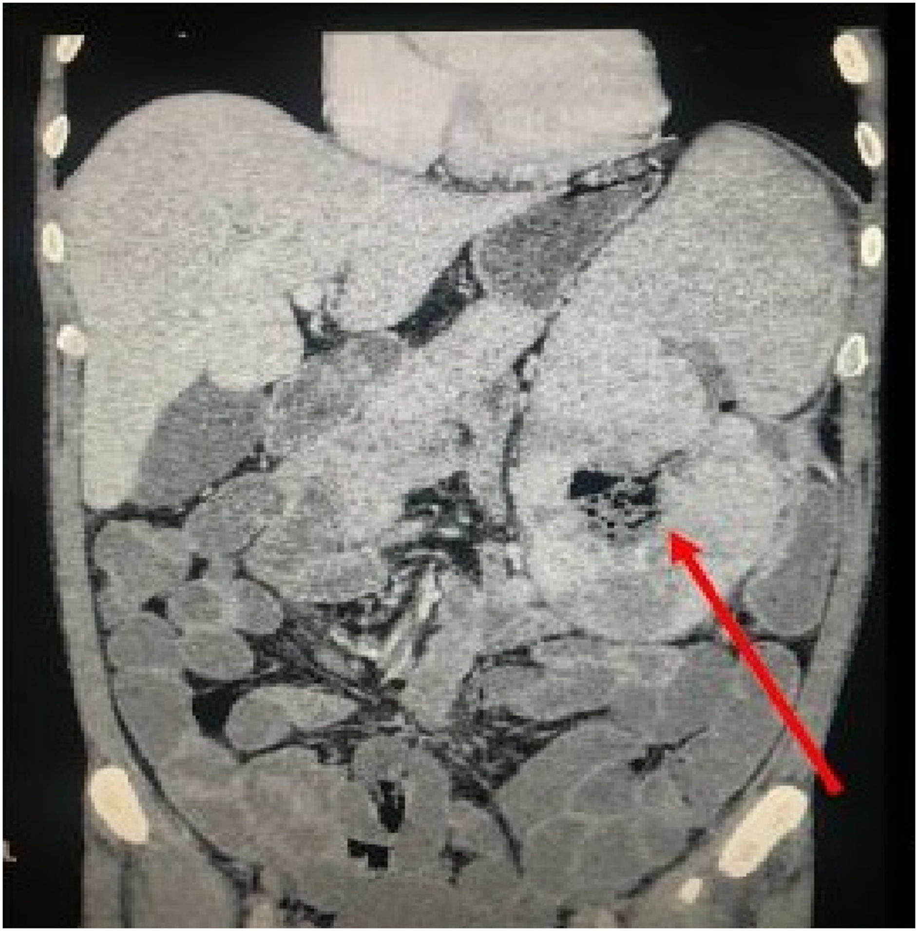

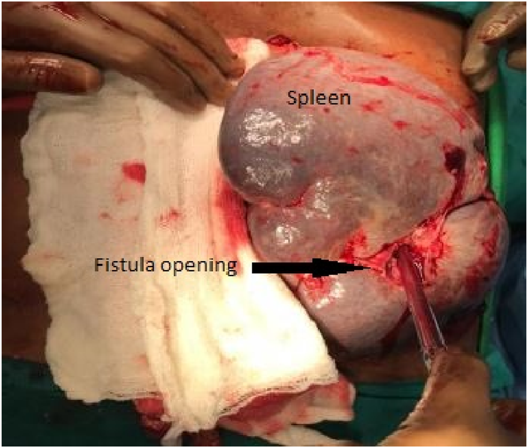
Surgical consultation was sought and open laparotomy with splenectomy and resection of colonic segment with end transverse colostomy was decided as the best management plan due to presumed pre-operative increased chances of disseminated tuberculosis and lymphoma. A massive spleen of around 17 cm with multiple grey-white necrotic areas with a fistulous tract connecting to the colon was revealed (Figures 3 and 4). Intra-operative gross image of colon and spleen with interconnecting, fistulous tract. intraoperative gross image of Massive spleen with the splenic end of the fistulous tract.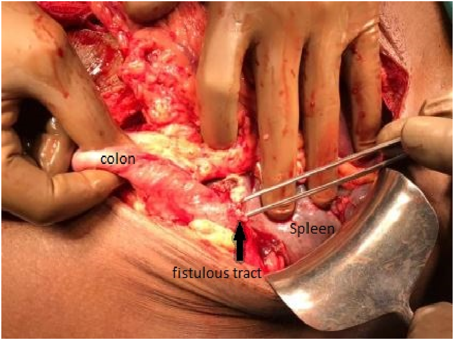
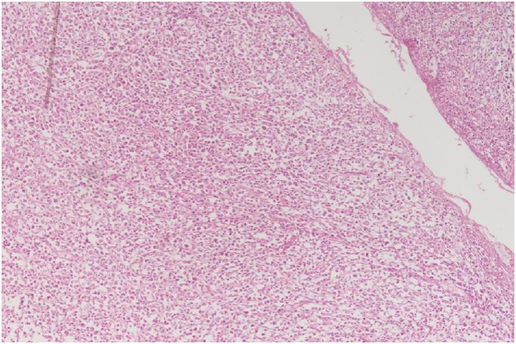
Histopathological evaluation (HPE) of the surgical specimen revealed diffusely infiltrative intermediate-grade NHL, with moderate anaplasia (Figure 5). On HPE, the tumor was seen arising from the mucosa, extending and breaching the serosa of the colon and forming a fistulous tract. The spleen showed multiple deposits of tumor cells of similar morphology as in colon. None of the 13 regional lymph nodes showed any abnormal cells. Lymphadenopathy could be attributed to the patient living with HIV. There were no lymph nodes present in the mediastinum. 10x view of the intestine, showing diffuse infiltration of tumor cells (right side).
The immunohistochemistry (IHC) showed diffuse positivity for leucocyte common antigen (LCA), CD20, and CD3 markers, while negative for CK7, chromogranin, HMB45, and CD117. The primary site was established on HPE, tumor was seen originating from the colonic mucosa, breaching serosa, and forming a fistulous tract to the spleen. (Figures 6 and 7). (10x view) Metastatic tumor cells deposited in the spleen on Hematoxylin and Eosin stain. (10x view) Fistulous tract, showing infiltration of anaplastic cells along the tract.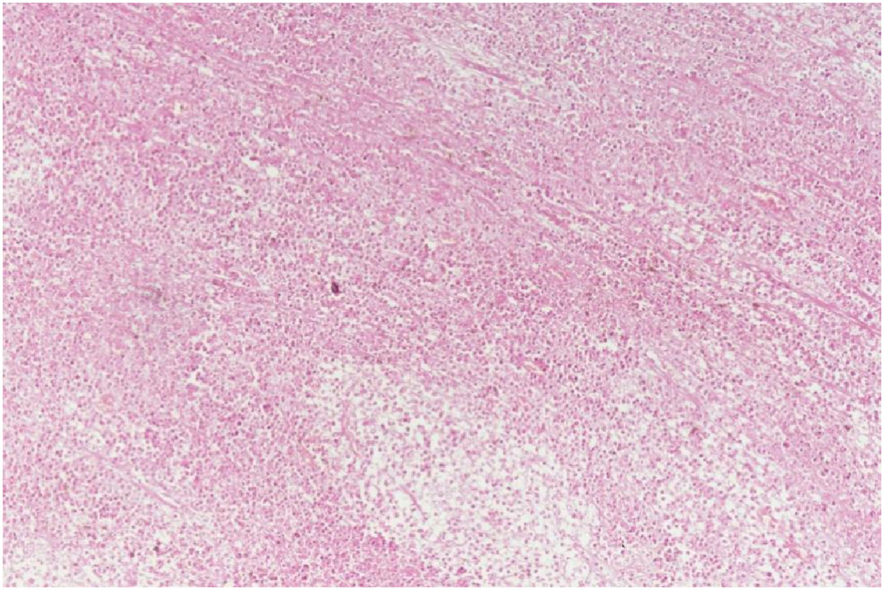
The patient was discharged on day 10 with advice to report for chemotherapy in 4 weeks after surgical wound healing on an outpatient basis. However, he presented to the emergency room 20 days later with pneumonia and acute respiratory distress due to his immunocompromised status and died.
Discussion
Diffuse large B-cell lymphoms (DLBCL) is an aggressive, AIDS-associated lymphoma that can occur at the nodal or extranodal site, with the gastrointestinal tract being the most common site. 1 The risk of developing non-Hodgkin’s lymphoma in PLWHA is 200 times higher than in the community with a predilection for extranodal sites, the central nervous system being the most common.3–5
HIV affects the development of lymphoma in numerous ways, including duration and intensity of immunosuppression, generation of cytokines that promote B cell growth, opportunistic infection with oncogenic viruses, and dysregulated immune response caused by memory cell loss. 5 Extranodal primary NHL originates from any site, wherever lymphatic tissue is found. White people, young or old, with a male predominance, experience it more frequently. 2 Primary colonic lymphoma is very rare, while the NHL is the usually found histological subtype approximating much over 90% of cases. 6 The ileocecal area, followed by the sigmoid and rectum, is the most frequent site of involvement. Disease severity, extranodal and bone marrow involvement, and CD4 lymphocyte count, are factors that affect a patient’s prognosis for AIDS-related lymphoma.
Intra-abdominal fistula is a rare phenomenon and most commonly associated with Crohn’s disease. The inflammatory process, ulceration, and extensive coagulative necrosis associated with malignancy appear to be precursors to fistula formation. 7 Other etiologies of fistula formation include diverticular disease, trauma, and post-surgical. It is known that splenic involvement occurs rarely in this process of fistulization and treatment includes resection of fistula and involved organs. 2
Our patient presented with atraumatic abdominal discomfort without any prior inflammatory disorder. Although his CECT abdomen in emergency had evidence of peritonitis the patient did not have any history of high-grade fever or positive findings suggesting acute abdomen or SBP on initial physical examination except a large spleen. If CECT abdomen was delayed it could have affected the patient’s prognosis significantly. Keeping a low threshold of suspicion lead to timely diagnosis and intervention salvaging a patient with rare disease presentation.
Conclusion
In any PLWHA with prolonged pain abdomen and an enlarged spleen, an emergency physician should think of lymphoma and request a CECT. Avoiding delay in diagnosis affects the prognosis of the patient and the chances of long-term survival.
Footnotes
Author contributions
All authors contributed to the study’s conception and design. The idea for the article was conceived by AKD,ZM, MG,PA and AD. Material preparation, data acquisition, and data collection were performed by ZM, MG, PA,AKD and AD. Data analysis and the first draft of the manuscript were written by ZM and MG. All authors commented on versions of the manuscript. AD, ZM and MG performed the literature search. ZM and MG critically revised the work. All authors read and approved the final manuscript.
Declaration of Conflicting Interests
The author(s) declared no potential conflicts of interest with respect to the research, authorship, and/or publication of this article.
Funding
The author(s) received no financial support for the research, authorship, and/or publication of this article.
