
Abstract
Background
Diabetes is an emerging health issue on a global scale, with increasing prevalence rates reported in many countries. Many mechanisms are proposed for diabetic nephropathy, with oxidative stress being the most significant. The effectiveness of gold nanoparticles (AuNPs) in the attenuation of nephropathy and oxidative stress in diabetic mice was assessed in this study.
Objective
The aim of this study is to synthesize AuNPs and assess their protective effect towards diabetic nephropathy by inhibition of oxidative stress.
Methods
The aqueous extract of Allium sativum was employed to synthesize AuNPs. The prepared AuNPs were characterized using a variety of microscopic and spectroscopic techniques. In-vitro studies were conducted using mice. Streptozotocin (STZ) was employed to induce diabetes in rodents. After 7 days of administration of STZ, anesthesia was given to all animals and blood was collected for the assessment of creatinine and Blood Urea Nitrogen (BUN) levels. Later, kidney tissue was removed at 4 °C and changes in pathology and oxidative stress were assessed.
Results
Nephropathy was confirmed in diabetic mice by the changes in the pathology of kidney tissue along with significant rise in the plasma levels of BUN and creatinine. Additionally, the peroxidation of lipids, formation of Reactive Oxygen Species (ROS), oxidation of glutathione (GSH), concentration of carbonyl protein was also increased in the tissue of kidney of diabetic mice. Oxidative stress in kidney tissue and changes in the pathology of diabetic mice were inhibited significantly (p < 0.05) with the treatment of AuNPs.
Conclusion
This study revealed the protective effects of AuNPs over diabetic nephropathy by inhibiting the pathway of oxidative stress. Since, the prepared AuNPs showed improvement over complications of diabetes, they may be believed as a potential gratuitous treatment next to other drugs for reducing blood glucose.
Introduction
Nanotechnology is the process of combining specific therapeutic compounds with nanoparticles, which are typically 1 to 100 nm in size, to improve their efficacy.1–3 Noble metallic nanoparticles have attracted substantial attention because of their exceptional fluorescence, electronic, catalytic, biological, and nanoprobe properties.4–6 Nanoparticles and nanostructured materials are of particular interest to researchers and the technology-economic sector due to their distinctive properties.7–9 These materials are distinctive in their physicochemical properties, which include conductivity, wettability, melting point, light absorption, and applications in the biomedical, pharmacological, and catalytic sectors.10,11 Consequently, they hold a unique position in technological advance.
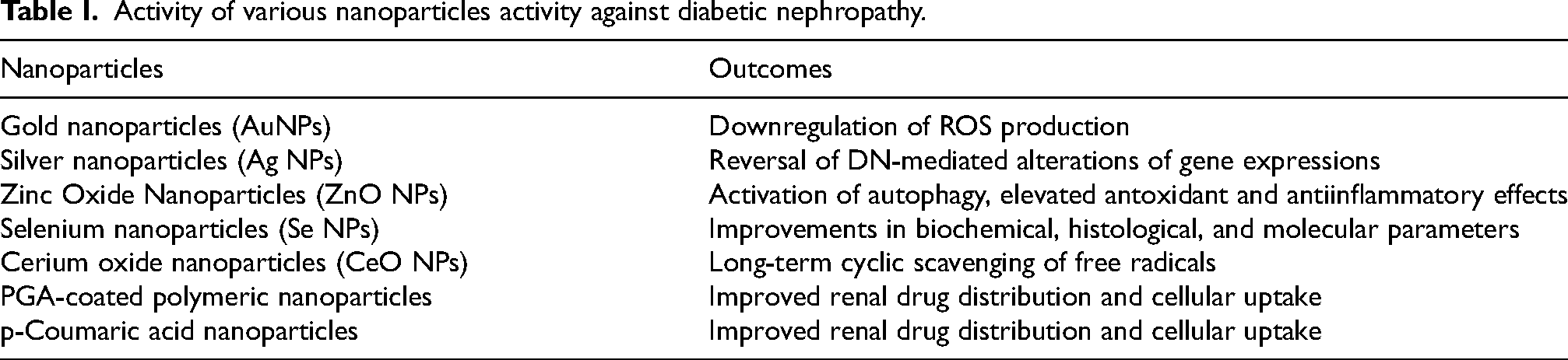
Gold nanoparticles (AuNPs) have been extensively investigated and are recognized for their medical applications. AuNPs are frequently synthesized using conventional chemical and physical procedures. Nevertheless, there has been global interest among researchers in the development of more cost-effective and environmentally favorable large-scale production methods, such as green synthesis, which employs biological processes. In response to this global emphasis on green nanotechnology, a variety of nanomaterials have been developed for applications that are both physiologically and environmentally acceptable. Green synthesis provides numerous advantages over traditional physical and chemical methods, such as biocompatibility, cost-effectiveness, energy efficiency, and simplicity. Additionally, it is a one-step synthesis process. These benefits have motivated researchers to investigate the green synthesis of AuNPs by utilizing the secondary metabolites of plants. The environmental and in vivo safety of metal nanoparticles, as well as the simplicity of the synthesis process, make plant-based synthesis an ideal approach. Various metal nanoparticles have been used in the treatment of diabetic nephropathy. Metal nanoparticles and their activity against diabetic nephropathy are depicted in Table I.12–17
Activity of various nanoparticles activity against diabetic nephropathy.
Diabetes mellitus (DM) is a main health concern marked by defects in either resistance or insulin secretion. DM is a metabolic disease that causes macrovascular complications which includes ischemic disease of the heart and microvascular complications like neuropathy, nephropathy and retinopathy. 18 Oxidative stress induced by high levels of glucose was shown as a main link between DM and their complications. The production of ROS by glycosylation of proteins and auto- oxidation of glucose was increased in chronic hyperglycemia. 19 After an increase in the generation of ROS, they react with the macromolecules which includes proteins, lipids, carbohydrates and DNA resulting in loss of their ability to function. 20 Additionally, various pathways of cell signaling were induced by ROS that resulted in the enhancement of diabetic complications. NF-kB (nuclear factor kappa-light-chain-enhancer of activated B cells) in answer to an elevation in the generation of ROS, improves pro-inflammatory cytokine transcription and chemokines like IL-1β, MCP-1, TNF-α and IL-6. 21 In 60% to 70% of patients with DM, a major common complication present may be Diabetic nephropathy (DN). Imbalance in between damage of nerve and its repair causes DN. Mechanism of DN involves pathway of polyol, oxidative stress, pathway of protein kinase C and nonenzymatic glycolation. Hence, it is predicted that the therapy of antioxidants helps in the delay or prevention of DN and other complications of diabetes. 22 NPs are ideal medicinal agents for therapy for several diseases because of possessing unique biological, optical, electrical, chemical and mechanical properties. 23 Free radicals were scavenged by some NPs having antioxidant properties.
Antioxidants are well-known for their advantageous effects in reducing complications associated with diabetes. Vitamin E is among the most thoroughly studied of these. It is essential for protecting lipids from oxidative damage induced by free radicals, as it is a fat-soluble vitamin. A lipid peroxidation chain reaction is initiated when reactive species target the lipids in cell membranes or lipoproteins. Vitamin E functions as a chain-breaking inhibitor of lipid peroxidation, thereby disrupting this process. 24 Hence, vitamin E was used as a control in this study.
Garlic (Allium sativum), a member of the Amaryllidaceae family, is highly regarded as a traditional condiment and as a critical element in folk medicine. The diverse phytochemical properties of garlic have been the subject of extensive investigation. 25 Numerous recent studies have illustrated the functional advantages of garlic in the treatment of cancer and cardiovascular disease. 26 Garlic possesses a diverse array of beneficial properties, such as anti-inflammatory, cardio-protective, anti-diabetic, antioxidant, reno-protective, anti-hypertensive, antimicrobial, immunomodulatory, and antiviral properties.26,27 In this work, AuNPs were prepared using aqueous extract of Allium sativum and characterized using microscopic and spectroscopic techniques. Also, the protective effects of AuNPs over DN by inhibiting the pathway of oxidative stress was studied.
Materials and method
Chemicals
99.9% of Hydrogen tetrachloroaurate (III) hydrate (HAuCl4·3H2O) was obtained from Sigma Aldrich (Shanghai, China) and each of the used glassware was washed and dried before the utilization.
Preparation of aqueous extract of Allium sativum
Allium sativum bulbs from Vellore, India, were meticulously divided into cloves without causing any damage. The cloves were frozen at −20 °C in plastic bags overnight. The cloves were heated to 90 °C for 1 h and then chilled to ambient temperature. The chilled cloves were grounded with distilled water in a 1:1w/v ratio, resulting in the extraction of water-soluble chemicals. The mixture was strained using muslin fabric to isolate the aqueous extract.
Biosynthesis of AuNPs using A. sativum Extract
Concentrated HAuCl4·3H2O was diluted with 100 mL of distilled water to initiate the synthesis. Subsequently, 40 mL of A. sativum extract was added to 70 mL of a 1 mM aqueous gold solution. A magnetic stirrer was used to continuously stir this mixture at 500 rpm for 90 min to facilitate the formation of AuNPs. The initial indication of effective AuNP synthesis was the appearance of a color change in the solution. Subsequently, 50 mL of the AuNP suspension was transferred to a petri dish and allowed to dry at room temperature.
Characterization
The UV spectra of a variety of colloidal AuNPs samples were measured in the 400–600 nm wavelength range using a UV-2600 Shimadzu UV–vis spectrophotometer. Spectral data was recorded between 4000 and 1000 cm−1 using the KBr pellet method, and Fourier transform infrared (FTIR) spectra were obtained using a PerkinElmer Spectrum 2 FT-IR spectrometer. The JEOL 2100F field emission transmission electron microscope (FETEM) was utilized to acquire high-resolution transmission electron microscopy (HRTEM) images at 100 kV. The same instrument was also used to conduct selected area electron diffraction (SAED) analysis. An X-ray diffractometer was employed to conduct X-ray diffraction (XRD) measurements.
Experiments
Animal handling
The mice of Swiss albino (male), each of around 30–35 g in weight were obtained from the Laboratory of Animal Research Center. All the mice were accommodated in a room, which was air-conditioned with temperature control (22 ± 2 °C), and within the reach of food and clean water and continued with 12/12-h light schedule.
Plan of the experiment
Five animal groups were formed with 6 mice in every group. AuNPs with a dose of 60 mg/kg were administered into the mice in the control group (without diabetes) and the same was administered to the diabetic mice for 28 days. Tocopherol (Vit. E) injection was also given to the diabetic mice for 28 days. Induction of diabetes in the mice was done by intraperitoneally injecting the citrate buffer (of 4.6 pH) diluted Streptozotocin (STZ) [65 mg/kg b.wt.]. 28 7 days after administering the STZ, blood was extracted from lateral veins in the tail part and glucose levels in the blood (BGL) were known by GOM with the help of a glucometer. Mice with BGL > 200 mg/dL were considered diabetic and further all the mice were given anesthesia followed by blood extraction from the heart with the help of a syringe. This extract was centrifuged for 5 min at 4 °C with an accelerative force (AF) of 3000 g and at a temperature of −70 °C, until the next use, the obtained plasma was frozen. Tissues of the kidney were cut on the ice and were homogenized in a solution of phosphate buffered saline (PBS) followed by centrifugation for 10 min with an AF 800 g at 4 °C. Assay for markers of the oxidative stress was done to the obtained supernatant.
Creatinine and Blood Urea Nitrogen (BUN) measurement
Commercial reagents have been used to determine creatinine and BUN, which have proved to be the markers of improper kidney functioning (kidney dysfunction).
Determining the ROS generation
The amount of ROS generated in the tissues of the kidney were determined with the help of dichlorofluorescin-diacetate (DCFH-DA), which was used as a marker. Around 2 mL of homogenate (1 mg/mL of protein) of the kidney tissue stocked with the indicator dye (DCFH-DA) was incubated with the same reagent at 37 °C for 15 min and then, monitored at 520 nm and 480 nm by using fluorescence spectrophotometer, Shimadzu RF5000U. 29
Lipid peroxidation measurement
One of the previously demonstrated literature was used to determine the content of 3,4-Methylenedioxyamphetamine (MDA). In brief, 0.05 M phosphoric acid of 0.25 mL was added to the homogenate of kidney tissue 0.2 mL along with adding 0.2%TBA of 0.3 mL. The samples are kept in a hot water bath for about half an hour. Then the tubes were transferred to an ice-bath and 0.4 mL of n-butanol was supplemented to every tube. Later the tubes were allowed to centrifugation for about 10 min at 3500 rpm. The quantity of MDA produced in every sample was evaluated by measuring the supernatant absorbance at a wavelength of 532 nm with an enzyme linked immunosorbent assay (ELISA) reader (Tecan, Rainbow Thermo, Austria). The content of MDA was expressed in nanomole mg−1 protein 30 and Tetramethoxypropane was utilized as a standard.
Glutathione content measurement
The content of GSH was evaluated with the help of a spectrophotometer along with Thermo Scientific Pierce Ellman's Reagent (DTNB) as an indicator. In brief, kidney tissue of 0.1 mL was supplemented to phosphate buffers of 0.1 M along with DTNB of 0.04% in a total volume of 3 mL (pH of 7.4). Using a spectrophotometer (Ultravoilet-1601 PC, Shimadzu, Japan), the yellow color developed can be read at 412 nm. The content of GSH was given in µg mg−1 protein. 31
Protein carbonyl measurement
Protein carbonyl was determined by using the method of spectrophotometric, in brief, 200 µL were required for kidney tissue homogenation. The Samples were obtained from 20% Tricholoroacetic acid solution (TCA) of 500 µL (w/v). And then, samples are stored for 15 min at 4 °C. The samples were administrated with 500 µL of Hydrochloric acid (HCL) 2N along with 500 µL of DNPH 0.2% for the animals in control group and the samples were incubated at room temperature for 60 min with an interval of 5 min. Then, on addition of 55 µL of 100% TCA, the proteins were precipitated. After centrifugation of micro tubes, they are washed thrice by using 1000 µL of ethanol ethyl acetate sample. Then in 200 µL of 6 M guanidine HCL, the micro-tubes were dissolved. By measuring the absorbance wavelength at 365 nm, the content of carbonyl was determined. 32
Catalase content measurement
The activity of Catalase was analyzed by calculating the decrease in absorbance in a reaction medium at 240 nm containing PBS (50 mM, pH: 7.0) and 10 mM of H2O2. 1 M of H2O2 as substrate used per 60 s is defined by 1 unit of enzyme and the specific activity is given in u mg−1 protein.
Protein concentration measurement
Bradford method was used to determine the protein content in the kidney tissue. 33 The standard used was Bovine Serum Albumin (BSA), and Coomassie blue was used to mix the homogenate samples. After 10 min absorbance was measured at 595 nm using spectrophotometer.
Pathological analysis
Initially, the animals are anesthetized by using chloroform, the kidney tissues were separated from tested and controlled mice followed by rinsing in physiologic serum and then placed in a solution of formalin for 18 h. Furthermore, they are desiccated in ethanol in a graded series and toluene was used to extract alcohol. Later, paraffin was used for saturating the tissues in oven and promptly, the tissues are saturated using paraffin followed by fixing the block samples on the microtome and the sections with 13 µm thickness were obtained after 240 min. Those sections were moved on to the slides and subjected for final assessment using a light microscope which was stained with eosin and hematoxylin. 34
Statistical analysis
The results are expressed as Mean ± Standard Deviation. 10th version SPSS software is used to perform all the statistical analyses. Analysis was done in triplicate, and we use mean for statistical analysis. One-way ANOVA test helps in determination of Statistical significance, succeeded by post hoc Tukey test. P < 0.05. was the fixed statistical significance.
Results
Ultraviolet (UV) / Visible (Vis) spectroscopy
The UV-Vis spectrum of the colloidal gold particles was demonstrated in Figure 1, which was recorded 38 min after the reaction process. The formation of AuNPs by reducing HAuCl4·3H2O was identified by the change of color to purple/ruby red from yellow. The exceptional surface plasmon oscillations with aqueous AuNPs were responsible for the color transformation. 35 Nearly, at a wavelength of 555.5 nm (λmax = 555.5 nm), the band of the surface plasmon resonance (SPR) 36 exhibited by the colloidal particles was observed. A band of SPR, which is always pointed and smooth clearly indicates that AuNPs is precisely spherical in shape.
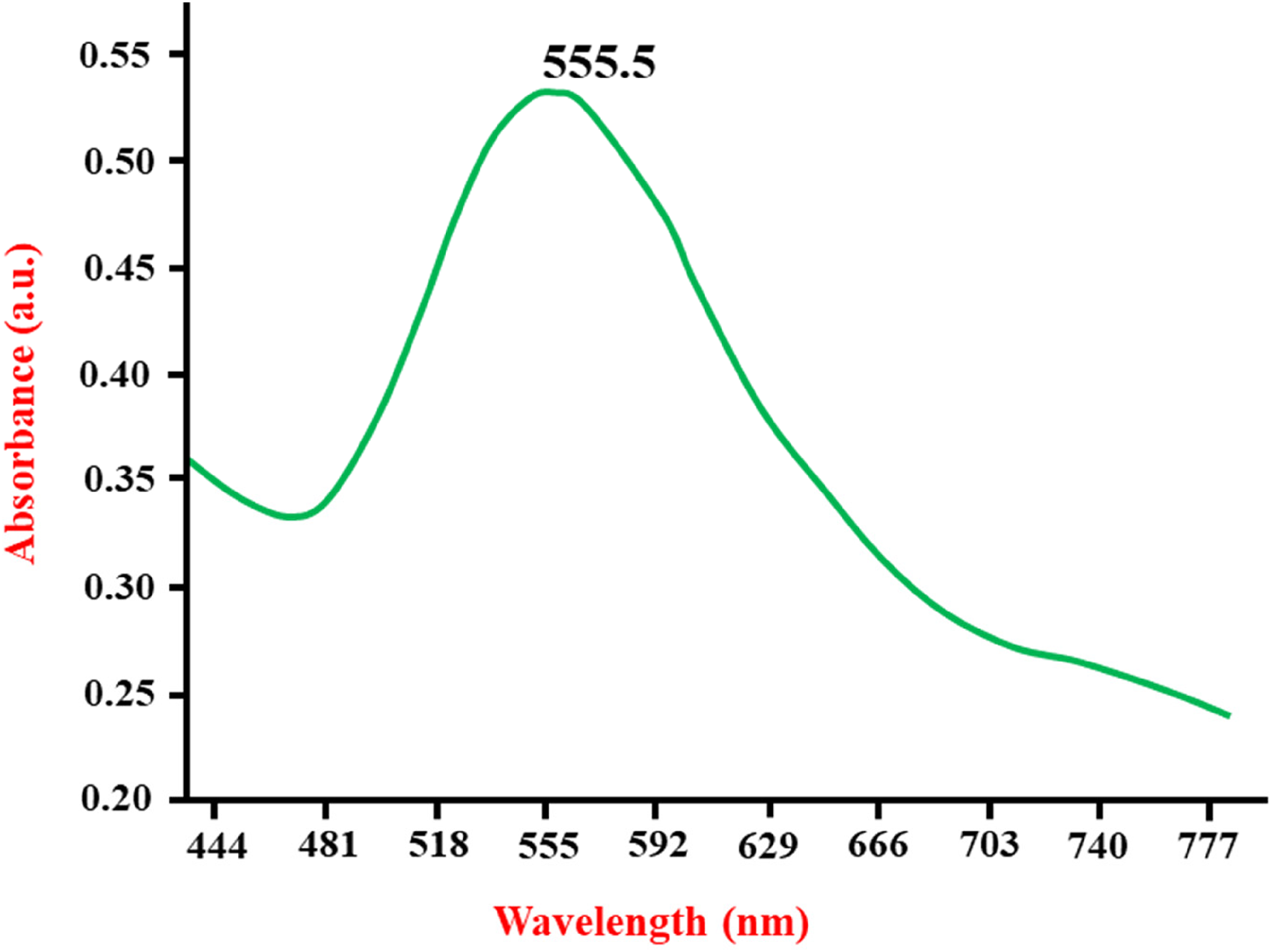
UV-Vis spectrum of the AuNPs bio-fabricated using Aqueous extract of A.sativum at a wavelength of 555.5nm.
Attenuated Total Reflectance-Fourier Transform Infrared (ATR-FTIR) spectroscopy
The spectroscopy of ATR-FTIR was performed to recognize the specific functional groups which mediated the HAuCl4·3H2O reduction to AuNPs and for determining specific biomolecules in aqueous extract of A. sativum which were actively responsible for the stabilization and capping of NPs. The comparison of the FTIR spectra of aqueous extract of A. sativum prior to and following the synthesis of AuNPs is clearly depicted in Figure 2. Band presence could be clearly seen at different wavelengths (1056, 1341, 1413, 1678, 2938 and 3117 cm−1) in the FTIR spectrum of aqueous extract of A. sativum. The presence of bands at 1256 and 1341 cm−1 were ascribed to the stretch C-O of the carboxylic group. 37 The C-C and C-O group oscillations of some compounds, especially the carbohydrates led to the formation of a strong and intense peak, which was observed at a wavelength of 1056 cm−1. Carbonyl and hydroxyl stretch oscillations were responsible for the bands at the wave lengths 1678 cm−1 and 3117 cm−1 separately. C-H bending and stretching oscillations are responsible for the formation of bands at wave lengths, 1413 cm−1 and 2938 cm−1. It was sorted out from the spectrum that existing bands in aqueous extract of A. sativum have similarities with the former reports on sugar 38 and proteins. 39 Following the HAuCl4·3H2O biological reduction to AuNPs by utilizing aqueous extract of A. sativum, noticeable shift in the bands was observed in the FTIR spectrum of the AuNPs (Figure 2B). Noticeable shifts were detected in the peaks from wave lengths 1056–1063 cm−1. One more shift was noticed among the bands 2938–2942 cm−1, 3117 −3126 cm−1 and from 1256 to 1263 cm−1. A sharp and medium peak was found at wavelength of 1678 cm−1 which further transferred to the band at the wavelength 1684 cm−1 and transformed into a small blunt band. Collectively, we recommend that protein molecules were involved in actively reducing the metal ions by the oxidization of aldehydes to carboxylic acids. 39 A fresh, medium and sharp peak was seen at the wavelength of 2890 cm−1, which could have resulted from the stretching oscillations of C–H group, On the other hand, the peak at the wavelength 1341 cm−1 had completely disappeared. The modified peaks in the nanoparticles at different wavelengths clearly imply that aqueous extract of A. sativum was the source from where nanoparticles have developed. On an obvious comparison, it was very clear that on increasing the wave numbers, the bands of absorption for AuNPs have increased. This could be highly possible because of the dative bonds (co-ordination bonds) which exist between the functional groups (C = O, O-H) of carbohydrates especially existing in aqueous extract of A. sativum and AuNPs. 40
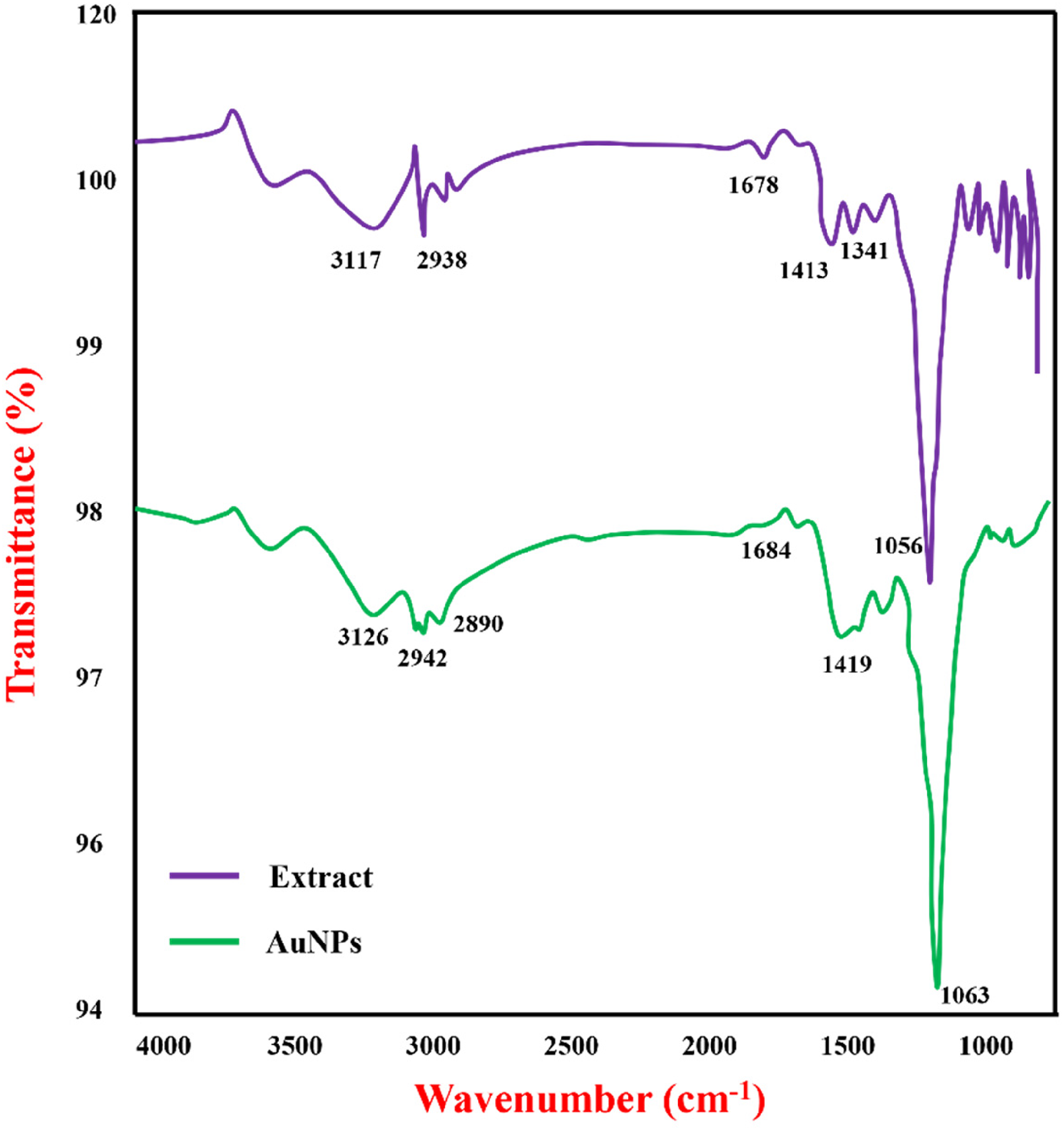
ATR-FTIR spectrum of Aqueous extract of A.sativum (A) and AuNPs, produced using Aqueous extract of A.sativum (B).
XRD analysis
XRD analysis was performed to analyze the crystalline nature of the formed AuNPs. The pattern of XRD for the AuNPs synthesized from aqueous extract of A. sativum is depicted in Figure 3. About four prominent peaks existed in the coupled θ range of 30–80. The consecutive peaks of diffraction 38.1 °, 44.5 °, 64.71 ° and 77.81 ° are analogous to (111), (200), (220) and (311) sides of the face centered cubic (FCC) crystal lattice, which is in acceptance with previous reports regarding the Phyto-synthesis of AuNPs41–43 and the lattice parameter was noticed to be 4.07860 °A. The four peaks which were spotted have a resemblance to the JCPDS card number 00-407-84. Near 38.1 °, the highest peak was observed which specified the appreciable progress along the Bragg's plane (111) 52 42 in the structure of FCC gold.
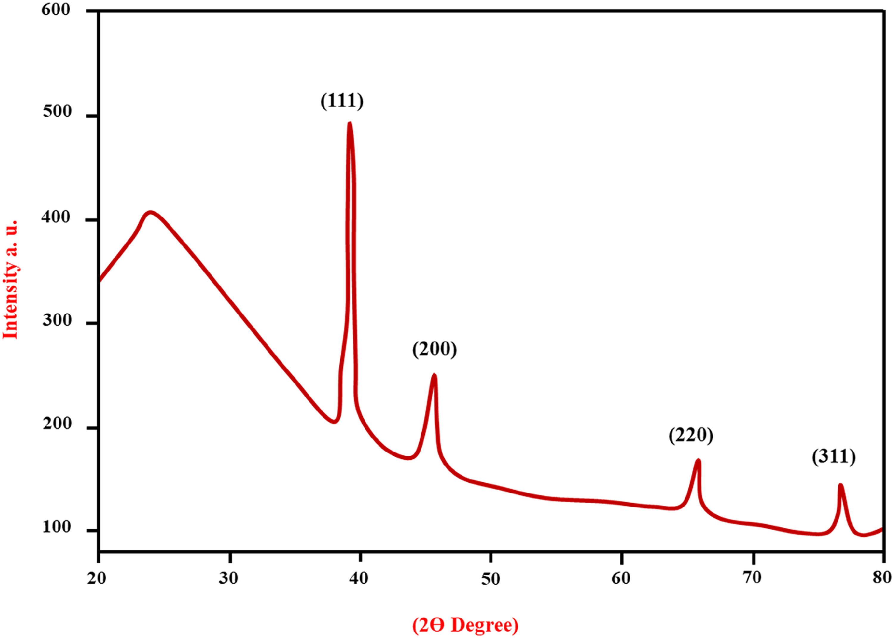
XRD pattern of AuNPs synthesized using A.sativum. The four peaks which were spotted have resemblance to the JCPDS card number 00-407-84.
Figure 4A shows the HR-TEM images of prepared AuNPs which showed the spherical NPs, and few are triangular with size existed in range from 30–50 nm. The crystalline nature of the AuNPs synthesized using aqueous extract of A. sativum was confirmed by the pattern amongst the bright cyclical rings of the selected area electron diffraction (SAED) and the observed lattice fringes (Figure 4B).
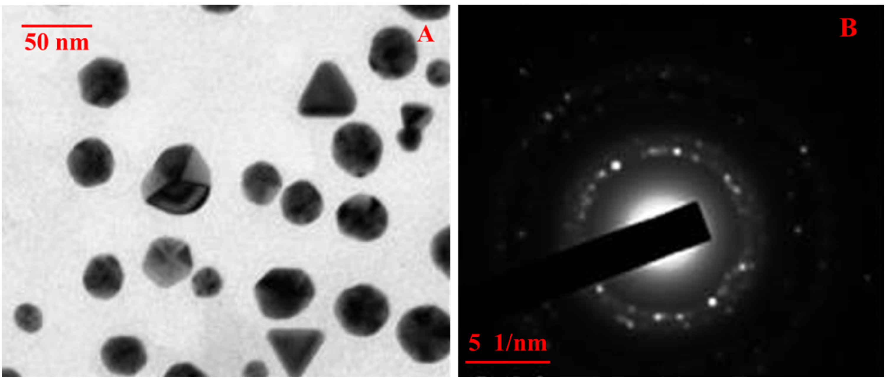
TEM image (A) of AuNPs which showed the spherical NPs range from 30–50 nm and SAED pattern (B) The crystalline nature of was confirmed by the pattern amongst the bright cyclical rings.
AuNPs effects on plasma glucose and body weight
Weight gained was noticed to be lower in diabetic mice than the animals in the control group as shown in Figure 5. Diabetic animals did not gain weight as significantly as control mice over the course of four weeks. In diabetic mice there was no significant reduction in blood glucose on administration of AuNPs injection and had similar effects to AuNPs, when diabetic mice received vitamin E (Figure 6).
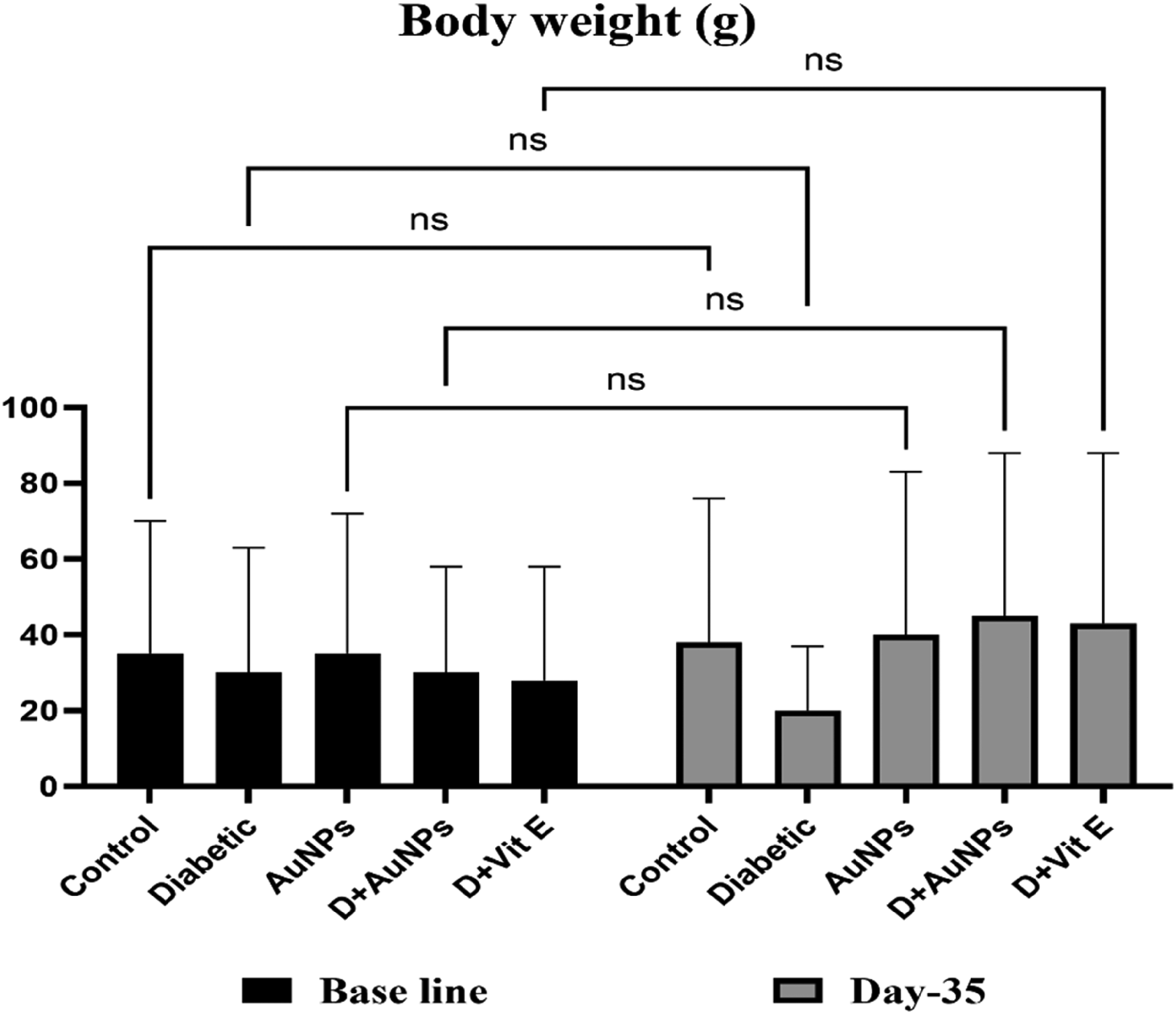
Impact of AuNPs administration on body weight of mice no obvious effect on the body weight was observed on treating AuNPs alone but, it inhibited weight loss in the diabetic animals.
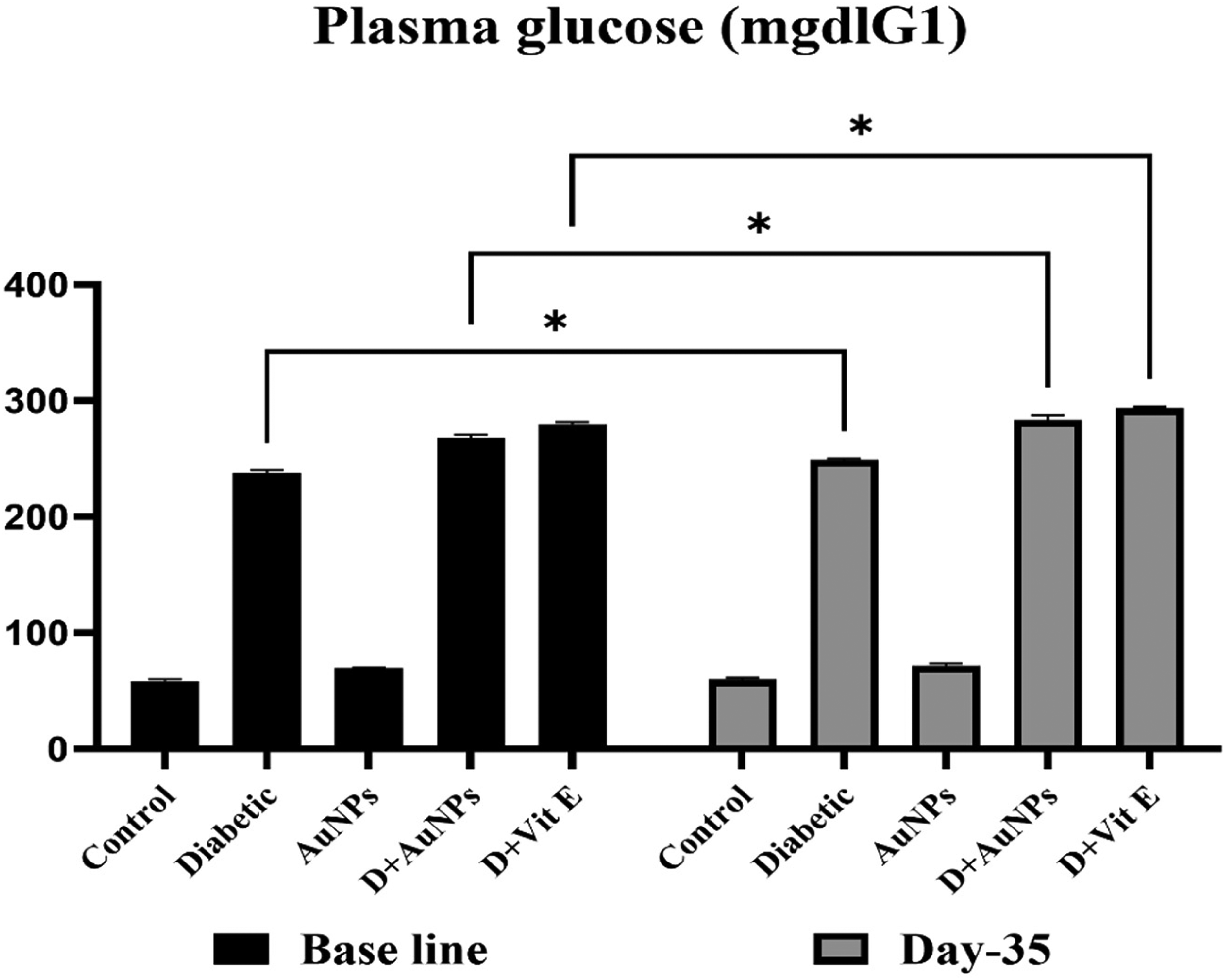
Impact of AuNPs administration on Blood Glucose Levels, there is no significant reduction in blood glucose on administration of AuNPs injection and has similar effects to AuNPs, when diabetic mice receive vitamin E.
The treatment of diabetic mice with AuNPs showed a time-dependent decrease in BGL. The reduction in BGL started to be significant by day 15 of treatment compared with the level before treatment. By day 35, the BGL decreased to 140 ± 6.84 mg dLG1, treated with AuNPs 1.5 mg kgG1 b.wt./day IP, which was non-significantly different from the value in the normal group 1. The diabetic mice treated with nanoceria (50 mg kgG1 b.wt. IP) for 25 days showed significant decreases in BGL 140.90 ± 6.74 compared with the diabetic saline group 2 (p > 0.05, ANOVA) (Figure 6).
In vivo administration effects of AuNPs on serum creatinine and BUN
Induction of diabetes was related with an obvious elevation in the serum levels of creatinine and BUN which indicates damage of kidney. In diabetic mice, the elevation of creatinine and BUN was prevented by administration of AuNPs as like Vitamin E effect (Figure 7a and b).
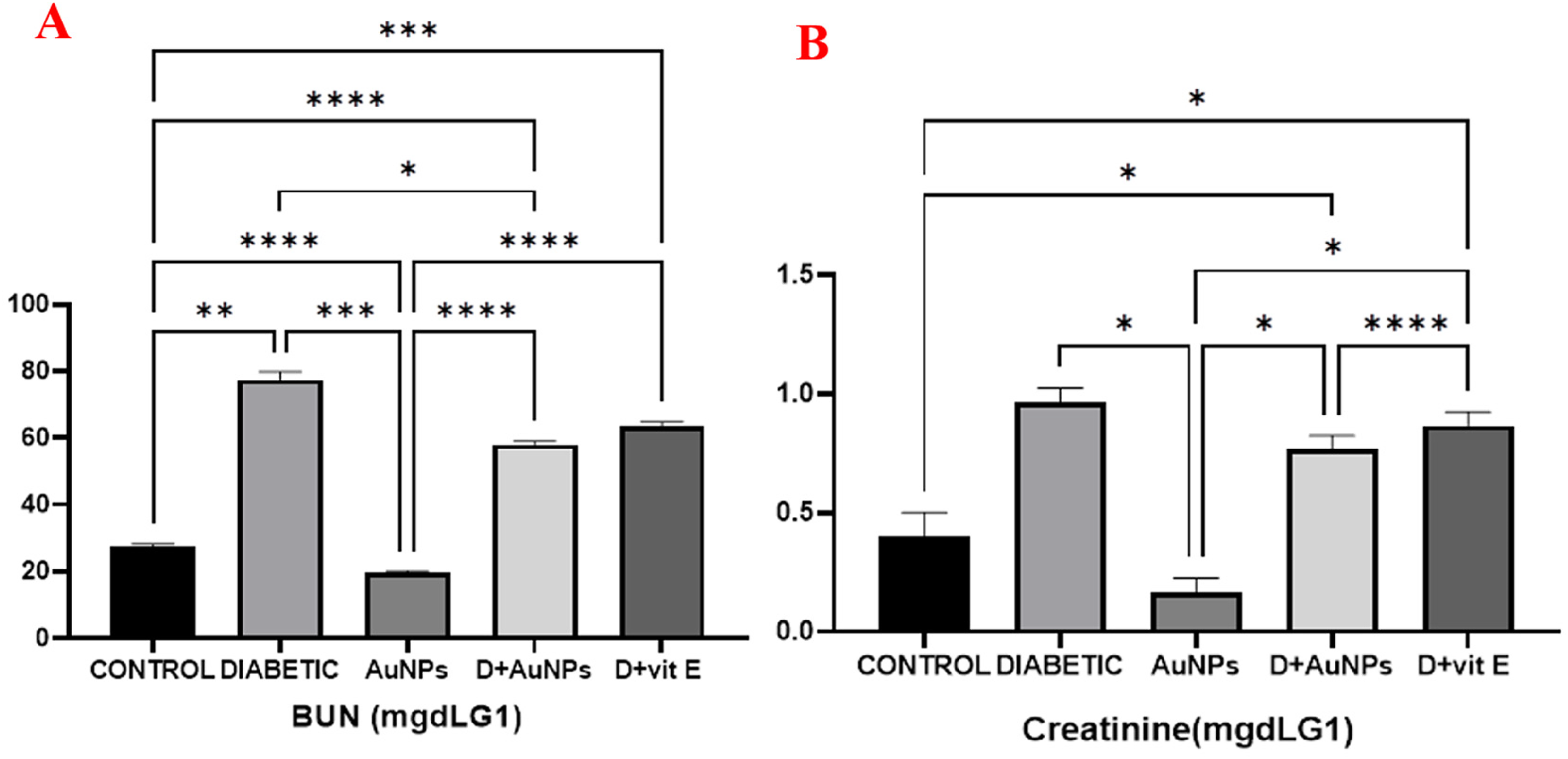
Impact of AuNPs and Vit E administration on (A) BUN and (B) creatinine. The elevation of creatinine and BUN was prevented by administration of AuNPs as like vitamin E effect.
Formation of Oxygen Species (ROS)
As shown in Figure 8, formation of ROS indicates oxidative pressure. It increases significantly in the diabetic mice and after the administration of AuNPs, the formation of ROS was reduced significantly but no apparent change was noticed in the formation of ROS when treated with vitamin E (Figure 8).
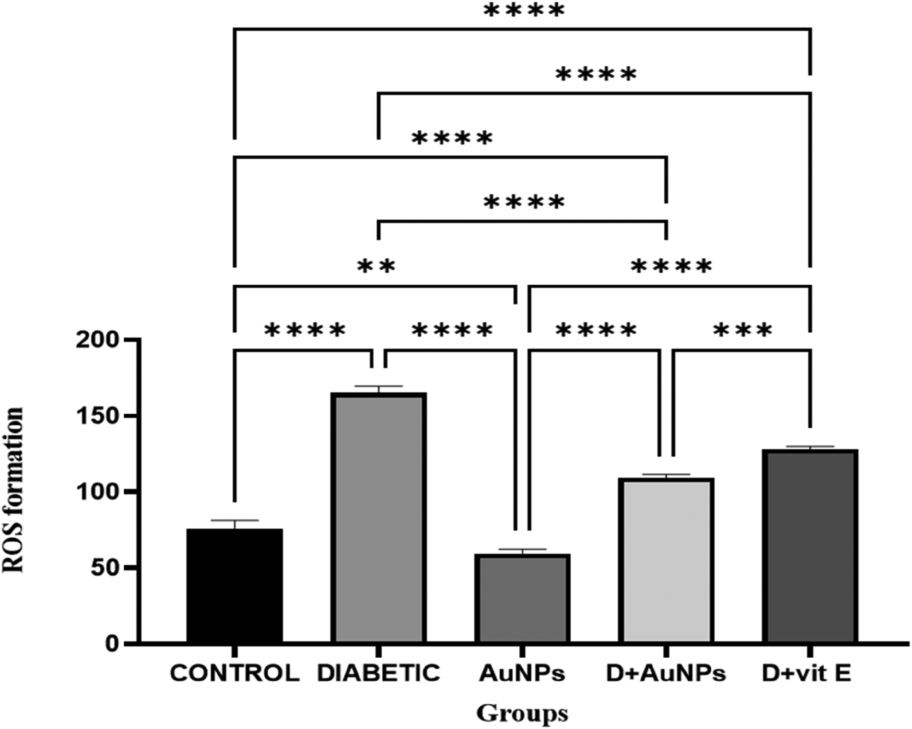
Effect of AuNPs on ROS formation in kidney tissue. It increases significantly in the diabetic mice and after the administration of AuNPs, the formation of ROS was reduced significantly but no apparent change was noticed in the formation of ROS when treated with vitamin E.
Peroxidation of lipids
Malondialdehyde (MDA) is one of the end products of Lipid Peroxidation (LPO) and a known indicator for oxidative pressure was MDA elevation. When compared with the control group, the level of MDA was elevated in kidney tissue of diabetic mice, as shown in Figure 9. AuNPs inhibits the lipid peroxidation significantly induced by diabetes and better protective effect was shown by AuNPs against lipid peroxidation than vitamin E (Figure 9).
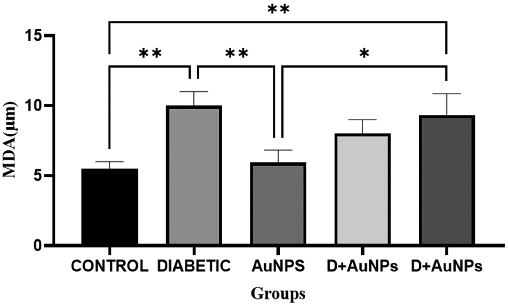
AuNPs exhibit significant inhibition of lipid peroxidation in kidney tissue induced by diabetes, demonstrating a better protective effect compared to vitamin E.
Concentration of glutathione GSH
Oxidative pressure-induced renal damage in patients of diabetes was generally due to an imbalance between ROS and levels of antioxidants like GSH in kidney. 44 The levels of GSH were reduced to 73.22 µm in diabetic mice kidney tissue when compared to that of control group (0.0381 µm) and the concentration of GSH was 3.2727 µm in the diabetic mice who were administered using AuNPs for one month, which was considerably greater than that of diabetic mice. The level of GSH in mice treated with vitamin E was 99 µm. The total AuNPs presented an enhanced effect when compared with vitamin E (Figure 10).
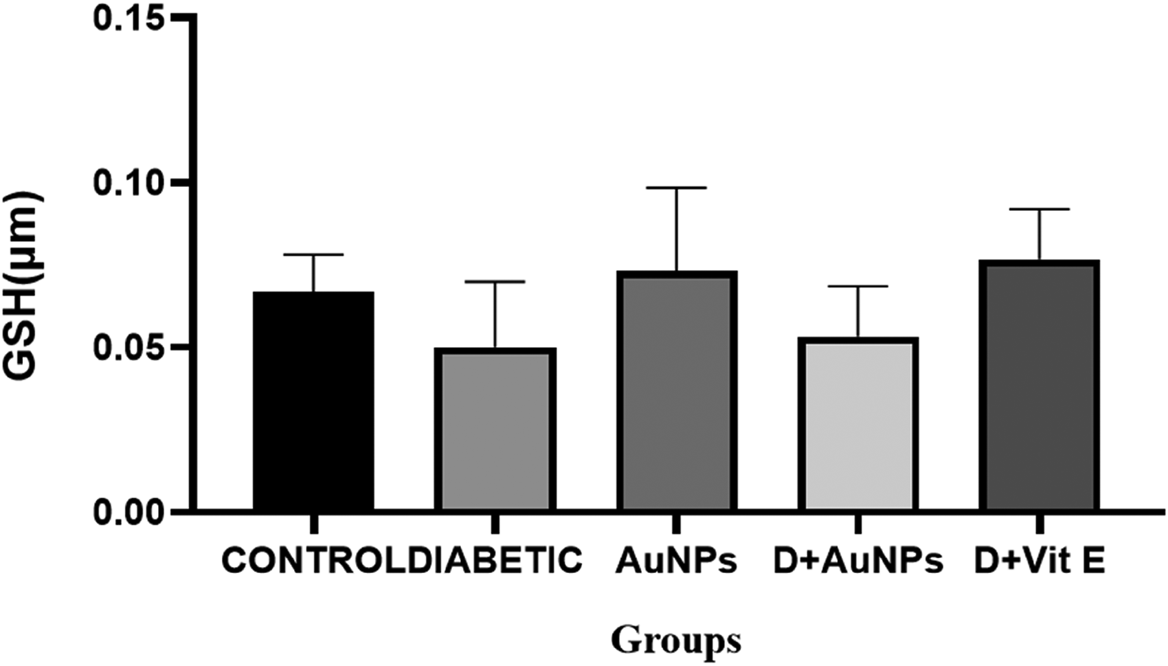
Effect of AuNPs treatment on GSH levels in kidney tissue shows an enhanced effect when compared with vitamin E.
Activity of catalase
In diabetic mice, the catalase (CAT) activity was increased as shown in Figure 11 and when compared with control group, AuNPs administration significantly reduced activity of CAT in diabetic mice and vitamin E has lesser effect than that of AuNPs (Figure 11).
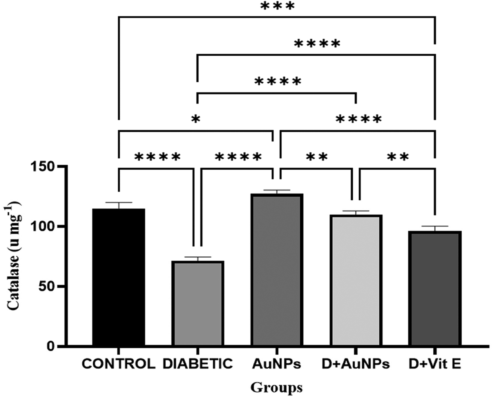
Effect of AuNPs treatment on CAT levels. When the administration of AuNPs significantly reduced activity of CAT in diabetic mice and vitamin E has lesser effect than that of AuNPs.
Protein carbonyl
In diabetic patients, protein oxidation is indicated by protein carbonyl which can be observed by absorbance changes at a wavelength of 365 nm. When compared with the diabetic group, decrease in carbonyl protein was due to administration of AuNPs (Figure 12).
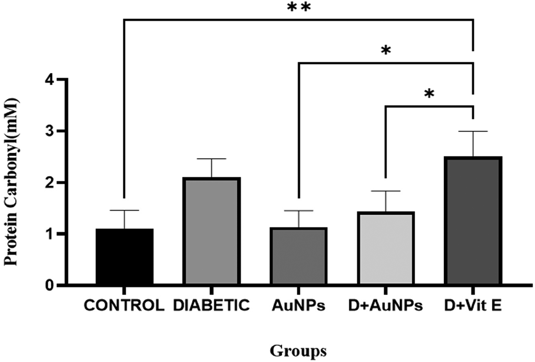
Effect of AuNPs treatment on protein carbonyl levels in kidney tissue. When compared with diabetic group, decrease in protein carbonyl was due to administration of AuNPs.
Histological examination
Increase in size of glomerulus in proximal convoluted tubules was observed in Streptozotocin -induced diabetic mice in the histological studies of kidney and after 1 month of treating with AuNPs these changes were reduced effectively (Figure 13 A-C).
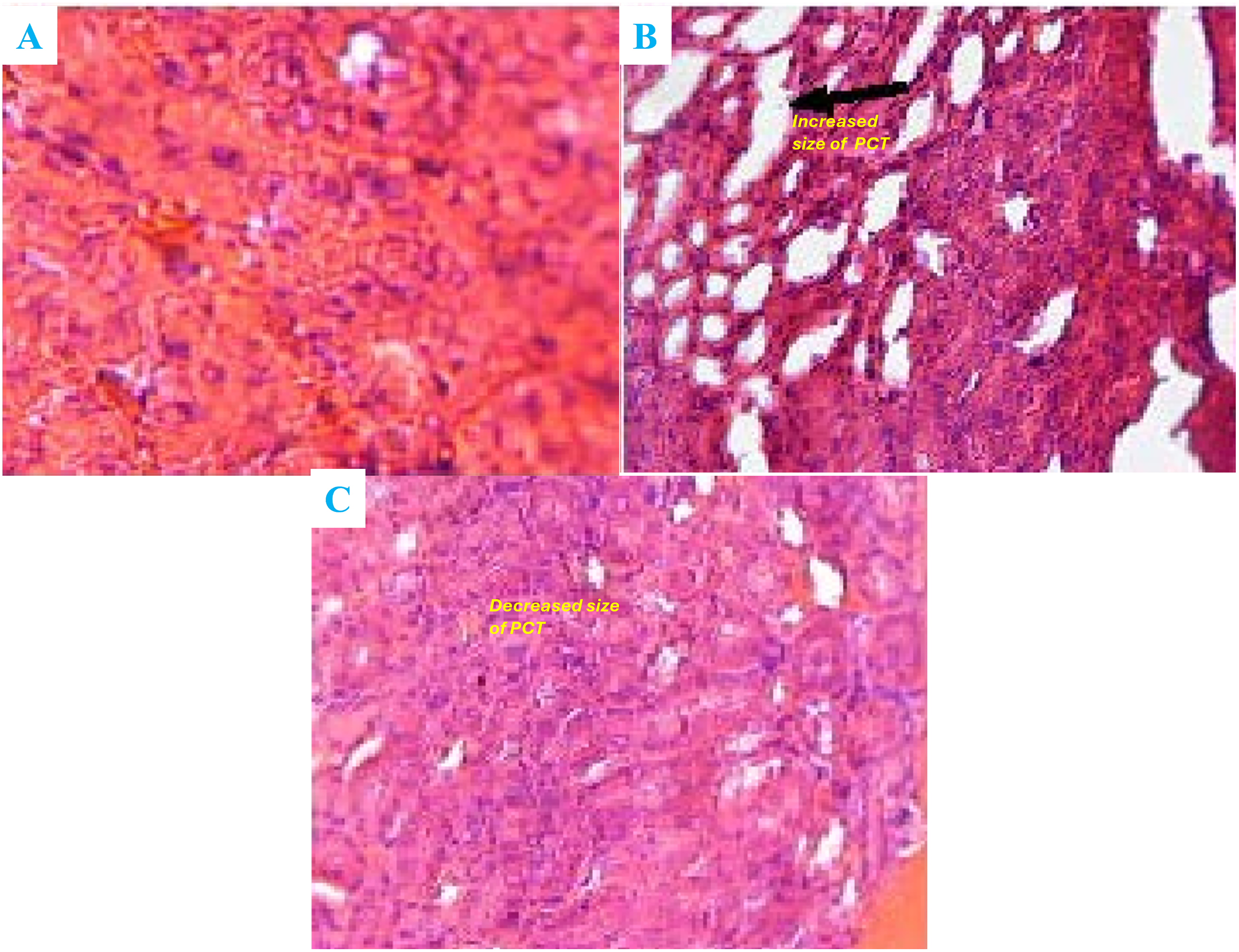
(A-C): Effects of AuNPs on diabetes induced-morphological change in kidney shows the tissue Increase in size of glomerulus in proximal convoluted tubules.
Discussion
In the present study, we evaluated the potential of AuNPs to prevent diabetic nephropathy in male mice that has been induced by streptozotocin. The results suggested that diabetic mice exhibited an increase in oxidative stress markers. Additionally, the administration of AuNPs appeared to improve both the pathological and biochemical indicators of kidney injury in these mice. Our findings indicated that the induction of diabetes in mice resulted in an imbalance in the production of ROS and the antioxidant system within kidney tissue. This was shown by a decrease in GSH levels and an increase in catalase activity in comparison to control mice. This imbalance was accompanied by pathological changes in kidney tissue, as well as elevated BUN and creatinine levels. Diabetic mice exhibited elevated serum creatinine and BUN levels, which were indicative of kidney injury. Oxidative stress also facilitated lipid peroxidation, as evidenced by an elevated MDA concentration. 45 This finding is consistent with prior reports of elevated lipid peroxidation during diabetic nephropathy. 46 In diabetic mice that were administered AuNPs, this parameter experienced a substantial decrease. Furthermore, protein oxidation is characterized by elevated protein carbonyl levels, which can result in ROS damaging proteins. 47 Our study showed that diabetic animals exhibited a substantial increase in protein carbonyl levels, while diabetic mice that were administered AuNPs experienced a decrease. Under normal conditions, the endogenous antioxidant system—which comprises glutathione peroxidase, catalase, superoxide dismutase (SOD), and glutathione—averts kidney injury caused by oxidative stress. In comparison to controls, diabetic mice exhibited increased catalase activity and decreased GSH content in their kidney tissue following streptozotocin administration, which was consistent with prior research. 48 The treatment with AuNPs prevented the depletion of the antioxidant system in diabetic mice thereby balancing the antioxidant system and reducing ROS production. At the conclusion of the experiment, histological analysis demonstrated that diabetic mice had higher kidney/body weight ratios, which may have been attributed to glomerular hypertrophy and increased kidney membrane thickness. 49 However, diabetic animals did not gain weight as significantly as control mice over the course of four weeks. The histopathological changes in kidney tissue were enhanced by the administration of AuNPs. Nevertheless, the diabetic group that received AuNPs did not exhibit any substantial variation in plasma glucose concentration when compared to the untreated diabetic group. This indicates that the antioxidant properties of AuNPs are likely responsible for the protective effects.
Conclusion
In conclusion, this study showed that AuNPs possess protective effects against diabetic nephropathy through oxidative stress. We have developed a simple and environmental-friendly method for the rapid production of AuNPs utilizing the aqueous extract of A. sativum. The nanoparticles produced had various shapes, mainly spherical, with a face-centered cubic (111) crystalline structure and 30–50 nm in size. This study also found that inducing diabetes in mice causes an imbalance in kidney tissues between the formation of ROS and the antioxidant system. This is shown by reduced GSH levels and improved CAT activity. This imbalance is associated with increased creatinine and BUN levels and pathological changes in kidney tissues. Oxidative stress plays a significant role in the advancement of DN due to the generation of ROS triggered by high blood sugar levels in several cell types. Therefore, in addition to careful glycemic control, using antioxidants could be an alternative approach to reduce the harmful consequences of high blood sugar. Thus, the antioxidant characteristics of AuNPs make them a promising addition to traditional glucose-lowering drugs for managing diabetic problems. Additional research is needed to determine the most effective dosage and length of treatment with AuNPs for therapeutic purposes. We would compare the therapeutic effects of AuNPs not with Vit E but traditional glucose-lowering drugs in our future studies.
Footnotes
Ethical approval
This research was carried out in line with the principles and standards outlined in the institutional Committee on Ethics of Animal Experimentation. All animal experiments were performed according to ethical committee guidelines of Chengdu University of Traditional Chinese Medicine (Approval number: 2022KL-052).
Author contributions
Ying Chun Bai designed the concept and wrote most of the manuscript. Bo Qu, QiQi Wang and Yuhua He contributed equally to this work by data interpretation and formal analysis. Shanlin Liu and Yulian Huang performed the validation and review. All authors read and approved the final version of the manuscript.
Funding
The authors received no financial support for the research, authorship, and/or publication of this article.
Declaration of conflicting interests
The authors declared no potential conflicts of interest with respect to the research, authorship, and/or publication of this article.
Data availability
The data associated with the findings of this study are available from the corresponding author upon reasonable request.