



Abstract
Background
Wound healing proceeds through a complex collaborative process. It has been shown that during the intermediate phase of the wound healing process, fibroblasts migrate into wound area and contract to contribute to the closure of the wound area. Moreover, previous studies have shown that fibroblast alignment was observed on the mature stage of wound scar. These studies clearly indicate that fibroblasts play a critical role in wound healing process, however, the whole mechanism of wound healing remains still unclear.
Objective
Fibroblasts are pre-aligned to evaluate the effect of cell alignment on cell migration rate.
Methods
The cell alignment was accomplished by PDMS microstamping with fibronectin and application of cyclic stretching. Wound was created by physical scratching and then the wound closure rate was measured.
Results
The pre-aligned cells perpendicular to the direction of scratched wound exhibited significantly higher migration rate, compared to non-aligned control cells. Moreover, pre-aligned cells with thick actin filaments by cyclic stretching migrated faster than those with less development of actin filament structures by microstamping.
Conclusion
The wound closure can be accelerated by the adequate alignment of fibroblasts as well as the development of actin filament structures.
Introduction
Cell migration is a central process in development and maintenance of multicellular organisms and is divided into single-cell migration and collective cell migration. In fact, collective migration plays an important role in tissue formation during embryonic development, such as cancer, wound healing and immune responses. As schematically shown in Figure 1, cutaneous wound healing in vivo is characterized by three sequential phases including early stage of inflammation, intermediate stage of cell proliferation and migration, and mature stage of barrier remodeling with collagen synthesis and neovascularization. 1 At the maturation phase, it has been observed that randomly-oriented myofibroblasts become aligned at a particular direction, migrate and contract to close wound,1,2 however, the detailed mechanism of myofibroblasts alignment is still unclear. One possible idea is that skins are more or less exposed to a stretch depending on the position of our body and the mechanical stimuli may thus induce myofibroblasts alignment. In fact, it is well known that many cells, such as smooth muscle cells, endothelial cells, and fibroblasts, exhibit alignment when they are exposed to a cyclic stretch.3–5 It can therefore be hypothesized that it would be more efficient to accelerate the wound repair rate if cells are pre-aligned to a particular direction prior to wound closure.
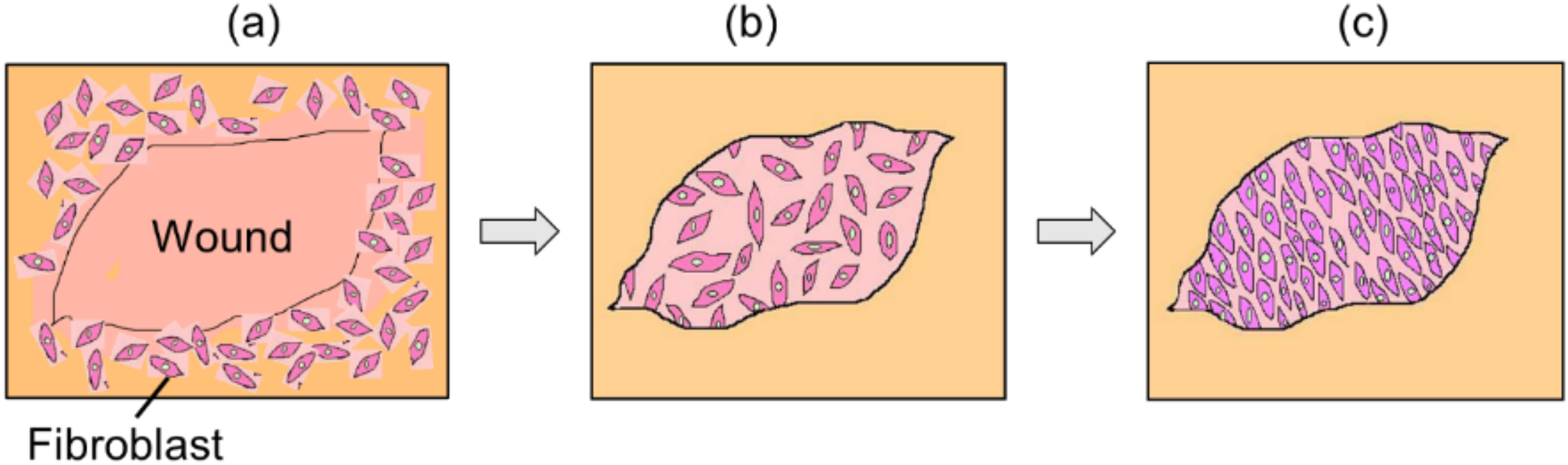
Potential process of wound healing process in vivo. (a) Early stage of inflammation, (b) intermediate stage of cell proliferation and migration, and (c) mature stage of barrier remodeling with collagen synthesis and neovascularization.
In this study, fibroblasts are pre-aligned to evaluate the effect of cell pre-alignment on wound closure rate. For cell pre-alignment, two experimental approaches, micropatterning technique with PDMS stamping of fibronectin and application of cyclic stretching were performed. A scratch assay was then performed to observe cell migration behavior, which is a conventional wound healing study where a cell-free region is created in a confluent cell monolayer by physically removing cells with a scraper and dynamic behavior of cell migration in the region is observed. 6
Materials and methods
Cell culture
Normal human male MJ90 fibroblasts (MJ90 fibroblasts) and normal human dermal fibroblasts (NHDFs) were used for experiments. The cells were grown in Dulbecco's modified Eagle's medium (DMEM) (Life Technologies, USA), supplemented with antibiotics (Penicillin-Streptomycin, Life Technologies, USA) at a concentration of 1% (v/v) and 10% (v/v) of fetal bovine serum (FBS) (Life Technologies, USA), in a humidified atmosphere containing 5% CO2 at 37 °C.
Pre-alignment by micropatterning
As shown in Figure 2(a), a silicon wafer mold was fabricated to create a PDMS (dimethylpolysiloxane, Sylgard 184, Dow Corning, USA) stamp for micropatterning. After the fabrication of the silicon mold using photolithography, PDMS prepolymer base and curing agent was mixed in a 10:1 ratio, poured over the silicon mold and cured in a 60 °C oven for 2 h. After cooling, PDMS was peeled off from the silicon mold and the first PDMS stamp was cut out. The double casting process was performed to produce the final PDMS stamp. The casted PDMS substrate has 20 µm height and 15 µm width of rectangular profile microgrooves and 15 µm spacing between the two adjacent microgrooves. Before seeding fibroblasts, O2 plasma-treated PDMS stamps were coated with 50 µg/mL fibronectin for 45 min and lowered face down over a dish surface and kept inside an incubator for at least 2 h. All substrate samples were then immersed in Pluronic F127 surfactant prill (BASF, Florham Park, USA) in order to block the adhesion of cells to uncoated regions. Control cells with non-micropatterned were also prepared.

Pre-alignment of cells. (a) Micropatterning technique with a PDMS stamp and (b) cyclic stretching technique with a PDMS chamber. (c) The wound creation method with a micropipette tip and a scratched wound area.
Pre-alignment by cyclic stretching
Cyclic stretching was performed to produce a uniform strain on culture substrates. Briefly, as shown in Figure 2(b), a rectangular PDMS stretching chamber with a cell culture area of 20 mm x 20 mm cells were treated with O2 plasma for 2 min, coated with 50 µg/mL of fibronectin solution and incubated for 1 h on MJ90 fibroblasts and 25 µg/ml for 4 h on NHDFs. Cells were then cultured and incubated for 24 h. After reaching a confluent condition, the PDMS stretching chambers was cyclically subjected to a 20% stretch at 1 Hz for 12 h using a stretching apparatus. In the apparatus, the PDMS stretching chambers were held at one end by a fixed clamp and the opposite end by a moving clamp. Control cells with non-stretched were also prepared.
Creation of wound and wound healing assay
To simulate wound, physical scratching by a micropipette tip was then performed to create a gap after cells reached a confluent condition (Figure 2(c)). For stretch-exposed cells, the gap was created both perpendicular (90°) and in parallel (0°) to the direction of cell alignment. MJ90 fibroblasts and NHDFs were used to compare cyclic stretch (90°) with micropatterning and cyclic stretch at 0° and 90°, respectively. The dimensions of the wound were about 300–500 µm wide and 5–7 mm long. Cell migration into the wound area were observed up to 50 h on a 37 °C hot plate in a 5% CO2 condition set on an inverted microscope (IX81, Olympus, Japan). After binarizing images, changes in wound area and cell migration rate were calculated based on the position of averaged front line of migrating cells (Figure 2(c)).
Morphological analysis
Cell elongation and orientation were quantitatively analyzed the pre-aligned cells and the control. A cell shape index (CSI) is a dimensionless quantitative measure of cell morphology acquired from images and determined based on the cell perimeter and area. The CSI were calculated from the following formula:

Fluorescence staining
Actin filaments and cell nuclei were stained with rhodamine-phalloidin (Life Technologies, USA) and with Hoechst (Dojindo Molecular Technologies, Inc., Japan), respectively. The complex of rhodamine-phalloidin stained actin filaments can be excited by blue light with a wavelength of 540 nm, emitting a light of a wavelength of 565 nm. Hoechst-DNA complex can be excited by light with a wavelength of 350 nm, emitting a light of a wavelength of 460 nm.
Statistical analysis
Statistical analysis was performed to compare the experimental results using an unpaired samples t-test in Microsoft Excel with the significance level at p < 0.05.
Results
Microscope images of pre-aligned and scratched cells
The changes in cell shape between control, micropatterned and stretched were observed as shown in upper panels of Figure 3. Cells were randomly oriented, exhibiting non-aligned shape for control (Figure 3(a)) while cells were completely aligned in the direction of the fibronectin stamp after being cultured for 12 h (Figure 3(b)) and oriented perpendicular to the direction of stretch after 12 h stretching (Figure 3(c)). Fluorescence images in the lower panels of Figure 3 showed that both stretched cells exhibited development of actin stress fibers while control and micropatterned cells exhibited less development. Figure 4 shows the creation of wound by scratching with a micropipette tip for the three culture conditions. Wound with 300–500 µm wide was clearly observed.
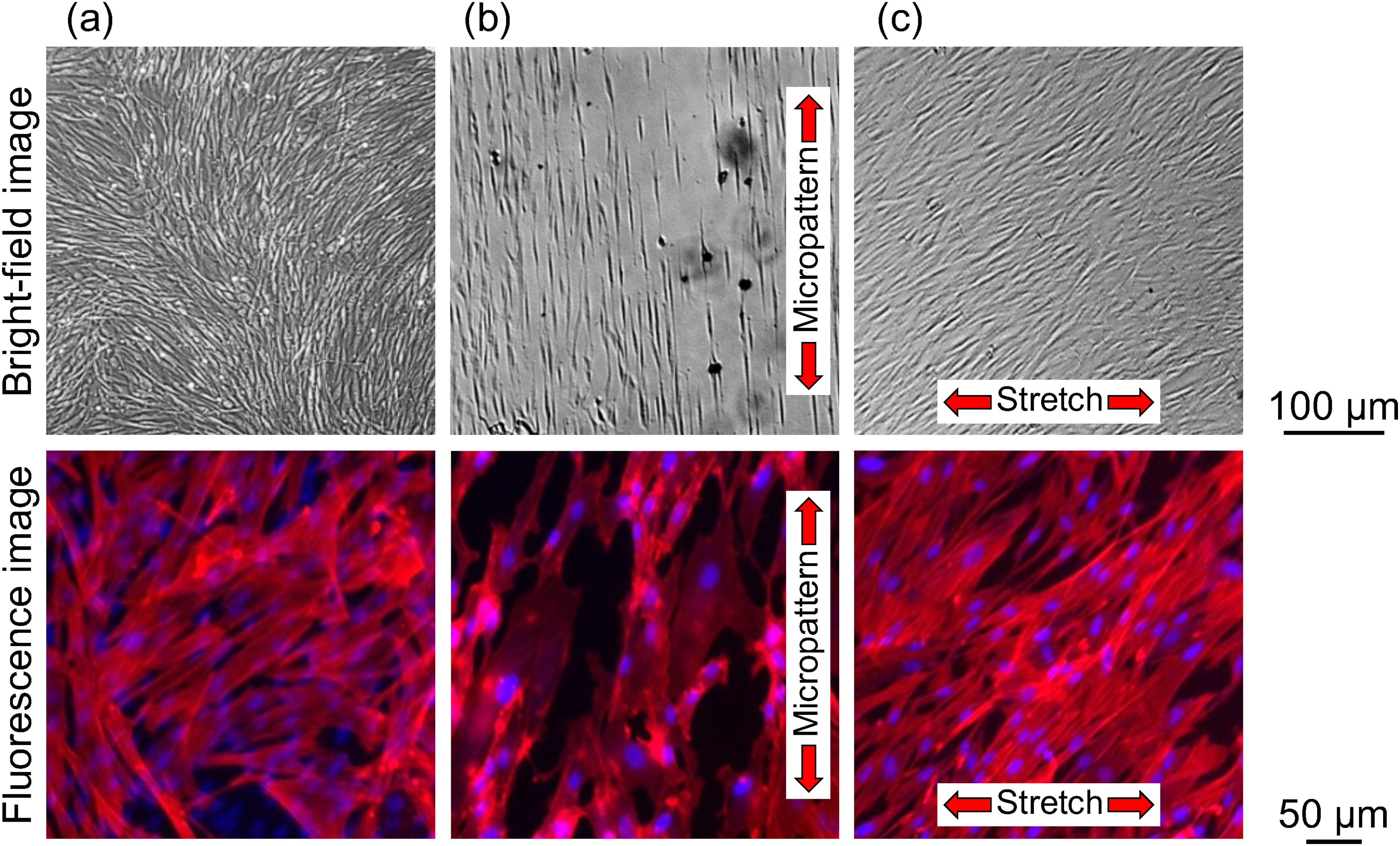
Bright-field (top) and fluorescence (bottom) images of cells. (a) Control, (b) micropatterned and (c) stretched. Bars = 100 µm (top), 50 µm (bottom).
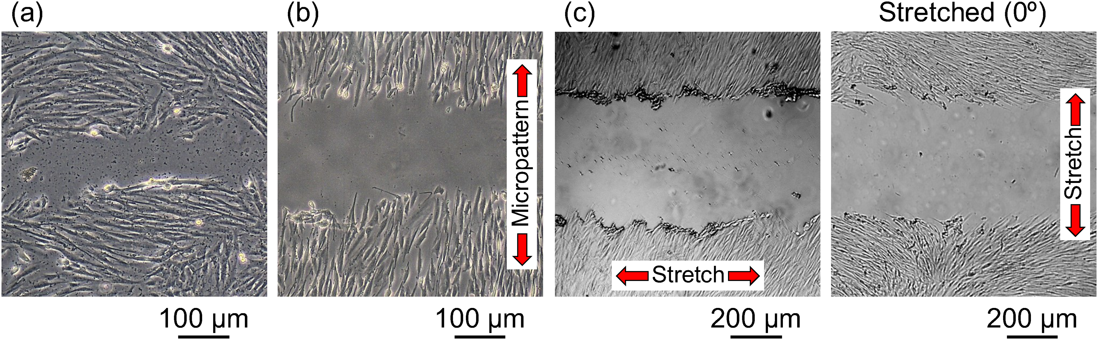
Creation of wound area by scratching with a micropipette tip. (a) Control, (b) micropatterned and (c) stretched with perpendicular (90°) and parallel (0°) wound. Bars = 100 µm (a)(b), 200 µm (c).
Measured parameters of pre-aligned cells
Figure 5 shows that the shape index (SI) of cells grown on the micropatterned substrate and exposed to cyclic stretch were significantly higher than that of control cells. This result indicates that micropatterning and cyclic stretching could elongate cells. The orientation angle of cells was randomly distributed for control (Figure 6(a)) while more distributed around 90 degrees for the stamping (Figure 6(b)) and stretching conditions (Figure 6(c)).
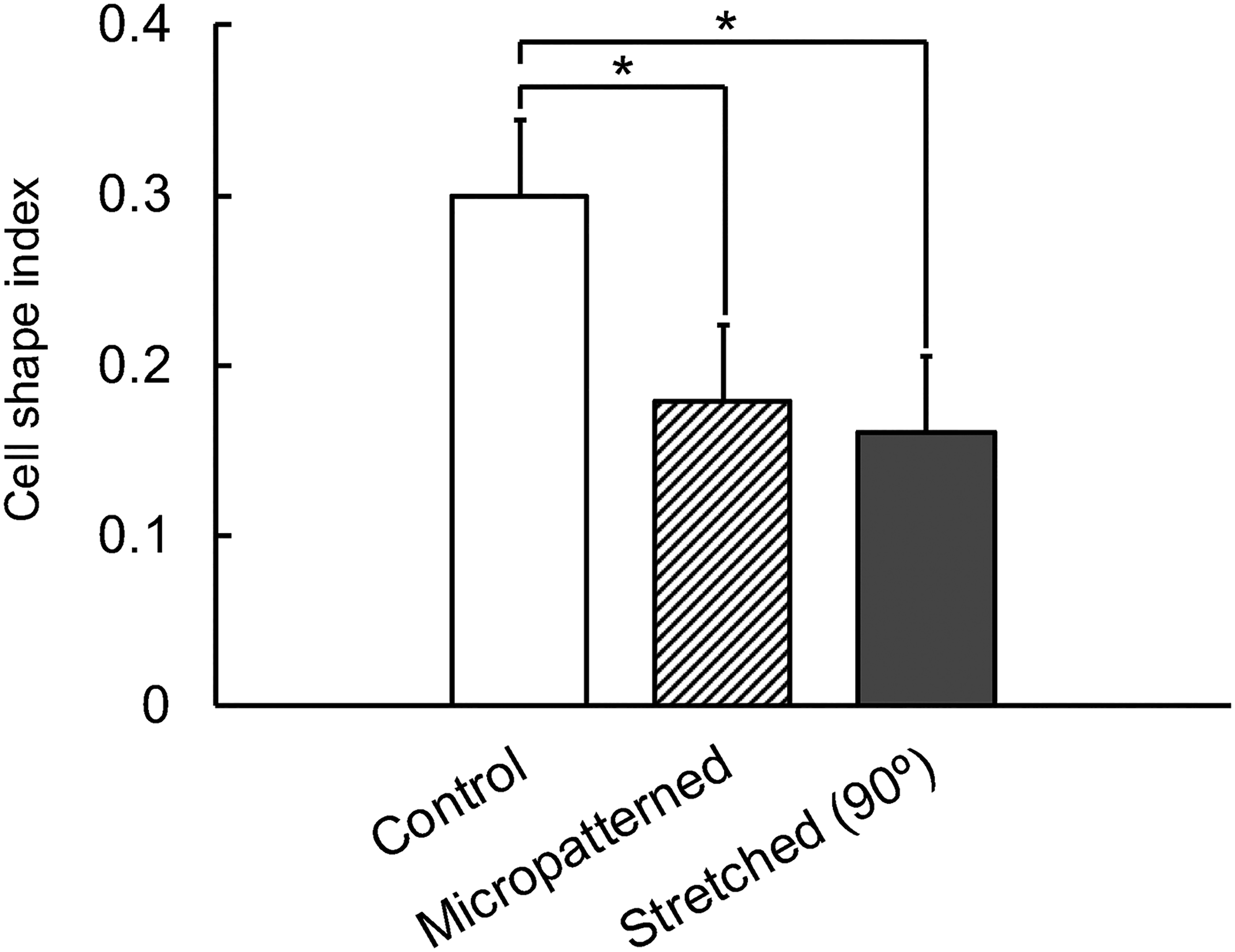
Cell shape index under the three different culture conditions (mean + SD, n = 5). * Statistical significance (P < 0.05).
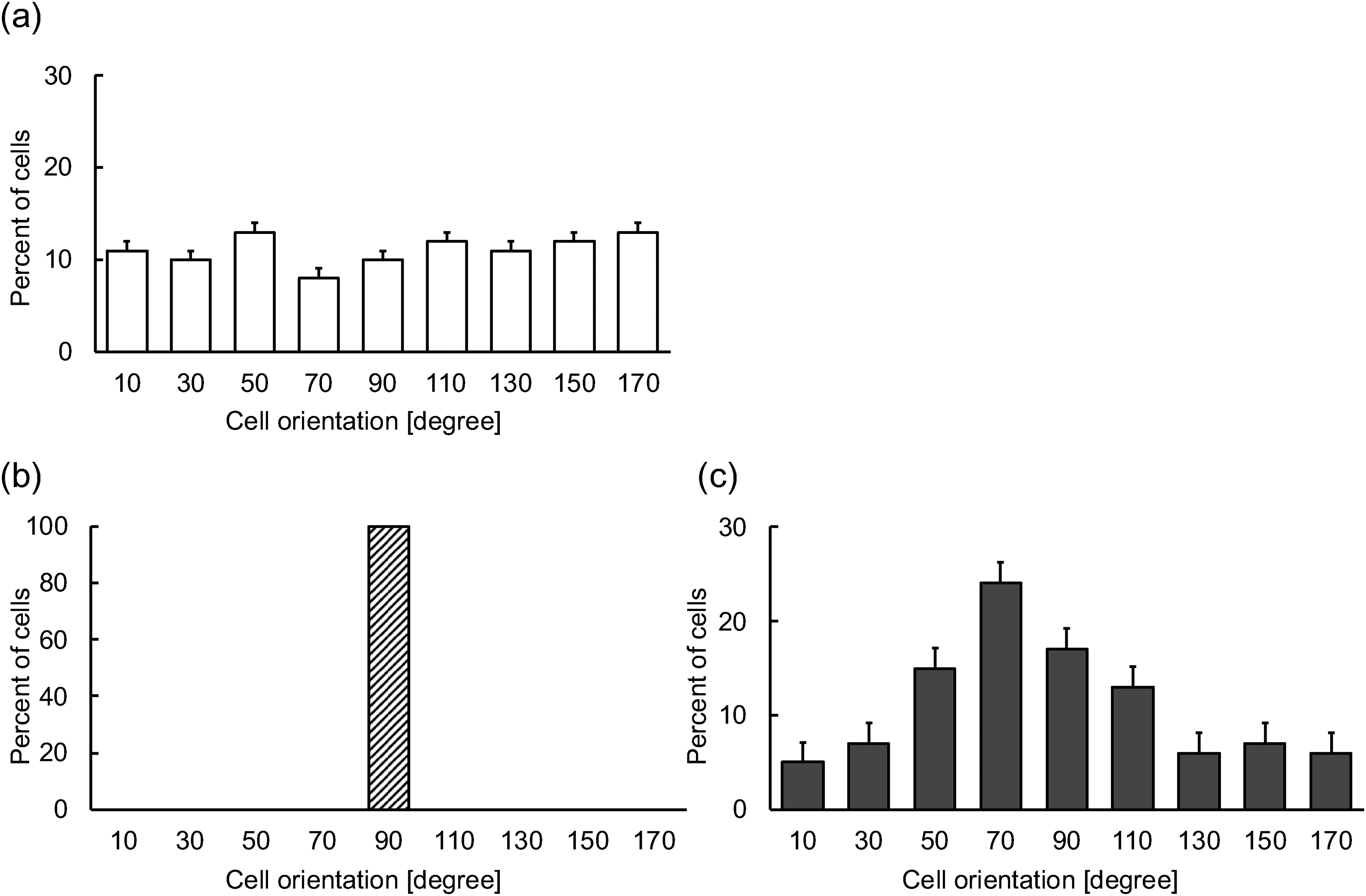
Cell orientation under the three different culture conditions, (a) control, (b) micropatterned and (c) stretched (mean + sd, n = 5 for control and stretched, n = 1 for micropatterned).
Cell migration rate
After creating wound, wound closure by cell migration was observed over time and analyzed using ImageJ software (Wayne Rasband, USA). Control cells initially migrated randomly and began to migrate together after hours while pre-aligned cells immediately migrated in a more directional manner. For all experimental groups, the wound area was completely closed within 20–50 h. As shown in Figure 7, changes in wound area were obtained, giving an averaged velocity of cell migration from the slope of the regression lines. Figure 8 showed that the migration rate of pre-aligned cells for micropatterned and stretched was significantly higher than that of control. Moreover, the migration rate for stretched was significantly higher than that for micropatterned. Figure 9 showed that migration rate of stretch-induced aligned cells perpendicular (90°) to the wound was significantly higher than that of the case of 0°.
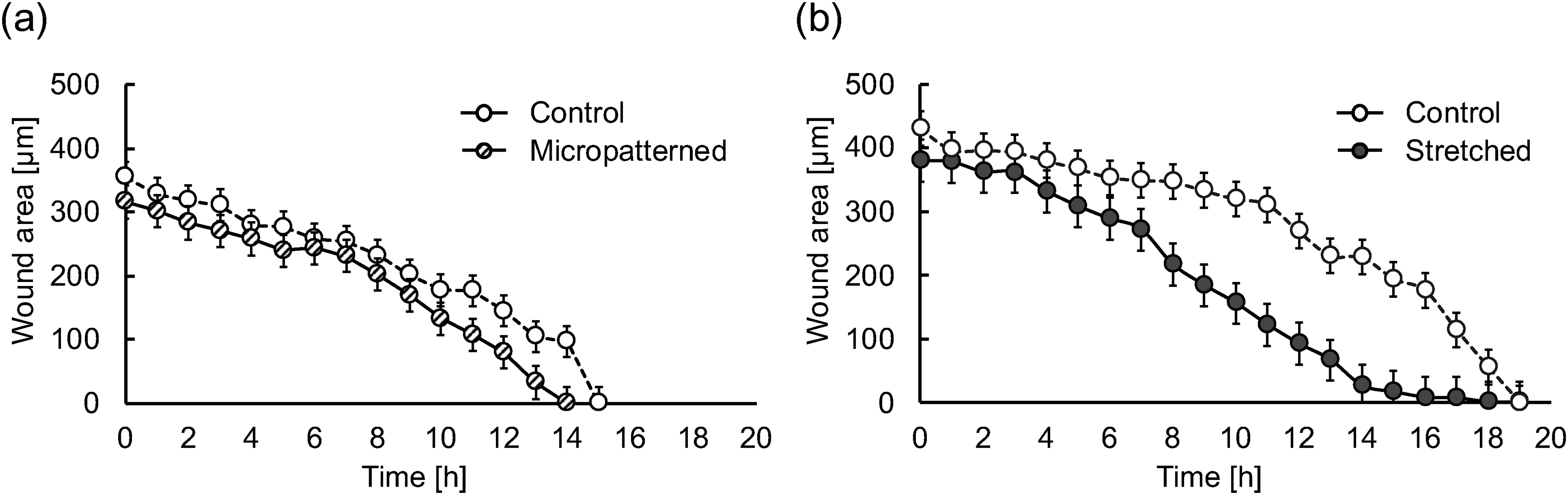
Time course of change in wound area for the three different culture conditions. (a) Control vs. micropatterned and (b) control vs. stretched (90°) (mean ± SD, n = 5).

Cell migration velocity of the three different culture conditions on normal human MJ 90 fibroblasts (mean + SD, n = 5). *Statistical significance (P < 0.05).
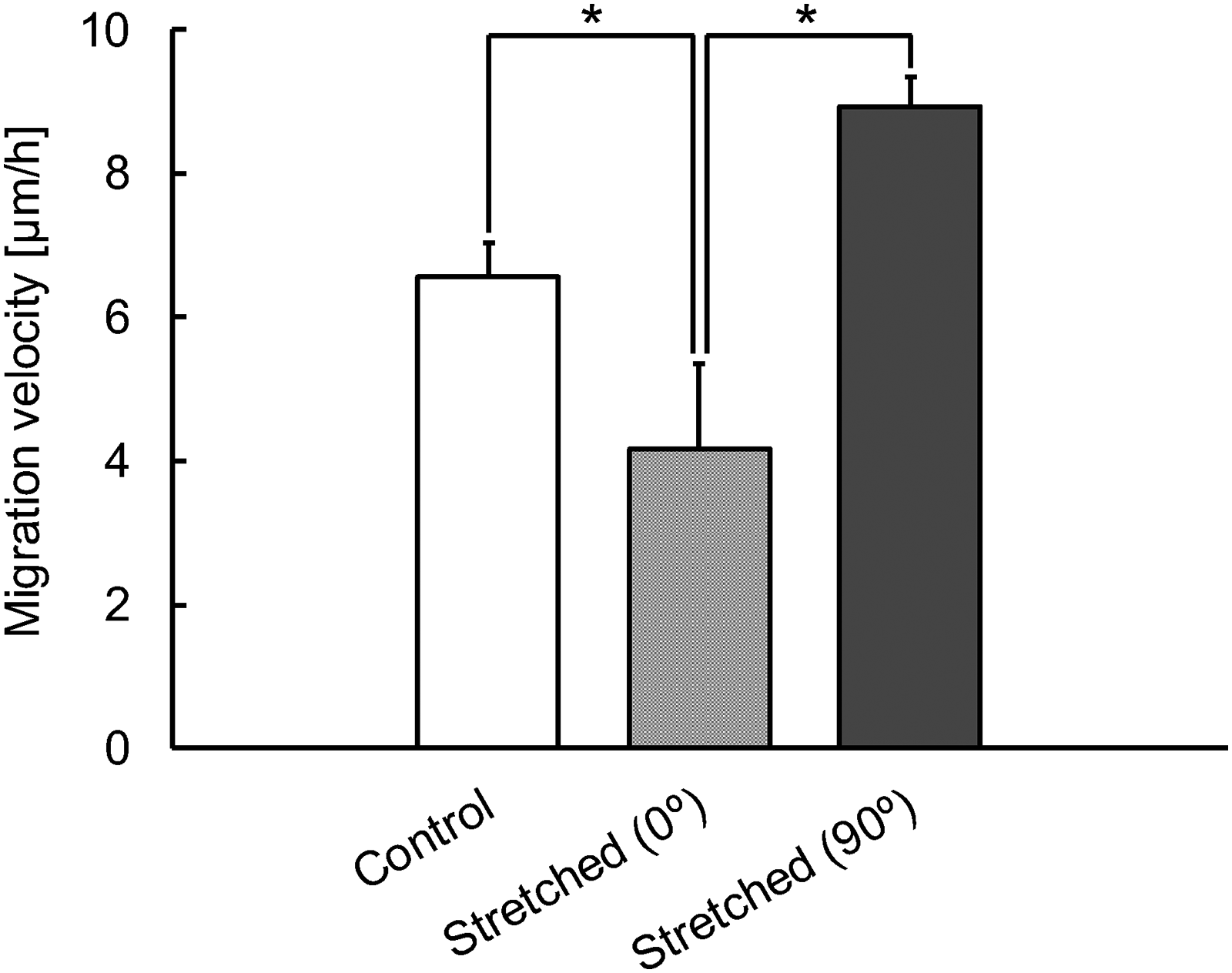
Cell migration velocity of stretch-induced aligned cells perpendicular (90°) and in parallel (0°) to the wound on normal human dermal fibroblasts (mean + SD, n = 3). *Statistical significance (P < 0.05).
Discussion
It is crucial to know underlying mechanism of wound healing process for a better understanding of the recovery of skin injury. In particular, as fibroblasts play a critical role in wound healing process controlled migration behavior of fibroblasts would contribute to an effective wound treatments. As many of previous studies reported, as shown in Figures. 3 and 6, fibroblasts could align with the fibronectin stamps and aligned perpendicular to the direction of stretch. Various techniques for cell alignment have so far been reported, in particular for tissue engineering application, including micropatterning of ECMs such as collagen, electromagnetic field, Electrospun nanofiber, microfabricated structures, mechanical strain, etc.7–10 Micropatterning technique with PDMS stamping and cyclic stretching used for this study are simple and effective approaches in light of equipments needed, experimental costs, and technical access for the experiments.
One of the most important findings in this study is that the migration rate of pre-aligned fibroblasts was significantly higher than non-aligned control (Figures 7 and 8). In particular, Figure 9 clearly shows that the pre-alignment of cells in the direction of wound (90°) could lead to faster wound closure. From the time-lapse observation, at the early stage of cell migration, the pre-aligned cells perpendicular to wound (0°) displayed a change in their orientation, resulted in the lower migration rate. As fibroblast alignment is in vivo observed at the mature stage of wound scar the present results could prove that pre-alignment of fibroblasts perpendicular to the wound would be effective for faster treatment of wound injury. As aforementioned, fibroblasts are in vivo observed proliferating from the early stage of inflammation to the intermediate stage, and aligning and migrating to close wound at the mature stage, together with secretion of numerous growth factors, cytokines and chemokines in association with the other cells involved in this process. 1 It can be suggested that it may take less time to cover the scratched space if fibroblasts can be aligned perpendicular to the wound earlier than the mature stage by for example mechanically or chemically minimally invasive manners. This could potentially be applied to artificial skin treatments: by creating artificial skin through cyclic stretching, it is possible to align fibroblasts within the skin and speed up healing after skin grafts.
Another important finding is that compared to the micropatterned cells, the stretched cells indicate a significantly higher migration rate (Figure 8). Previous studies have demonstrated that actin stress fibers of cells are developed specifically in response to cyclic mechanical stretching. 11 Fluorescence observation, as shown in Figure 3, fibroblasts aligned with fibronectin coated by PDMS stamping exhibits less actin organization while fibroblasts aligned by cyclic stretching exhibit the development of actin stress fibers. This result may indicate that the development of thick actin stress fibers might greatly play a critical role in the faster migration rate.
Conclusions
In this study, the chemically-patterned cell culture substrates with fibronectin-coated PDMS stamps and mechanically-stretched cell culture substrates with cyclic stretching were used to pre-align fibroblasts to investigate the hypothesis whether pre-aligned cells perpendicular to the direction of scratched wound could migrate faster than non-aligned cells to accelerate wound closure. It was indicated that the chemically- and mechanically-treated substrates aligned cells perpendicular to wound, contributing faster closure of wound. Moreover, it was found that cells with the thick actin filaments migrated faster. It can be concluded that the wound closure is greatly influenced by the adequate alignment of fibroblasts as well as the development of actin filament structures.
Footnotes
Funding
The authors received no financial support for the research, authorship, and/or publication of this article.
Declaration of conflicting interests
The authors declared no potential conflicts of interest with respect to the research, authorship, and/or publication of this article.