
Abstract
Background
Monopolar Radiofrequency uses high-frequency waves to generate heat for skin tightening and tissue repair. However, individual fat layer thickness variation causes uneven radiofrequency (RF) penetration and heat thresholds, compromising personalized results.
Objective
The purpose of this study is to analyze the temperature distribution of tissues with different fat thickness after radio frequency treatment and the experimental temperature distribution and tissue changes of pork tissues in vitro by finite element analysis and in vitro experiment verification, so as to achieve appropriate energy parameters for different individuals.
Methods
A two-dimensional bio-thermal model including epidermis, dermis and subcutaneous tissue was developed in COMSOL Multiphysics 6.2. Four fat thicknesses (2, 4, 6, and 8 mm) were simulated to assess their impact on dermal temperature distribution during 6.78 MHz, 120 W radiofrequency exposure. The electromagnetic-thermal coupling effects were validated through in vitro experiments.
Results
Experimental results validate the simulations, demonstrating consistent thermal trends across fat thicknesses (2–8 mm). Post-treatment intratissue temperatures reached 69 °C (2 mm), 60 °C (4 mm), 55 °C (6 mm), and 45 °C (8 mm), all within epidermal safety limits.
Conclusion
The results show that the energy parameters need to be adjusted according to the thickness of adipose tissue during radiofrequency therapy, and higher energy or longer treatment time may be needed for the treatment site with thicker adipose tissue to achieve the expected effect.
Indroduction
Radio frequency technology is widely used in cosmetic medicine, as a non-invasive treatment, monopolar radiofrequency has been proved to be effective in promoting collagen remodeling and fat metabolism.1,2 The principle of radio frequency therapy is to use high-frequency alternating electromagnetic waves, when radio frequency current acts on skin tissue, charged particles (mainly water molecules) in skin tissue oscillate and rub rapidly under the action of radio frequency electric field, thus generating heat.3–5 Radio frequency energy can heat the collagen in the deep skin, make it shrink and stimulate the production of new collagen, thus firming the skin.
At present, the research mainly focuses on the mechanism and clinical effect evaluation of RF technology. Tunnell et al. 6 put forward a mathematical model of monopole radio frequency heat conduction in skin tissue, which can simulate the propagation and heat distribution of monopole radio frequency energy in different layers of skin, and use R-134A coolant to cool the epidermis, through this model, the temperature distribution of human skin under RF heating and coolant cooling can be better predicted, which is convenient for understanding the action mechanism of monopole RF and provides theoretical basis for optimizing the parameter setting of RF equipment. 7 In clinical application, many medical and aesthetic institutions have carried out evaluation research on the effect of monopolar radiofrequency on facial rejuvenation, 8 covering people of different ages and skin types. By measuring the depth of facial wrinkles, skin firmness and elasticity, it was found that after monopolar radiofrequency treatment, facial wrinkles were improved in most patients, and skin firmness and elasticity were improved.9–11 Thermage system is a monopole RF device that provides non-ablation treatment. Thermage CPT uses monopole capacitively coupled RF to tighten the skin and reduce relaxation. The device was approved by the US Food and Drug Administration (FDA) in 2002 to treat periorbital wrinkles, facial wrinkles in 2004 and all wrinkles in 2005, and the system has been developing continuously. 12 Although there are other RF devices on the market, Thermage has published the most documents and clinical trials so far, supporting monopolar radiofrequency as an effective model for rejuvenation. 13 Bora et al. 14 confirmed the influence of different RF frequencies on skin tissue, the impedance of skin decreases with the increase of frequency, and the penetration depth of RF energy is inversely proportional to frequency.
When the dermal temperature reaches 55 °C–65 °C, it can cause collagen contraction and regeneration and remodeling. 15 Although higher temperature may bring more obvious tightening effect, too high temperature will cause damage to the epidermal tissue of the skin, and the temperature of the epidermal tissue should not exceed 45 °C, 16 once it exceeds this temperature, it may cause thermal burns of the epidermis, leading to adverse reactions such as redness, blisters, etc., which may seriously affect the normal function of the skin and even leave scars. Among the patients with complications, 49% had blisters/vesicles(17), 33.3% had erythema for more than 24 h, 51% had edema for 24∼48 h, and 23.5% had severe pain, most dermatologists believe that high energy, pulse beating and problematic tips are the causes of complications. 3.9% dermatologists think that fat atrophy is a complication. The methods to prevent fat atrophy are to reduce the treatment energy, avoid treating patients with less facial fat and increase the treatment interval. Suh et al.(18) suggested that skin thickness is still the most important determinant in the reactivity to plastic beauty pole. However, the systematic study on how subcutaneous fat, a key thermal barrier, modulates the temperature field distribution is still blank. The significant difference of subcutaneous fat thickness in different individuals leads to different treatment responses, which directly affects the predictability and safety of clinical efficacy. It is of great clinical significance to clarify the quantitative relationship between fat thickness and temperature field distribution for individualized parameter setting and curative effect optimization.
The purpose of this study is to quantitatively reveal the modulation effect of fat thickness on surface temperature and deep temperature by constructing a multi-physical field coupling model and combining with in vitro tissue experiments. Firstly, the heat conduction model of monopolar radiofrequency in skin tissue will be established by COMSOL Multiphysics 6.2, and the cooling effect of tissues with different fat thickness after radio frequency treatment and optimization of cooling parameters will be evaluated. By observing the temperature distribution at different stages, the transfer and diffusion of thermal energy and the influence of cooling measures on temperature can be evaluated. Because pig skin is closest to human skin in characteristics, the subcutaneous heating experiment of isolated pork will be carried out to verify the results of finite element analysis(19,20), and the changes of collagen and overall tissue reaction of pork tissue after radiofrequency treatment will be evaluated.
Methods
Numerical simulation
Geometric model construction
In this study, COMSOL Multiphysics 6.2 was used to construct the probe of RF therapeutic instrument and adipose tissue models with different thickness to perform monopolar radiofrequency action, and to explore the influence of different thickness of fat on the temperature change of subcutaneous temperature field (Figure 1).
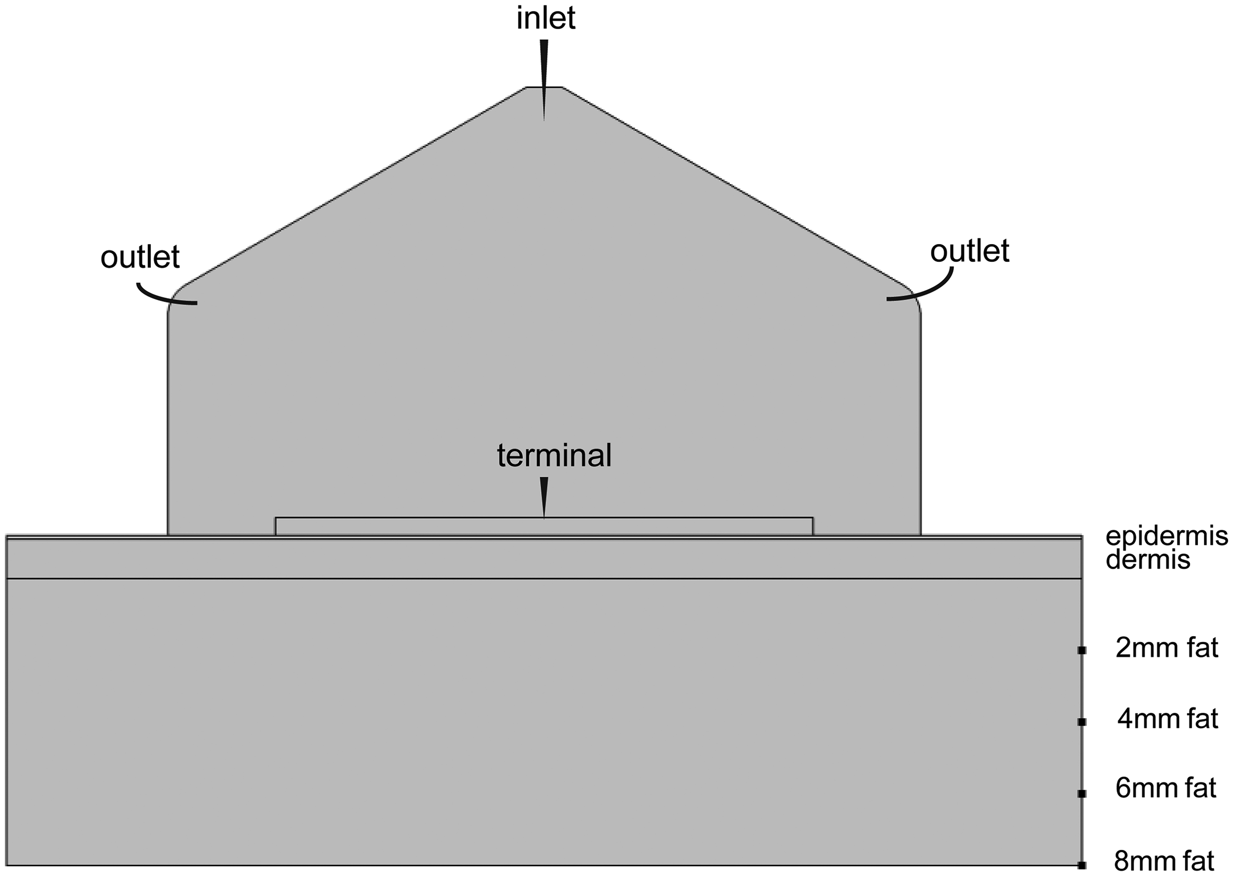
Model geometry.
From top to bottom, there are cooling system, electrode, epidermis, dermis and fat layer(21).
The cooling system model has a height of 12 mm, a width of 21 mm, a cooling fluid inlet width of 1 mm and a fillet radius of 1 mm at the outlet. The electrode thickness is 0.5 mm and the width is 15 mm; The thickness of epidermal layer is 0.1 mm, the thickness of dermal layer is 1.1 mm, and the thickness of fat layer is set to 2 mm, 4 mm, 6 mm and 8 mm respectively according to the experiment, and the width is 30 mm.
The cooling system sets the cooling fluid as R-134A, which enters from the top of the cooling system model and flows out from the rounded corners on both sides of the cooling system model to carry out the fluid heat transfer process. The initial temperature Tustr is 263 K, and the fluid flow is incompressible. It is assumed that R-134A is an ideal liquid. The axisymmetric 2D model is used to reduce the calculation amount, and the non-isothermal flow interface is used to couple heat transfer and fluid flow, without sliding boundary conditions.
The electrode material is copper for current transmission(6).
The physical parameters of skin tissue and adipose tissue are shown in Table 1, assuming that the tissue is an isotropic material, and the density, electrical properties, thermal properties and blood perfusion rate are constant values, and have nothing to do with temperature.
Physical attribute parameters of tissue.

Control equation
Radio frequency electromagnetic field and heat conduction(24) equation are simultaneous:
Energy acts on skin tissue through electrode output, and the electrostatic field in skin tissue is expressed as 
Calculation of temperature distribution in skin tissue by Pennes(26) biothermal equation, which is given by
Electrical boundary conditions: in practical applications, high-frequency sinusoidal voltage is usually used for RF processing, but in finite element analysis, this sinusoidal voltage is usually converted into the effective value of the corresponding DC voltage, and the RF voltage can be expressed as
In the finite element analysis, in this study, the electrode is set as the terminal, the bottom of the fat is set as the ground, the initial zero-time voltage is set to zero (when t = 0, V = 0), and the other epidermis areas and two sides are set to zero electric flux conditions. The electric potential in the tissue was controlled by the Laplace equation given by
Thermal boundary conditions: In order to effectively simulate skin heat dissipation, the corresponding surface heat dissipation coefficient is set at the interface between air and skin, and the boundary convective heat flux is set at 10 W/(m2 K), and the other boundaries are zero heat flux conditions. When t = 0, it is assumed that the temperature of biological tissue T0 is 297 K and the ambient temperature Text is 293 K.
Grid division
Mesh generation is a key stage in finite element analysis, in which the finite element mesh divides the simulation model into many small areas, and equations and polynomials are solved in these areas to approximate the simulation results. COMSOL software has an adaptive grid division tool, which can automatically divide the simulation model into domains as needed, and use the error estimate of the solution at each time interval as a measure to re-divide the grid of the model and resize the domain. In this study, the mesh adaptive segmentation tool is used to segment the model, and the conventional mesh size is built inside the cooling system, with the largest cell being 0.706 mm and the smallest cell being 0.0314 mm; And the boundary of the cooling system is refined.
Temperature acquisition
The distribution of RF and cooling parameters in this study is that the skin tissue is pre-cooled for 200 ms at first, and the pulse energy emission mode is adopted. After the voltage is applied for 220 ms, five pulse voltages are applied. At the beginning of the application of the last four pulse voltages, synchronous cooling is carried out for 50 ms, and finally, after cooling is carried out for 200 ms, so as to maintain the safety of the epidermis. The principle of this mode is based on the absorption and heat dissipation characteristics of the skin tissue(12), and during the pulse interval, the skin tissue has time to dissipate heat.
As shown in the schematic diagram of simulation evaluation in Figure 2, the midpoint of the skin surface is taken as the origin of coordinates, and the local point probes are placed at the skin surface and 2 mm under the skin at coordinates (0,0 mm) and (0,-2 mm) respectively(27). The device is used to collect the data of local temperature change during the treatment of RF therapeutic instrument, and draw the collected data into a curve.

Simulation model domain point probe.
In vitro experiment
The RIMGE 100–002 monopolar RF system (Ningbo Ruisirui Medical Technology Co., Ltd, China) was employed for all treatments. The device delivered RF energy at a fixed frequency of 6.78 MHz,the pulse duration is 1 s. In this experiment, the device is set to 120 W, and uses a 4cm2 needle tip, including a liquid nitrogen cooling system. Calibration was performed before each session following the manufacturer's instructions. The subcutaneous heating experiment of isolated pork was carried out by using this equipment, pig abdominal tissue including skin layer and fat layer was used. Pig tissue samples were collected from slaughtered pigs in slaughterhouse, and the whole experiment was processed by cold chain: the samples were stored in a constant temperature refrigerator at 0∼4 °C immediately after slaughter, and were quickly transported to the laboratory console within 2 h after slaughter by special transport tools(28). The experimental group was divided into four groups, the thickness of pork fat was 2 mm, 4 mm, 6 mm and 8 mm respectively under the intact skin layer. As shown in Figure 3(b), four parallel samples were set in each group, and the control group did not receive any treatment. The initial temperature of pork tissue was 23 °C before the experiment. The pork tissue was precooled by RF therapeutic apparatus, and the pork tissue was cooled at the same time and after cooling, and the bottom of the pork tissue was connected with a negative plate. The treatment area is 4cm2, and the treatment time of each point is 0.2 s before pre-cooling, 1.5 s after RF and simultaneous cooling, and 0.2 s after additional cooling.
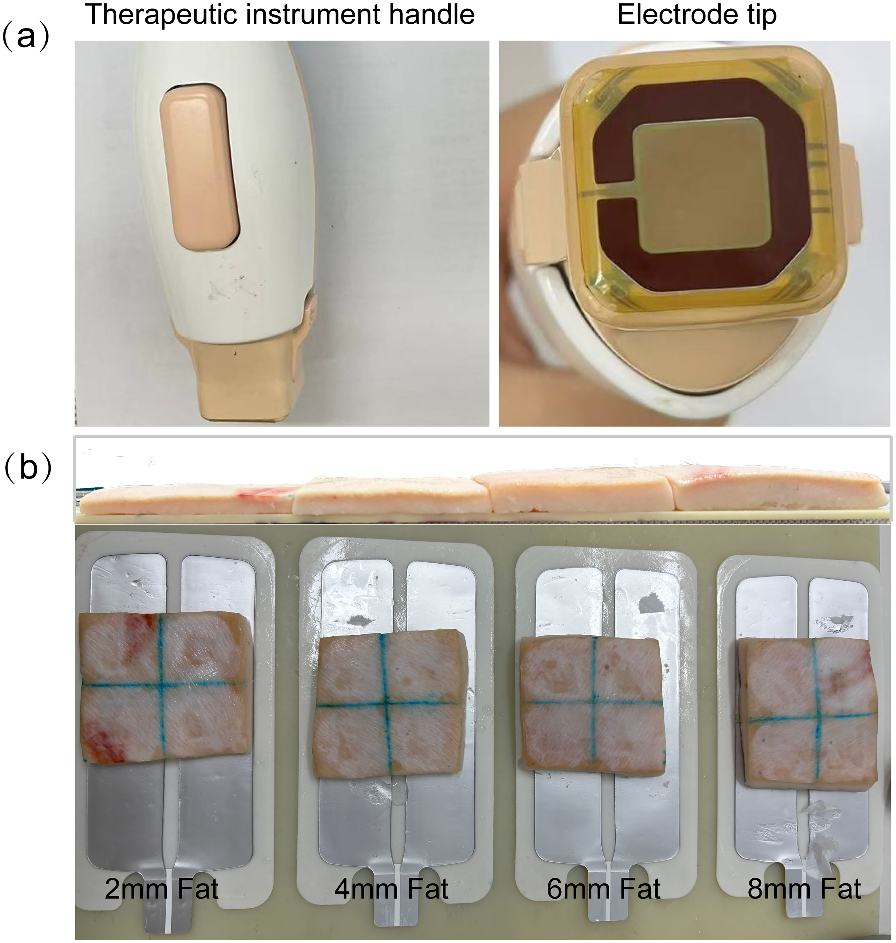
Experimental materials.(a)Radio frequency therapeutic instrument: handle and electrode tip.(b)Pork tissues with different fat thickness.
Three K-type thermocouple probes were used to detect the temperature change. The three thermocouple probes were placed at the center of the pork tissue surface, 2 mm under the pork tissue skin and at the epidermis of the electrode edge. The thermocouple is connected with the signal conditioning module, and the microvolt temperature difference potential is converted into a voltage signal that can be recognized by the oscilloscope, and the oscilloscope is statically calibrated, and the voltage-temperature corresponding relationship is established with ice-water mixture and boiling water, based on which the temperature change of pork tissue can be obtained.
Masson staining is a multifunctional and high contrast staining method, which is widely used in histological and pathological research. It can clearly display the tissue structures such as collagen fiber, muscle fiber and nucleus, and help researchers evaluate the degree of fibrosis and observe the process of tissue repair and regeneration(29). In this study, after radiofrequency treatment, samples of pork tissues in the control group and four groups were selected for Masson staining to observe the changes of collagen in pork tissues. After the pork skin tissue was fixed with 4% paraformaldehyde, the tissue was trimmed, dehydrated, embedded, sliced, dyed and sealed. Finally, the qualified samples were examined by microscope, and the changes of collagen fiber content in the skin were observed and measured by Masson staining. After Masson staining, collagen fibers exhibited dense, wavy bundles, while muscle fibers showed denser, elongated structures.
Results
Finite element simulation results
The cooling fluid cools the tissue surface
The vector arrows in Figure 4 represent the flow direction of the cooling fluid. It can be seen that in the middle area at the top of the graph, the arrow flows vertically downward, indicating that the cooling fluid is injected from the top. At the two sides, the fluid flows downward along the wall of the cavity in an arc shape. The velocity of the cooling fluid at the top fluid inlet is relatively large, about 1.5 m/s, while the velocity of the cooling fluid at the bottom and parts of the two sides is relatively small, about 0.6 m/s.
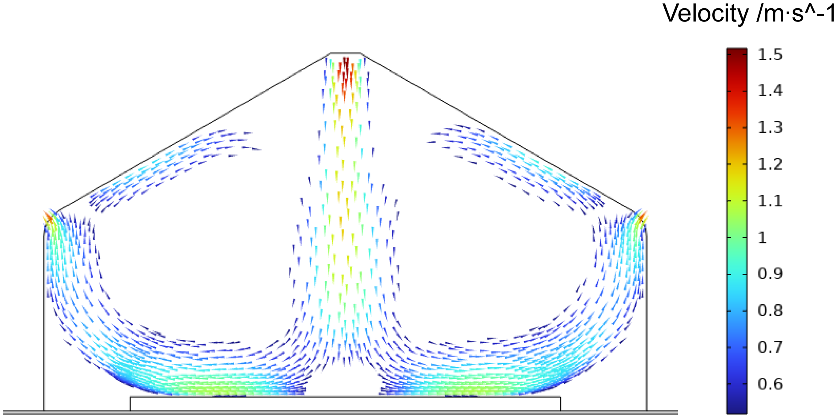
Flow velocity distribution of cooling fluid.
The cooling fluid is injected at high speed from the top inlet, and then gradually spreads to both sides during the downward flow. The high-speed injection is helpful to quickly transfer the cooling effect to the lower area, an obvious circulation structure is formed on both sides of the graph. The cooling fluid flows along the wall of the cavity, turns at the bottom area and flows to the middle or both sides, forming a vortex-like flow pattern. This circulation can make the cooling fluid contact the cooled skin more fully, enhance the cooling effect, and help to distribute the cooling effect evenly.
As can be seen from the figure, the cooling fluid has a certain flow distribution in the skin area, which can directly cool the area. Different fluid velocities at different locations mean that the cooling intensity at different locations is different, and the cooling efficiency in high-speed areas is higher.
Changes of tissue temperature
Under the same power and cooling parameters, the tissues with different fat thickness were treated by radiofrequency. After the treatment, it can be seen from the figure that in Figure 5(a), there is an obvious high-temperature area in the middle of the tissue area, the temperature is close to 70 °C, and the temperature on both sides gradually decreases, indicating that the heat is mainly concentrated in the middle of this area, and there is a significant temperature gradient. There is a high temperature area in the middle of the area below Figure 5(b), but the range of the high temperature area is relatively expanded. The high temperature area in the lower part of Figure 5(c) spreads further, and the higher temperature area extends downward, indicating that heat is continuously transferred and diffused. In Figure 5(d), the high-temperature part in the lower region is weakened, and the overall temperature is reduced. After radiofrequency treatment, the highest temperature of tissues with different thickness is at the level of 2 mm under the skin, which is the deep part of dermis(30). Collagen is rich in providing skin elasticity and toughness, maintaining skin structural integrity, reducing wrinkles and relaxation, and is a key component of skin rejuvenation.
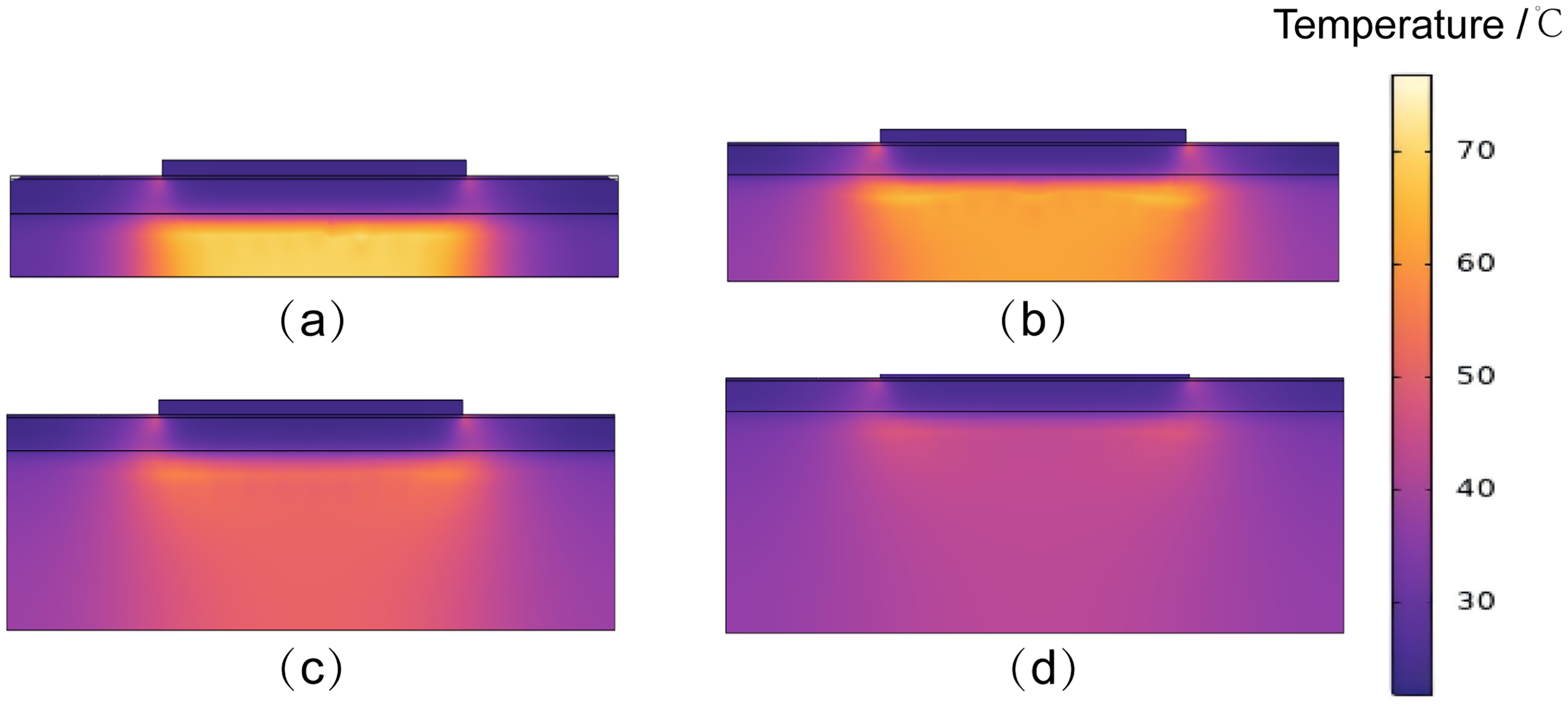
Temperature distribution after radiofrequency therapy. (a) Temperature distribution of 2 mm fat thickness after radiofrequency therapy.(b) Temperature distribution of 4 mm fat thickness after radiofrequency therapy. (c) Temperature distribution of 6 mm fat thickness after radiofrequency therapy. (d) Temperature distribution of 8 mm fat thickness after radiofrequency therapy.
Figure 6(a) shows the changes of tissue surface temperature during the whole treatment. The initial surface temperature is 23 °C, and after 200 ms pre-cooling, the surface temperature drops below 22 °C. After RF voltage was applied, the tissue surface temperature increased, but under the condition of synchronous cooling, the tissue surface temperature remained below 30 °C during RF therapy, maintaining the safety of epidermis. As can be seen from the figure, the surface temperature of 2 mm adipose tissue and 4 mm adipose tissue increases rapidly when they are subjected to radio frequency treatment, while the surface temperature of 6 mm adipose tissue and 8 mm adipose tissue increases slowly.
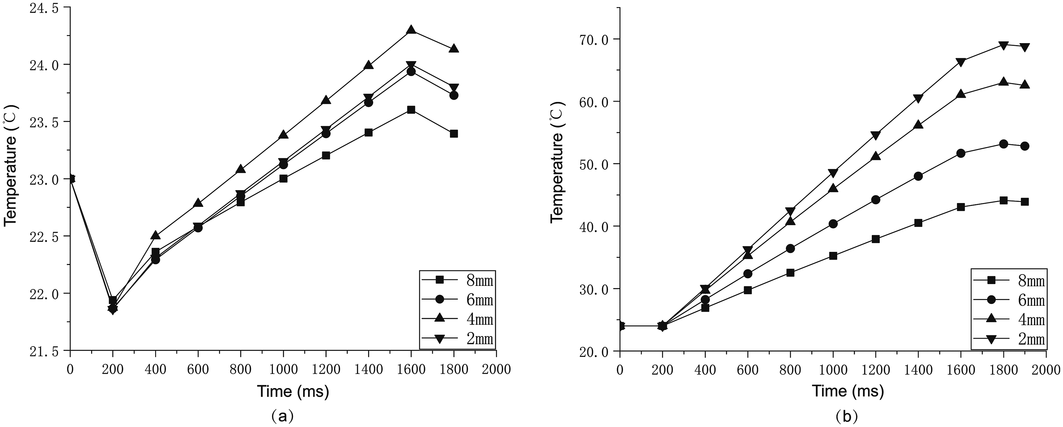
Temperature-time curves of surface and subcutaneous during radiofrequency treatment. (a) Surface. (b) 2 mm under the skin.
Figure 6(b) shows the change of the temperature at 2 mm under the skin during the whole treatment process. When no RF treatment was performed for 0–200 ms, the initial temperature in the tissue was kept at 24 °C. After RF voltage was applied for 200 ms, all four curves began to rise with the passage of time, and the rising rates were close at the initial stage, indicating that the tissues began to be heated by heat. However, it can be seen that the temperature of the curve corresponding to the tissue with 2 mm fat thickness rises the fastest. At the end of radiofrequency treatment, the temperature inside the tissue is about 69 °C, that of the tissue with 4 mm fat thickness is about 63 °C, that of the tissue with 6 mm fat thickness is about 53 °C, and that of the tissue with 8 mm fat thickness is the slowest, and that of the tissue with 45 °C. After 1700 ms, after RF treatment and post-cooling, the rising trend of the internal temperature of the tissue slowed down and gradually stabilized, and then the internal temperature of the tissue gradually decreased.
In vitro experimental results
Pork tissue temperature change
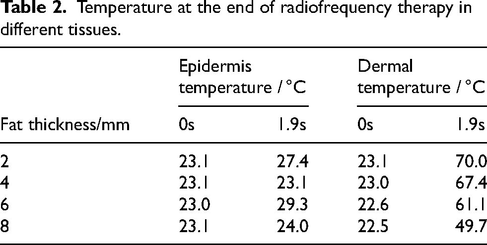
By recording the temperature before RF and the temperature at the end of RF treatment, the following temperature changes can be obtained: on the whole, the surface center temperature is below 40 °C after the end of a treatment, and the subcutaneous 2 mm temperature decreases with the increase of fat thickness.
According to the experimental data, as shown in Table 2, the temperature changes of pork tissues with different fat thicknesses are significantly different after RF treatment. When the thickness of fat layer is 2 mm, the tissue center temperature reaches 70 °C; With the thickness increasing to 4 mm, the central temperature drops to 67.4 °C; When the thickness is further increased to 6 mm and 8 mm, the central temperature drops to 61.1 °C and 49.7 °C respectively. The experimental results show that the radio frequency heating efficiency is negatively correlated with the thickness of fat layer, which may be related to the dielectric properties and heat conduction efficiency of fat tissue.
Temperature at the end of radiofrequency therapy in different tissues.
Masson staining analysis results
Masson staining is a commonly used staining method in histological research, which is mainly used to distinguish collagen fibers, muscle fibers and nuclei. Masson staining can help to observe and analyze the tissue structure of isolated pork tissues, especially when studying the distribution, morphology and pathological changes of muscle fibers and collagen fibers. Positive area ratio refers to the percentage of the stained area of specific tissue components (such as collagen fibers) in the whole tissue section area in Masson staining, which is often used to quantitatively analyze the content or distribution of collagen fibers in tissues, especially when studying fibrosis, tissue repair or pathological changes. In this study, the positive areas of Masson stained pork tissue samples are shown in the following table: Table 3
Positive area ratio.
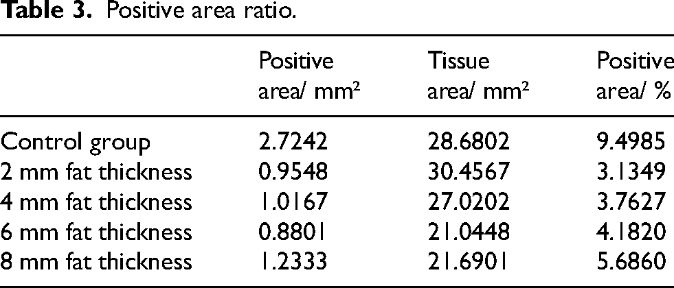
In the Figure 7 of the control group, the structural level of pork skin tissue is relatively clear, and the muscle fiber and cytoplasmic components are uniformly stained with a deep, homogeneous tint, which indicates that the structure and components of pork skin are dyed in a normal state, and the arrangement of muscle fiber and cytoplasmic components shows a certain regularity. The collagen fibers are evenly stained with a dense, fibrillar pattern and are distributed evenly, forming a network structure between muscle fibers that provides mechanical support for the muscle tissue. There is no obvious abnormal aggregation or breakage, and the positive area ratio of collagen fibers is 9.4985%.
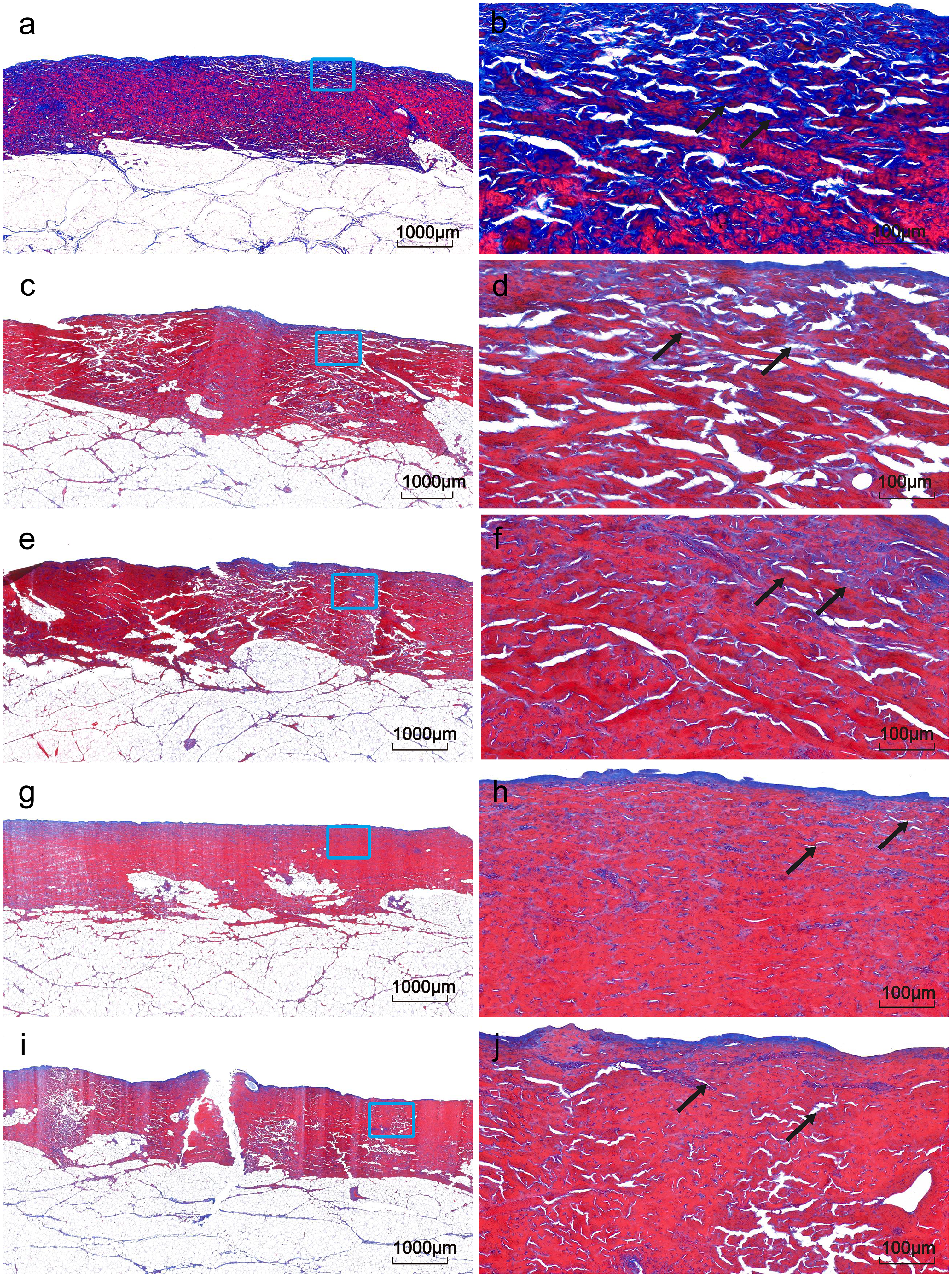
Masson stained sections of adipose tissue with different thickness. (a, c, e, g and i were the control group, 2mm fat thickness, 4mm fat thickness, 6mm fat thickness, 8mm fat thickness, Masson ×1.2; b, d, f, h, j are the boxed areas of a, c, e, g, i respectively, Masson ×10.0.).
However, after radio frequency treatment, the number of collagen fibers in dermis decreased, combined with the analysis of temperature change. The denaturation temperature of collagen fibers is about 55–65 °C, the tissues with 2 mm, 4 mm and 6 mm fat thickness all reach the denaturation temperature of collagen fibers at 2 mm under the skin after radio frequency treatment. The structural changes of collagen fibers can be seen from the arrows in Figure 7. the triple helix structure of collagen fibers is destroyed by radio frequency treatment, the fiber network is relaxed, and the muscle bundle gap is enlarged. The phenomena of loose arrangement, reduced diameter and fracture of collagen fibers are observed under the electron microscope. The positive area ratios of collagen fibers in tissues with 2 mm, 4 mm and 6 mm fat thickness after radio frequency treatment are 3.1349%, 3.7627% and 4.1820%, respectively, so the higher the temperature of dermis, the higher the degree of destruction of collagen fibers and the less the content. Therefore, when the fat thickness is 8 mm, the content of collagen fiber is the highest compared with the other three fat thickness of tissues, and the positive area ratio of collagen fiber is 5.6860%.
Discussion
In this study, the finite element modeling and analysis of biological tissue were carried out, and the in vitro experiments were carried out to systematically explore the effect of adipose tissue thickness on subcutaneous temperature field distribution during monopolar radiofrequency therapy. Comparing the results of finite element analysis with the experimental results, it was found that the temperature distribution of tissues with different fat thickness was similar to that of pork tissues in vitro. With the increase of fat thickness, the temperature increase of dermis decreased. After Masson staining analysis in vitro, it was found that the higher the temperature of dermis, the higher the degree of collagen fiber destruction, so the less collagen content after radio frequency treatment. From the four Masson stained sections of adipose tissue with different thicknesses in Figure 7, it can be seen that with the increase of adipose thickness from 2 mm to 8 mm, the content of collagen fibers in adipose tissue is gradually increasing, the distribution is gradually dense and the structure is gradually complicated, and more collagen fibers can provide better mechanical support for thicker adipose tissue(31). Therefore, when the fat thickness increases from 2 mm to 8 mm, the RF energy will be absorbed by the collagen fibers in the deep dermis, resulting in the decrease of the energy in the dermis. Therefore, with the increase of the fat thickness, the temperature at 2 mm under the skin of pork tissue will gradually decrease. Abraham et al.(32) put forward another mechanism of monopolar radiofrequency action, which can explain the contour improvement immediately after treatment. 10%-30% of subcutaneous tissue is composed of collagen-based fibrous intervals, which form a staggered connective tissue network among fat bodies. When radio frequency energy is transmitted to the skin surface, it tends to the path of least resistance, so it is preferentially transmitted through these collagen fiber intervals, which leads to the selective heating of fibrous intervals prior to fat(33), thus leading to subcutaneous tissue.
The experimental results show that the surface temperature is kept at 40 °C, which maintains the safety of the epidermis(34). Adipose tissue is a poor conductor of heat, the greater the thickness, the longer the path of heat transfer from RF energy source to the surface, which leads to the slower rate of surface temperature increase. The thicker adipose tissue absorbs more energy, but the energy distribution is more dispersed, which leads to the slower surface temperature increase.
Simulations and experiments reveal that in monopolar radiofrequency therapy, despite energy being delivered across the entire electrode contact surface, the skin temperature directly below the electrode edge rises significantly faster and reaches a higher peak than the temperature below the central area. This localized overheating at the electrode periphery creates a significant burn risk and requires special attention. To mitigate this edge effect, therapy requires electrode design optimization (such as increasing contact area or modifying edge curvature) alongside careful treatment protocols: avoiding prolonged edge contact on a single skin area and implementing continuous epidermis temperature monitoring.
Conclusion
In this study, the finite element analysis method was used to explore the changes of subcutaneous temperature of tissues with different fat thickness during radiofrequency therapy, and then the finite element analysis results were verified by in vitro pork experiments. Comparing the results of finite element analysis with those of in vitro experiments, they were basically consistent. The results show that the energy parameters need to be adjusted according to the thickness of adipose tissue during radiofrequency therapy, and higher energy or longer treatment time may be needed for the treatment site with thicker adipose tissue to achieve the expected effect. Thicker adipose tissue may warm up slowly, which may reduce the risk of overheating of the surface skin, but it is necessary to monitor the temperature of deep tissue to avoid overheating. Thin adipose tissue may be easier to achieve ideal therapeutic effect, while thick adipose tissue may need multiple treatments.
Footnotes
Acknowledgments
None.
Ethical considerations
This article does not contain any studies with human or animal participants.
Consent to participate
Not applicable.
Consent for publication
All authors have read and approved the final version of the manuscript and consent to its publication in Bio-Medical Materials and Engineering.
Author contributions
(I)Conceptualization: All authors; (II)Formal analysis: All authors; (III)Investigation: Jiawen Zong; (IV)Methodology: Jiawen Zong; (V)Visualization: Qinghua Kang; (VI)Writing–original draft: Ping Ye; (VII)Writing–review & editing: All authors.
Funding
The authors disclosed receipt of the following financial support for the research, authorship, and publication of this article: This work was supported by a grant from Shanghai Collaborative Innovation Centre for Tumor Energy Therapy Technology and Devices and Xinjiang Production and Construction Corps 2024 Corps Science and Technology Program (Key R&D Achievement Transformation Innovation Environment Construction and Capacity Enhancement Project) (No. 2024AB065).
Declaration of conflicting interests
The authors declared no potential conflicts of interest with respect to the research, authorship, and/or publication of this article.
Data availability
Data used are available throughout the manuscript text.