
Abstract
Objective
The popularity of brushite cement (BrC) in bone regeneration is related to its biocompatibility and favorable resorption properties. Nevertheless, it has poor clinical performance due to quick settling, lack of mechanical strength, and anti-bacterial activity. This paper offered the research on the impact of adding chitosan-selenium nanoparticles (CS-SeNPs) into BrC to improve its mechanical, physical, and biological characteristics.
Methods
CS-SeNPs were added to BrC at 1 and 2 wt.% concentration. Scanning electron microscopy (SEM), energy-dispersive X-ray spectroscopy (EDX), X-ray diffraction (XRD) and fourier transform infrared spectroscopy (FTIR) were used to characterize the modified materials. Physical and mechanical properties were evaluated through mass-loss investigations, setting time, and mechanical properties, shear bond strength [SBS], diametral compressive strength [DCS] and biaxial flexural strength [BFS]. The antibacterial activity, and cytocompatibility were also tested.
Results
The use of CS-SeNPs enhanced BrC crystallinity, mechanical strength and antibacterial activities. SBS, DCS as well as BFS improved considerably with nanoparticles inclusion. The highest mass loss was observed at 1 wt.% CS-SeNP after 48 h and the setting times were longer in comparison with the control. The zone of inhibition and decrease in optical density were seen significant in 1 wt.% group, which indicates superior antibacterial activity. The biocompatibility tests showed moderate cytotoxicity at the higher concentrations.
Conclusion
The use of up to 2 wt.% CS-SeNPs increases the structural, mechanical, and antibacterial behavior of brushite cement considerably without deteriorating its fundamental features. This engineered formulations promises to be applied in the regeneration of bones that need a greater mechanical strength and antimicrobial coverage.
This is a visual representation of the abstract.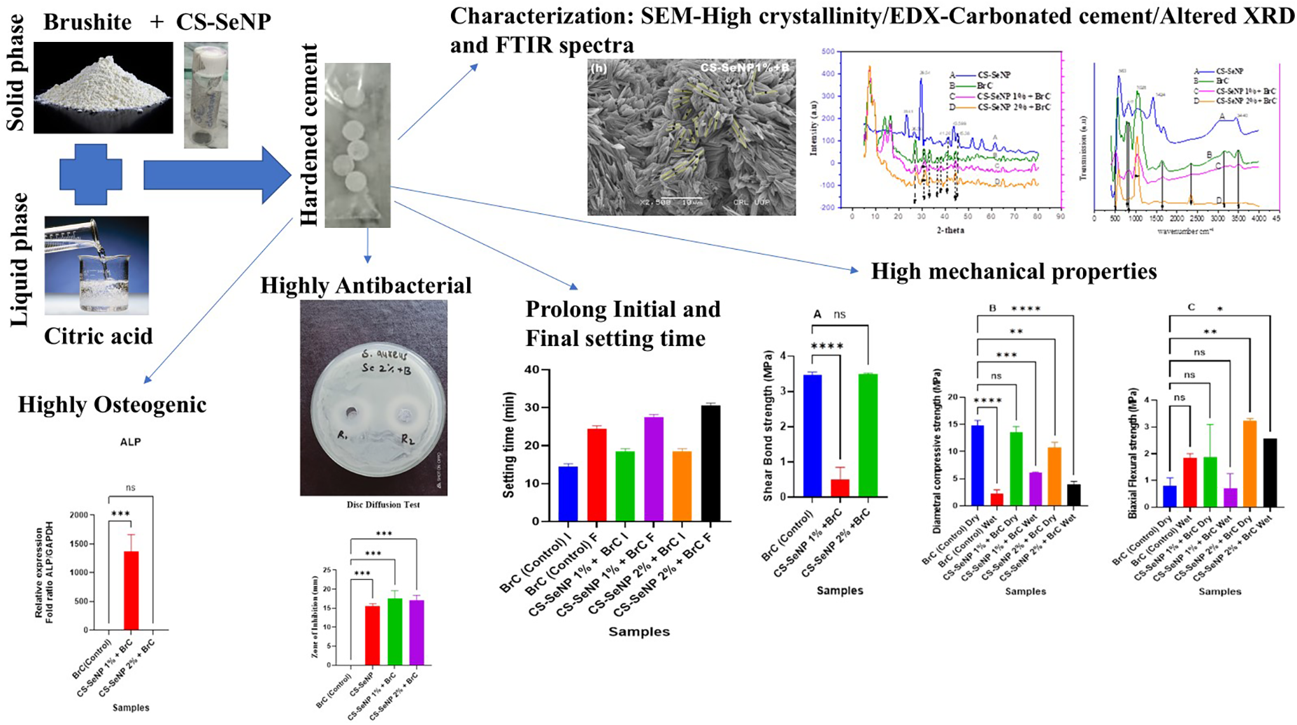
Keywords
Introduction
The existing drawbacks of autologous bone grafts in trauma induced hard tissue defects have necessitated the search for alternative bone regeneration products. The challenges of harvesting autologous bone include the morbidity of the donor site, a scarce amount and inconsistent resorption, which promotes the creation of artificial substitutes. Over the last several decades, biomaterials as scaffolds for bone regeneration have been designed, including ceramics, hydroxyapatite (HA) and tricalcium phosphate (TCP), which are major inorganic bone substitutes. 1
Calcium phosphate cements (CPCs) have attracted special interest because they can be chemically similar to bone. They also offer excellent biocompatibility and biodegradability. 2
However, their inadequate mechanical properties has limited their clinical application primarily to non-load-bearing areas, such as craniofacial or skeletal defect sites, often requiring external stabilization. Therefore, it is crucial to improve the mechanical properties of CPCs to further explore their therapeutic potential.3–5
CPCs are generally classified into the alkaline and acidic groups and the hydroxyapatite-based cements belong to the former. 6 Brushite cement (BrC) which was first introduced by Mirtchi et al. 1989 is an acidic CPC that is predominantly made of monocalcium phosphate monohydrate (MCPM) and β-tricalcium phosphate (β-TCP). 7 Brushite cements offer several advantages such as rapid setting, room temperature stability, excellent biocompatibility and resorption profile.8,9 However, their clinical use is hindered by drawbacks including poor mechanical strength, difficult manipulation due to rapid hardening, limited osteoconductivity, and lack of intrinsic antimicrobial properties, thereby highlighting the ongoing need for improved bone repair materials.7,10,11
Selenium is a trace element important in human health that has special antimicrobial, antioxidant and tissue repairing effects. 12 The size of selenium nanoparticles is nanoscale, which enhances its biological activity and biocompatibility compared to other selenium compounds. 13 SeNPs aid the formation of selenoproteins, which counter the free radicals caused by oxidative stresses.14,15 Nevertheless, the aggregation of SeNPs in aqueous conditions occurs readily and minimizes its bioavailability and biological effectiveness. In order to handle this shortcoming, stabilizers like polysaccharide (chitosan) are thus widely used to sustain SeNPs dispersion and activity. 16
Chitosan (CS) is a biocompatible, biodegradable, and polysaccharide polymer, that is a derivative of chitin. It is a substance with low toxicity and is highly biologically advantageous, and is widely applied in biomedical practice.17–19 The positive surface charge of SeNPs interacts with the chitosan in an electrostatic manner, to create chitosan-selenium nanoparticles (CS-SeNPs) that are more stable and have better antioxidant potential. 20
To address the greatest shortcoming of brushite cement, this research paper presents a new strategy to improve it with CS-SeNPs. Therefore, this study aimed to optimize the addition of CS-SeNP into the BrC cement and determine its effects on the mechanical, physical and biological characteristics of the brushite cement. We hypothesized that such a change would help to improve the mechanical, physical, and biological performance of BrC, and thus, its clinical applicability. The CS-SeNPs were added to BrC at 1 and 2 wt.% concentrations, and the obtained modified cements were compared in terms of their structural, mechanical, and biological properties using SEM, XRD, FTIR, mechanical tests, analysis of the setting time, degradation of cements, antibacterial test, and cytotoxicity test. To our knowledge, this is the first comprehensive investigation of how CS-SeNP incorporation influences the overall performance of brushite cement.
Methodology
Materials
All reagents were used as received without further purification.
Calcium phosphate monobasic monohydrate (C0295MA) and calcium phosphate tribasic (C0058UE1) were purchased from DAEJUNG Reagents & Chemicals, Korea. Citric acid anhydrous (C0155VK1) was also obtained from DAEJUNG. Chitosan-selenium nanoparticles (CS-SeNPs) were synthesized locally following previously reported methods.21,22
Preparation of Cs-SeNPs
CS-SeNPs were synthesized according to the previously reported method by Zhang et al. 21 Briefly, a 0.25 M selenium dioxide solution was prepared in deionized water, and a 0.5% (w/v) chitosan (78% DDA; Qingdao BZ Oligo Biotech Co. Ltd, China) solution was prepared in 2% (v/v) acetic acid. Both solutions were stirred continuously for homogeneity. Subsequently, 0.05 M L-ascorbic acid (Sigma-Aldrich) was added dropwise under constant stirring to initiate nanoparticle formation.
A visible color change from clear to red indicated SeNP formation. The reaction mixture was stirred for 30 min at room temperature, after which 1 M NaOH was added to adjust the pH above 6.0, allowing chitosan to coat the SeNPs. The suspension was centrifuged, and the CS-SeNP precipitate was washed repeatedly with deionized water and dried for 12 h. The prepared nanoparticles were stored under sterile conditions until further use. 22
Preparation of modified brushite cement
BrC was prepared by mixing monocalcium phosphate monohydrate (MCPM, 1.00 g) and β-tricalcium phosphate (β-TCP, 1.23 g) with 1 mL of 800 mM citric acid solution. CS-SeNPs were incorporated at 1 wt.% and 2 wt.% (0.023 g and 0.046 g, respectively) to ensure uniform dispersion.23,24
Characterization
Scanning Electron Microscopy (SEM) (JSM5910, JEOL, Japan; 30 KV, 2.3 nm resolution, magnification 200× to 30,000×) analysed surface morphology of the samples.
Energy Dispersive X-ray (EDX) (JEOL JSM-IT100) was used to examine the elemental composition. Specimens were soaked in phosphate buffer saline (PBS; 1 pill in 100 mL deionized water according to the user manual) and stored in sterile Petri dishes. Cylindrical specimens (10 mm diameter × 3 mm height) were fully immersed in 50 mL PBS, and incubated at 37 °C for 28 days.25,26
Crystalline phase identification was performed using X-ray Diffraction XRD (JDX-3532, JEOL, Japan) at 20–40 kV. Scans were obtained over a 2θ range of 20°–50° with a step size of 0.01°/min. Data were analysed using XRD Origin software.
Fourier Transform Infrared Spectroscopy (FTIR-990 LABOR, China) conducted functional group analysis in the range 4000–500 cm−1 to confirm chemical interaction between CS-SeNPs and BrC.
Mechanical testing
Mechanical performance was evaluated using a Universal Testing Machine (UTM; AG-IS Autograph Shemadzu Japan) with a 5 kN load cell and a crosshead speed of 1 mm min−1. The shear bond strength (SBS) tests were performed on 5 specimens following ISO 29022 standards using 20 extracted human teeth with RCT, segmented, and retro-cavities. The formula used was (1):
Where, N is the applied force (Newton), p = 3.14, r = 2 mm, and h = 3 mm. 27
Diametral compressive strength (DCS) was measured according to ASTM C749-14 (Brazilian test), using 3 cylindrical specimens (4 mm × 6 mm). DCS was calculated based on Della Bona,
28
using the formula (2):

Where, σ is stress, P is load, and d = 4 mm (specimen diameter).
Biaxial flexural strength (BFS) for 3 specimens were assessed using 3 ball-on-piston test (ASTM F394-78),
29
and calculated using Timoshenko's formula (3):

Where, σ is BFS, P is load (Newtons), a = 4 mm (radius of support) t = 6 mm (specimen mean depth/mold thickness), Ω = 0.3 (Poisson's ratio) in accordance with Charriére et al. Cylindrical (10 mm × 6 mm) dry specimens were stored at room temperature; wet specimens were incubated at 37 °C for one-day. 23
Physical properties
Triplicate specimens were immersed in deionized water at 37 °C for 48 h. Mass loss was recorded at 1, 3, 6, 12, 24, 48 h intervals using formula (4):
Where, m0 is the initial mass, mt is the mass at each time point, and Δm is the mass difference. 23
Initial and final setting times were determined for 3 cylindrical (10 mm × 2 mm) specimens; at 37 °C using the Gillmore needle method. The initial and final needles weighed 113 g (2.12 mm tip) and 453.5 g (1.06 mm tip), respectively (ASTM C266-99), where outer layer showed no indented markings.30–33
Antibacterial activity
Triplicate specimens for antibacterial performance were tested against Staphylococcus aureus using the disc diffusion test (DDT) and direct contact test (DCT).
In DDT, specimen discs as per ISO 10993-12 were 10 mm × 2 mm, placed on nutrient agar plates inoculated with Staphylococcus aureus; ATCC 6538 (106 CFU/mL) and incubated at 37 °C for 24 h as per protocol of Fazal, 2011. The CS-SeNP utilized was 12 mg/mL, and each 50 μl experimental samples were placed into the wells of inoculated plates. 50 μl involved the control and antibiotics Azithromycin/ciprofloxacin (positive control) with DMSO (negative control) were used for standardization in DDT. Zones of inhibition were measured. 34
In DCT, specimen discs as per ISO 10993-5 and ISO 10993-12 were 5 mm × 2 mm, Staphylococcus aureus growth suppression was quantified by measuring optical density (OD) at 600 nm for 1 h incubation utilizing spectrophotometry analysis as per protocol of Anumula, 2012. Staphylococcus aureus inoculum was made by corresponding with 0.5 McFarland solutions (1.5 × 10⁸ CFU/mL). The 200 μl inoculum suspension was placed to samples individually. 35
Biocompatibility
Cytotoxicity was evaluated using NIH3T3 mouse fibroblast cell lines via the Alamar Blue assay. Cells (2 × 10⁴ cells/well) were seeded in 24-well plates and exposed to varying concentrations of CS-SeNP for 24 h. Alamar Blue reagent was added to each well, and fluorescence was recorded to determine cell viability. Experiments were performed in triplicate. 36
Optical profilometry
White light interferometry (WLI) was used to analayse surface roughness “Sa, µm” (Profilm 3D-200 model; Filmetrics; KLA, USA), featured 100 mm × 100 mm XY stage range, probe tip-tilt ±5°, 500 µm piezo range, step heights 1 nm–10 mm, thickness range 50 nm to 10 mm, 2592 × 1944 (5 megapixels), and a scan speed of 12 µm/sec.
Statistics
Data were analysed using one-way and two-way ANOVA followed by Tukey's multiple comparison test (GraphPad Prism 10.6.1). The statistical comparison of the data at the same time point between the two groups, the Mann-Whitney U-test (Multiple T tests and non-parametric tests) have been utilized. The normality of data distribution was assessed utilizing the Shapiro-Wilk test, and the homogeneity of variance was verified utilizing Levene's test. Differences were considered statistically significant at p < 0.05. The average results were illustrated as mean ± standard deviation.
Results
Characterization
SEM images of MCPM, TCP, Brushite, CS-SeNP, 1 and 2% CS-SeNP with brushite cement have been shown in Figure 1. Figure 1(a) of MCPM shown particles of planer with irregular morphology in the range of 5–10 µm. Figure 1(b) of TCP depicted spherical particles of agglomerate nature having particle size around 40 nm. The brushite cement shows porous texture with void spaces in Figure 1(c). The prepared CS-SeNP nanoparticle shows irregular shape with particles in the range of 50–70 nm in the form of clusters (Figure 1(d)). While adding 1% and 2% of CS-SeNPs in brushite cement change its morphology to needle like structure as presented in Figure 1(e) and (f).
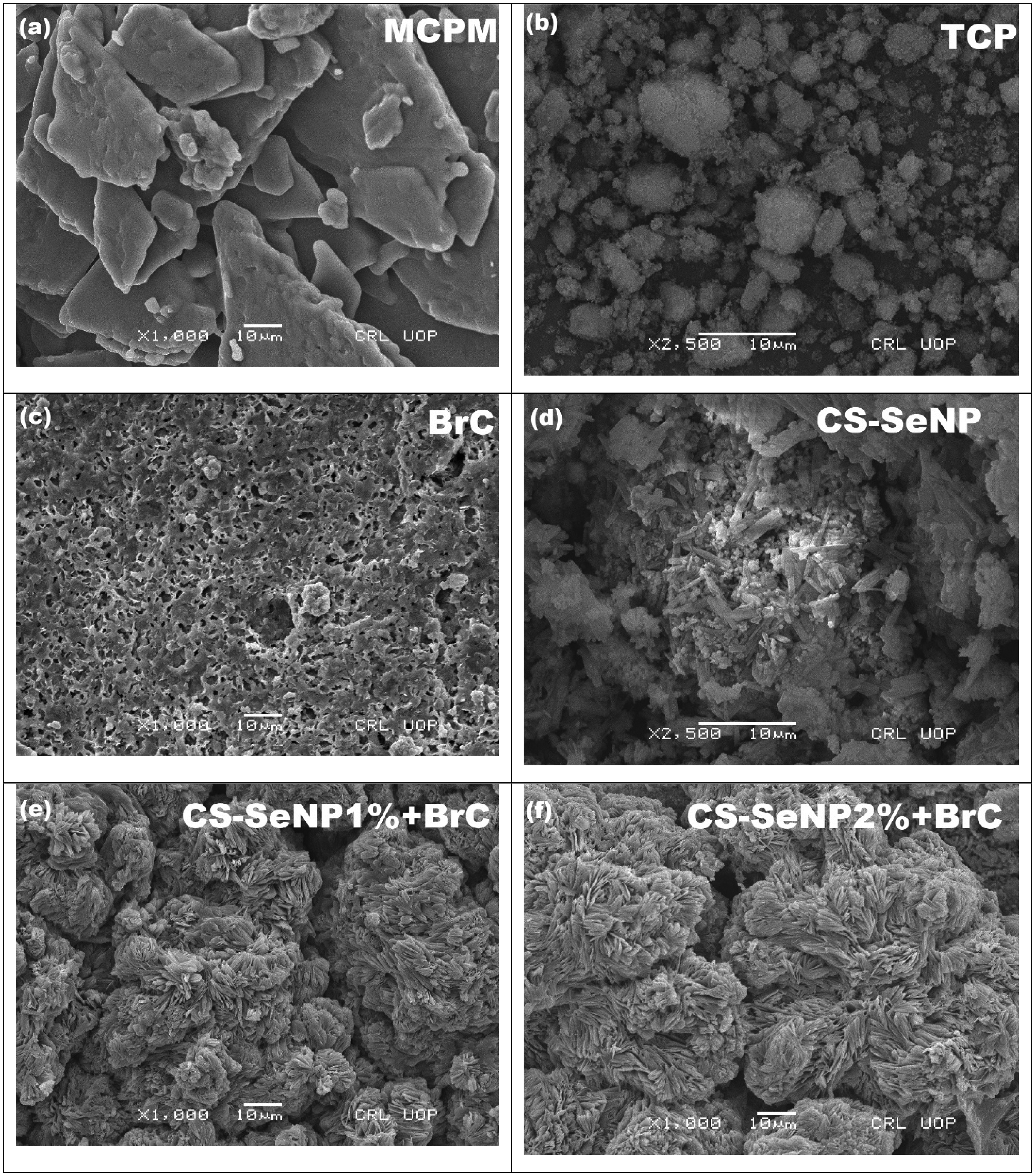
SEM images of (a) MCPM ×1000, (b) TCP ×2500, (c) BrC ×1000, (d) Pure CS-SeNP ×2500, (e) CS-SeNP 1% + BrC ×1000, (f) CS-SeNP 2% + BrC ×1000.
EDX results (Table 1) confirmed the elemental presence of O, P, and Ca corresponding to brushite (CaHPO4·2H2O). CS-SeNP 1 and 2 wt.% additionally presented carbon (C) and selenium (Se), indicating successful incorporation.
EDX comparison between control and experimental groups.
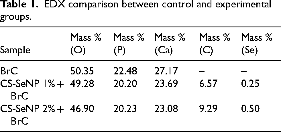
XRD relative spectra (Figure 2A) confirmed the crystalline phases of all formulations. BrC confirmed characteristic peak at 2θ ≈ 30.74°, consistent with reported BrC values (30.79°). 37 Chitosan exhibited a peak at 2θ ≈ 20.08°, consistent with reported value (20°). 38 Similarly, selenium peaks appeared at 2θ ≈ 23.45°, 26.67°, 29.59°, 41.28°, 43.59°, and 45.28° confirmed by reports Kalishwaralal et al. 38 and Pei et al. 39 between 20° to 80°. Addition of 1 wt.% and 2 wt.% CS-SeNPs caused distinct peak shifts towards higher angles (rightward shift) at 2θ ≈ 27–46°, (leftward shift) at 2θ ≈ 44° and amplified peak intensity at 2θ ≈ 46°, attenuated peak intensity at 2θ ≈ 27–36°, indicating lattice distortion and enhanced crystallinity. These modifications validated the successful integration of CS-SeNPs into BrC lattice structure.
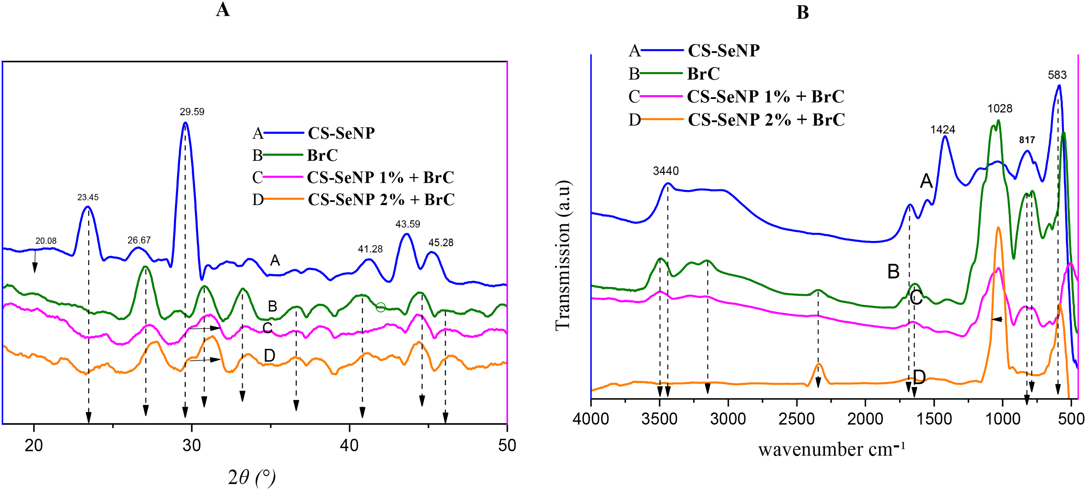
XRD (A) comparison between control and prepared samples, and FTIR (B) comparison between control and prepared samples.
FTIR relative spectra (Figure 2B) confirmed the presence of CS-SeNPs into BrC. CS characteristic peaks were observed at 817 cm−1 (
Mechanical properties
Relative shear bond strength (SBS) values in Figure 3(A) increased significantly with CS-SeNP incorporation. For 2 wt.% CS-SeNP, SBS increased maximum from 3.14 ± 0.01 MPa to 3.49 ± 0.02 MPa (p = 0.0004), and for 1 wt.% to 3.46 ± 0.08 MPa (p = 0.0012).
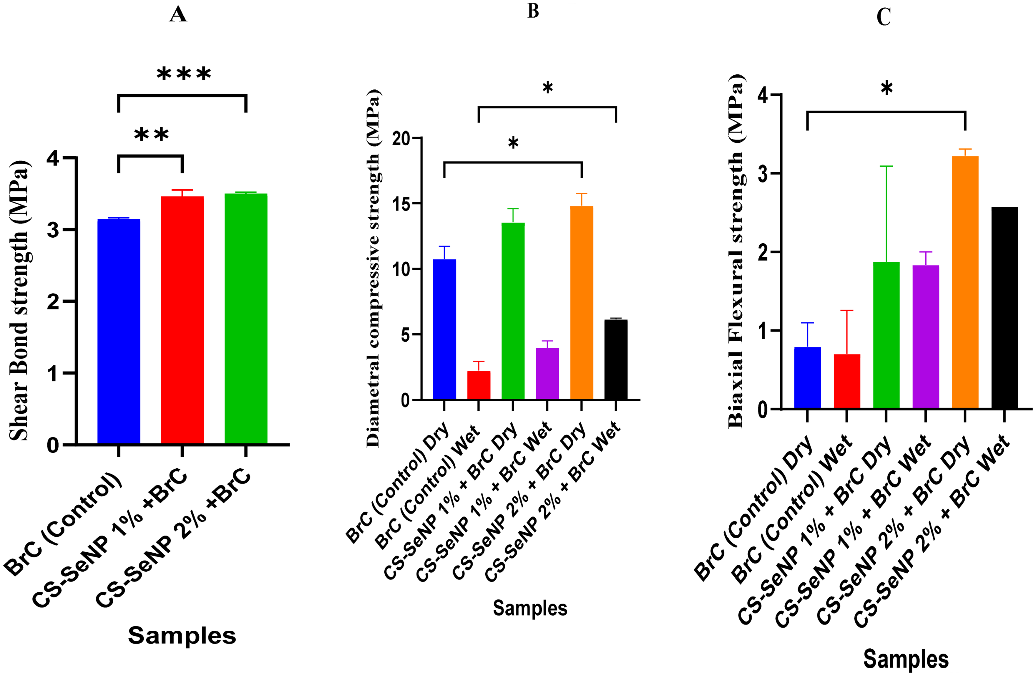
Shear bond strength (A), diametral compressive strength (B), and biaxial flexural strength (C) between control and experimental groups collectively showed significant difference (p < 0.05). The asterisk * denotes significant (p < 0.05) and ** (p < 0.01) highly significant difference.
Relative diametral compressive strength (DCS) values in Figure 3(B) also improved notably. The BrC (control) displayed 10.75 ± 0.96 MPa (dry) and 2.23 ± 0.70 MPa (wet), while 2 wt.% CS-SeNP increased strength maximum and significantly to 14.83 ± 0.92 MPa (dry) and 6.14 ± 0.08 MPa (wet) (p = 0.0149 dry and p = 0.0181 wet). The 1 wt.% group exhibited moderate enhancement to 13.55 ± 1.05 MPa (dry) and 3.97 ± 0.52 MPa (wet).
Relative biaxial flexural strength (BFS) values in Figure 3(C) increased significantly and maximum for 2 wt.% from 0.79 ± 0.30 MPa (dry) and 0.70 ± 0.55 MPa (wet) for BrC to 3.22 ± 0.08 MPa (dry) (p = 0.0129), and 2.57 ± 0.00 MPa (wet) and improved to 1.87 ± 1.21 MPa (dry) and 1.83 ± 0.16 MPa (wet) for 1 wt.%.
These results confirm that CS-SeNP addition increases the mechanical strength of BrC, with 2 wt.% achieving the maximum improvement.
Physical properties
Percentage mass loss profiles (Figure 4) demonstrated that 1 wt.% CS-SeNP displayed the significant and maximum degradation, reaching 2.35 g after 48 h (p = 0.0246). The 2 wt.% CS-SeNP group demonstrated less degradation (0.63 g) and minimum for BrC (−0.630 g). These values indicate that 1 wt.% CS-SeNP promotes faster degradation and resorption, and 2 wt.% maintains stability.
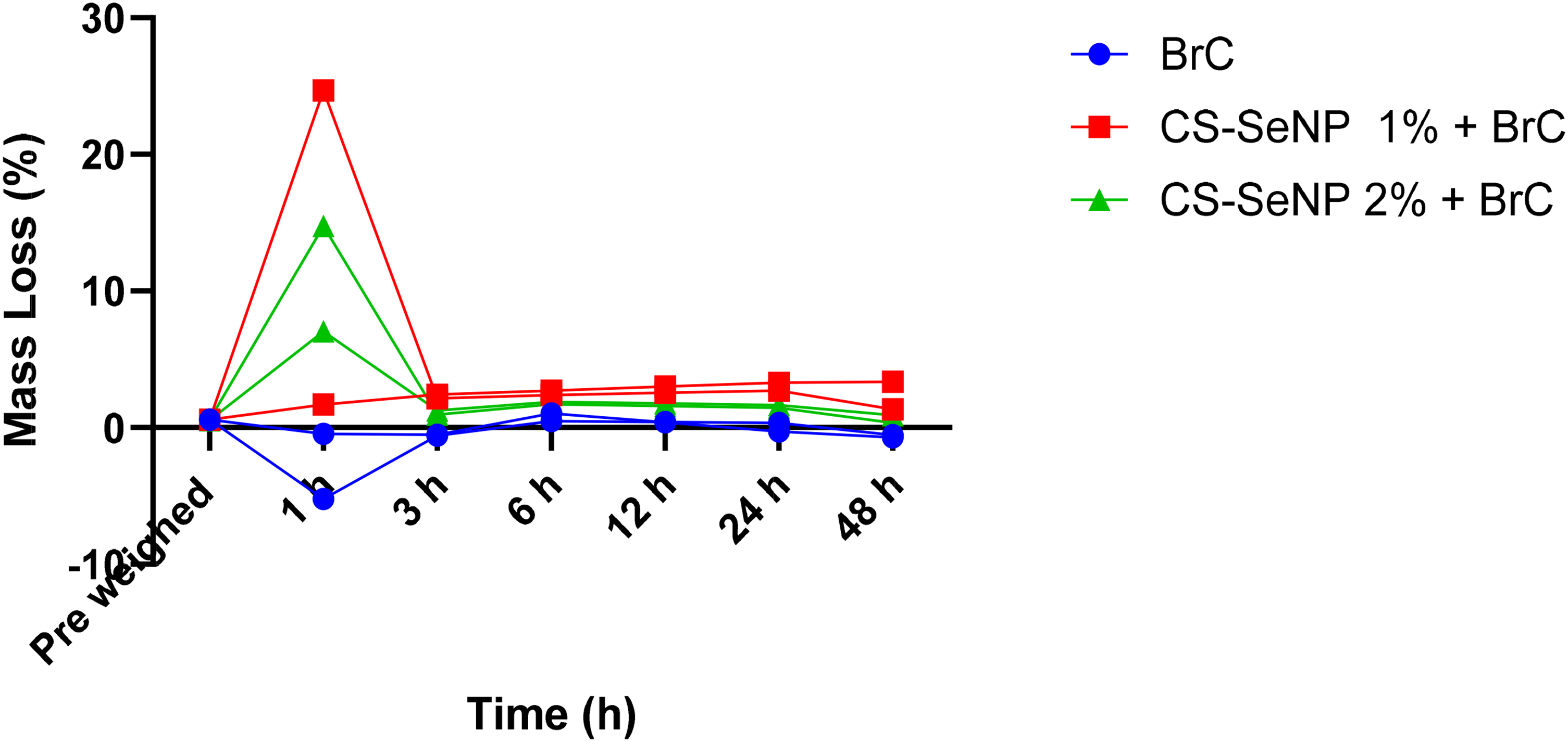
Degradation study comparison between control and experimental samples.
The initial (I) and final setting times (F) in Figure 5, were prolonged by CS-SeNPs compared to BrC. For 1 wt.% CS-SeNP, initial and final setting times significantly enhanced from 14. 5 ± 0.70 min to 18. 5 ± 0.70 min (p = 0.0098) and from 24.5 ± 0.70 min to 27.5 ± 0.70 min (p = 0.0380), respectively. For 2 wt.% CS-SeNPs, they further enhanced to 18. 5 ± 0.70 min (p = 0.0098) and 30.5 ± 0.70 min (p = 0.0012).
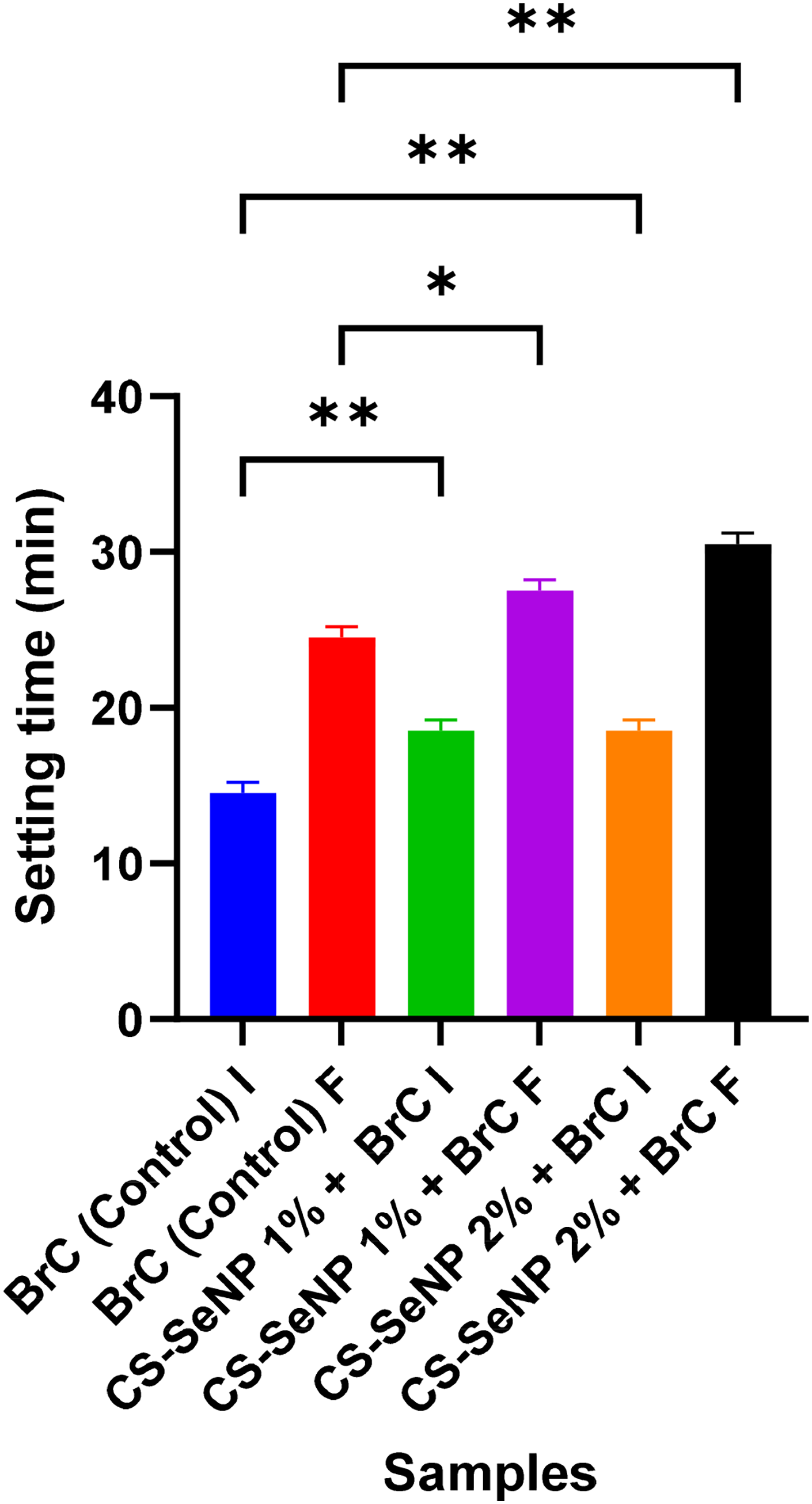
Setting time (I and F) comparison between control and experimental groups collectively showed significant difference. (p < 0.05). The asterisk * denotes significant (p < 0.05) and ** (p < 0.01) highly significant difference.
Anti-bacterial activity
Disc diffusion test (DDT) presented in Figure 6, 1 wt.% CS-SeNP group exhibited the significant and maximum zone of inhibition of (17.5 ± 2.1 mm; p < 0.0001), followed by 2 wt.% (17 ± 1.4 mm; p < 0.0001). Pure CS-SeNP alone displayed a 15.5 ± 0.7 mm ZOI, whereas BrC showed none.
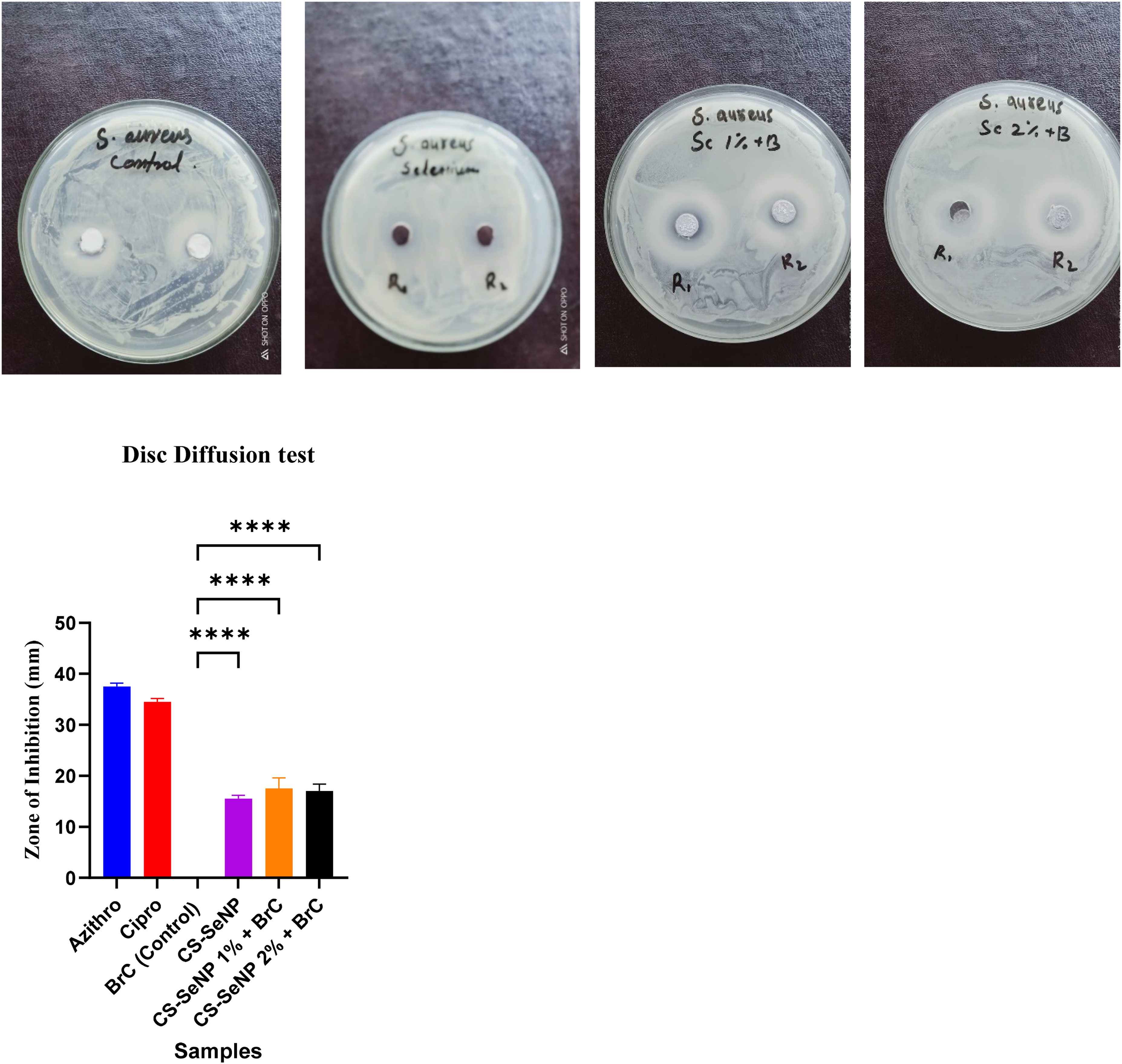
DDT images and graph comparison between control and experimental groups for Stap aureus. ZOI collectively showed significant difference (p < 0.05). The asterisk **** (p < 0.0001) showed highly significant difference.
Direct contact test (DCT) results presented in Figure 7 illustrates optical density (OD) changes pre and post incubation. The 1 wt.% CS-SeNP displayed a maximum drop in OD (0.222 to 0.059 = 0.163), and a smaller drop for 2 wt.% CS-SeNP (0.140–0.097 = 0.043) (Collectively p = 0.333; & nd on Mann-Whitney U) was observed, whereas BrC remained unchanged.
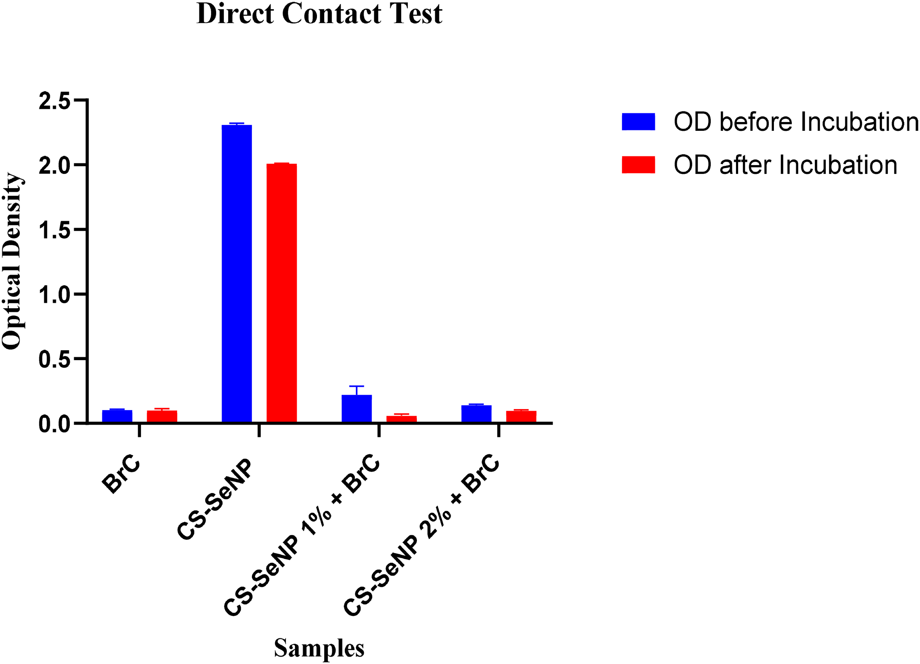
DCT comparison between control and experimental groups showed ns (p > 0.05) & nd (not a discovery) on mann-whitney U test.
Collectively, these values confirm that CS-SeNP added BrC displays potent antibacterial activity, most marked for 1 wt.% group.
Biocompatibility
Cytotoxicity analysis in Figure 8, showed reduced cell viability after CS-SeNP addition compared to BrC. Cell viability significantly reduced for 1 wt.% (from 0.852 ± 0.008 to 0.446 ± 0.029; (p < 0.0001), and for 2 wt.% (to 0.554 ± 0.067; p < 0.0001). The cell viability remained within acceptable biocompatibility limits.
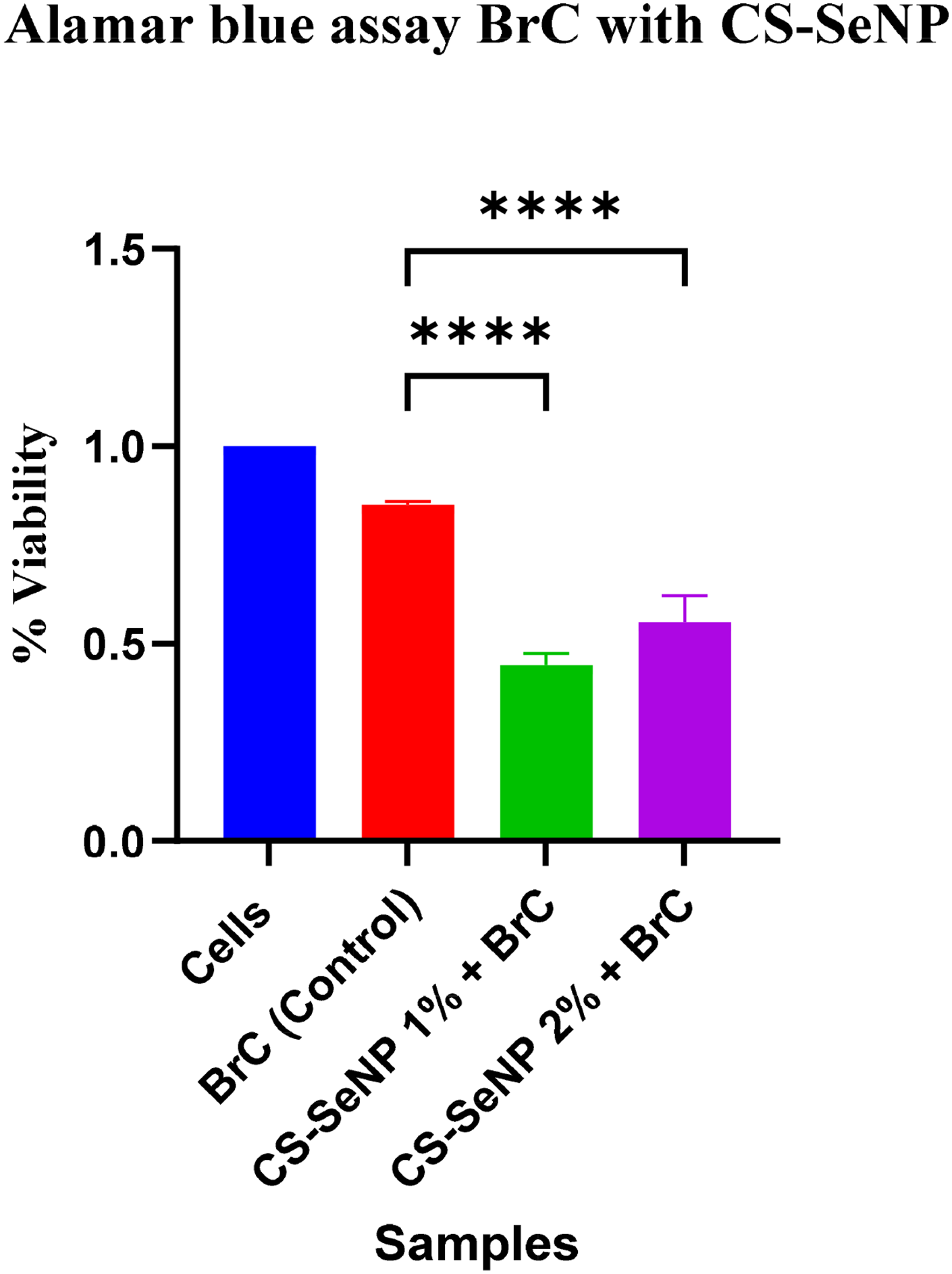
Cytotoxicity comparison between control and experimental groups collectively showed significant difference (p < 0.05). The asterisk **** (p < 0.0001) showed highly significant difference.
Optical profilometry
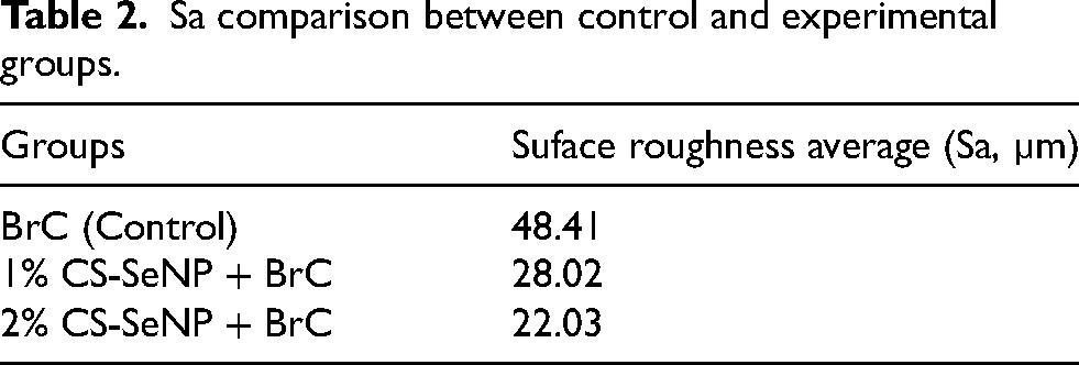
Surface roughness average (Sa) were the surface roughness parameters analyzed for control and experimental samples in Table 2. Figure 9 displayed optical surface profilometry images both top view and 3D view for prepared samples were collected.

Top view and 3D view surafce roughness of control and experimental group.
Sa comparison between control and experimental groups.
Discussion
CS-SeNP-modified materials have not been previously explored as bone cements for hard tissue engineering applications. This study investigates the effects of incorporating CS-SeNP into brushite cement (BrC) to evaluate its potential as a modified bone cement and its interaction with hard tissue.
SEM analysis confirmed that CS-SeNP incorporation altered BrC surface morphology, making it rougher and more porous. Rougher surfaces enhance cellular adhesion and osteoblastic growth that is essential in bone regeneration. 23 The same findings were presented by Alajmi et al. who stated that the incorporation of selenium nanoparticles increased the porosity and the interconnectedness, which facilitated cell growth and nutrient exchange. Better crystallinity and plate like organization observed in modified BrC is in line with results reported by Rattanachan et al., 37 which proves the hypothesis that selenium plays a part in superior structural organization.
XRD data showed peak shifts and changes in intensity, indicated lattice incorporation of CS-SeNPs into brushite matrix. Similar results by Krishnaraj et al. and Pei et al.,39,42 confirmed that incorporation of selenium nanoparticles triggers phase transformation and strengthening of structure. FTIR spectra also established chemical bonds and integration between CS, Se, and BrC matrix, which were in line with the results of the research conducted by Chen et al.43,44 These structural differences justify the effective integration of CS-SeNPs and their great adherence to brushite lattice.
CS-SeNPs increased the shear bond strength, Diametral compressive strength and biaxial flexural strength particularly at 2 wt.%. This improved performance may be explained through the appearance of entangled plate-like crystal structure that can offer internal reinforcement consistent to the mechanisms reported by Della Bona et al. 27 and Rattanachan et al. 37 Also, it was similarly found that CS-SeNPs extended the setting period of brushite, probably due to the retardation of ionic interactions in the early setting phase by chitosan. 39 Such enhancements indicate that the modified brushite could be suitable in the work with increased handling and mechanical performance.
The ability of brushite cement to be colonized by staphylococcus aureus increases a clinical difficulty. In this research, disc diffusion and direct contact tests confirmed significant antibacterial performance for modified brushite cement, with the largest effect at 1 wt.%. This value is likely to generate the best selenium release to generate antibacterial activity, yet it is not likely to produce excessive CS-SeNPs aggregation. Peng et al. and Krishnaraj et al. reported a similar dose effect influence, a bactostatic property of chitosan and efficacy of selenium in relation to S. aureus.42,45 The self-sterilizing nature of CS-SeNP-modified BrC presents a promising strategy to prevent infection, without the need for exogenous antibiotics and potentially mitigating the risk of antibiotic resistance. 39
Cytotoxicity assays indicated that CS-SeNP inclusion exhibited mildly reduced cell viability, specifically 1 wt.%, though values stayed within acceptable limits. This aligns with the identified dose-dependent cytotoxicity of selenium, balancing antimicrobial activity with cytocompatibility. 45 Further in vivo investigations are warranted to calibrate selenium concentrations to maximize therapeutic benefits.
Mass loss analysis showed that CS-SeNPs regulate brushite cement degradation in a dose-dependent manner. The 1 wt.% concentration degraded more rapidly, whereas 2 wt.% displayed more gradual and controlled resorption profile. Pei et al., 39 reported a stabilizing effect of chitosan on selenium nanoparticles that mitigated rapid degradation. Controlled degradation profile is essential for bone regeneration, as it maintains the structural integrity of the material to better facilitate the healing process, before gradual resorption. 41 The current results align with earlier research by Lewin et al. and Alajmi et al.,26,41 verifying the need of bioresorbable scaffolds providing mechanical support during bone healing and that nanoparticle does not compromise stability.
To analyse the surface topography of the experimental samples qualitatively and quantitatively, 3D optical surface profilometer are utilized. 46 In the current research, a qualitative assessment of surface topography through profilomteric images, and a quantitative assessment was achieved through computation of the surface roughness of experimental specimens via utilizing a 3D profilometer.
An enhanced average surface roughness and porosity of the control shown as an unevenness that may induce cell attachement and osteogenic growth. 47 Moreover, our study also showed reduced surafce roughness (Sa) and porosity for SeNPs added brushite cement, but still reports have mentioned an improved osteogenic cell attachement and osteogenesis. 48 The reduced Sa and porosity for CS-SeNPs added brushite cement can be contributed to the inclusion of nanoparticles into the interconnecting pore spaces as per reports of Lin et al. 49 Thus, it can be concluded that nanoparticles would merge within the brushite cement, and are miscible. As per Kadir et al., 50 nanoparticles are predicted to facilitate rapid ionic conduction, and provide an amorphous structure.
Conclusion
Incorporating chitosan–selenium nanoparticles (CS-SeNPs) into brushite cement (BrC) markedly enhanced its structural, mechanical, and antibacterial performance while maintaining acceptable biocompatibility. The modified cements exhibited improved crystallinity, higher shear, compressive and flexural strength, extended setting times, and strong antibacterial efficacy particularly at 1 wt.% CS-SeNP concentration. Although mild cytotoxicity was observed, it remained within clinically acceptable limits.
These findings suggest that CS-SeNP incorporation up to 2 wt.% produces a multifunctional cement with superior mechanical integrity and antibacterial protection, making it a promising candidate for use in load-bearing and infection-prone bone defects.
Further in vivo studies are recommended to optimize selenium dosage and evaluate long-term biological performance before clinical translation.
Footnotes
Funding
The authors received no financial support for the research, authorship, and/or publication of this article.
Declaration of conflicting interests
The authors declared no potential conflicts of interest with respect to the research, authorship, and/or publication of this article.