



Abstract
Background:
Bone grafts are commonly employed for the reconstruction of bone defects, and dentin has been reported as a promising bone graft material that supports early graft vascularization. However, clinical applications typically involve a prolonged demineralization process prior to the use of dentin samples.
Objective:
This in vitro study aimed to evaluate the biological properties of dentin samples without demineralization procedure.
Methods:
Dentin extract (DE) was obtained by mechanically crushing dentin samples, dissolving and filtering the mixture. Enzyme-linked immunosorbent assay (ELISA) was first performed to identify cytokines released from DE, revealing that TGF-β was notably enriched. Subsequently, cell viability, wound healing, and tube formation assays were conducted to assess the effects of DE on cell proliferation, migration, and angiogenic potential in human umbilical vein endothelial cells (HUVECs).
Results:
The in vitro results demonstrated that DE significantly enhanced HUVEC proliferation, migration, and tube formation capabilities. These effects were markedly attenuated by treatment with the Notch pathway inhibitor DAPT and the tyrosine kinase inhibitor N-Desethyl Sunitinib. RT-qPCR and Western blot analyses further revealed that DAPT and N-Desethyl Sunitinib, downregulated the mRNA and protein expression of markers associated with cell migration and angiogenesis signaling pathways, and these effects were significantly reversed by treatment of DE without demineralization.
Conclusion:
In conclusion, this study demonstrates that DE, without demineralization, promotes cell proliferation, migration, and angiogenesis in HUVECs via the Notch and VEGF/VEGFR2 signaling pathways. These findings suggest that dentin, without the need for demineralization, could serve as a viable alternative to conventional bone graft materials, offering a more streamlined process for regenerative surgery.
Keywords
Introduction
Bone tissue engineering is a rapidly evolving field in regenerative medicine and dentistry, requiring the integration of cells, scaffolds, and growth factors. 1 Materials that serve as both osteoinductive agents are highly desirable for effective bone regeneration. 2 Since autogenous bone grafts possess osteoconductivity, osteoinductivity, osteogenesis, and bone binding properties and good biocompatibility, they have always been regarded the gold standard for bone grafting.3,4 Recently, dentin has gained significant attention as a potential material for bone tissue engineering due to its dual function as a structural scaffold and a reservoir of growth factors. 5 Similar to bone, dentin is an organic-inorganic composite composed of proteins and minerals, characterized by high fracture strength and a chemical composition closely resembling that of bone.6,7 Furthermore, dentin contains bone morphogenetic proteins (BMPs), which promote osteogenesis and dentinogenesis through Smad signaling pathways. 6
Clinical studies have explored the use of tooth-derived materials, including dentin, as viable alternatives to autogenous bone grafts for treating bone defects and reconstruction.8,9 The demineralization of dentin has been reported to enhance osteogenic potential by facilitating the release of growth factors.10,11 Animal studies conducted by Kim et al. demonstrated that the bone-inductive properties of autogenous teeth are primarily attributed to the dentin component. 10 Various forms of dentin, such as noncollagenous proteins, dentin particles, deproteinized dentin, and demineralized dentin, have shown promise in promoting bone regeneration.12,13
Despite the widely reported osteoinductive advantages of demineralized dentin matrix (DDM), its clinical translation remains challenging. Demineralization has been shown to affect the mechanical integrity of dentin, increase the complexity of material preparation, and potentially introduce variability in the release profiles of bioactive factors. 14 In addition, extensive chemical processing raises concerns regarding standardization, cost-effectiveness, and chairside applicability in routine clinical practice. Currently, there is also a lack of standardized protocols guiding the selection of specific dentin forms for defined clinical indications. 15 Collectively, these limitations underscore the need to re-evaluate alternative dentin-based materials that preserve structural integrity while retaining biological activity.
Therefore, this study aims to systematically evaluate the biological characteristics of dentin samples without undergoing demineralization, with particular emphasis on its cellular compatibility and regenerative potential, with particular emphasis on angiogenic activity. By focusing on untreated dentin, this work addresses existing controversies regarding the necessity of demineralization and proposes a simplified and potentially more clinically practical alternative to conventional dentin-derived graft materials.
Methods and materials
Cell culture
The human umbilical vein endothelial cells (HUVECs) were purchased from the American Tissue Culture Collection (ATCC; Manassas, VA, USA) and cultured in Endothelial Cell Medium (ECM, ScienCell; California, USA), supplemented with 1% Endothelial Cell Growth Supplement (ECGS, ScienCell; California, USA), 10% fetal bovine serum (FBS, Gibco, Grand Island, NY, USA), 100 U/mL penicillin, and 100 μg/mL streptomycin. Cells were maintained at 37°C in a 5% CO2 incubator, and cells from passages 3 to 6 were used in all experiments.
Preparation of dentin extract (DE)
Intact and non-infected human teeth with no caries, fractures, or resorption were obtained following protocols established in previous studies.7,16 Briefly, soft tissues, the enamel, dental pulp, cementum, and dentin matrix were removed to obtain dentin blocks. These dentin blocks were then mechanically crushed, and the resulting dentin powder was sieved through a 1600 mesh stainless-steel filter (Suzhou Jinyuan Mesh Co., Ltd, China). Approximate 15 g of dentin powder was transferred to a 50 mL centrifuge tube and mixed with 30 mL of sterile PBS. The mixture was placed on a shaker for 24 h and then filtered through filter paper to separate solid and liquid components. Finally, the dentin extract (DE) was obtained by filtering through a sterile 0.22 μm disposable filter (Millipore, USA) and stored at 4°C for further use.
Enzyme-Linked immunosorbent assay (ELISA)
Cytokines, including TGF-β and VEGF, released from the DE was quantified using a Quantikine Human ELISA kit (ZelBio, Germany) according to the manufacturer's protocol. Briefly, the DE was transferred to a 96-well plate of ELISA kit, and cytokine release was measured by comparing the absorbance to the calibration curves. The experiment was performed in triplicate.
Cell viability assay
The HUVECs were placed in a 96-well plate at a density of 5000 cells per well and cultured overnight for cell adhesion. Then cells were treated with DE and culture medium at a 1:5 ratio, the Notch pathway inhibitor DAPT (Beyotime, China) at 10 µM, and/or tyrosine kinase inhibitor N-Desethyl Sunitinib at 10 ng/mL. Cells were incubated at 37°C for 24 and 48 h, and cell viability was assessed using the Cell Counting Kit-8 (CCK-8) reagent (Dojindo Molecular Technologies, Japan) according to the manufacturer's protocol. Briefly, 10 µL of CCK-8 reagent in 100 µL of culture medium was added in each well and incubated for 1.5 h at 37°C. Absorbance at 490 nm was measured using a microplate reader (ELx800; BioTek Instruments). The cells without treatment were used as the control group, and the relative cell viability was calculated with the viability of the control group set as 100%.
Wound healing assay
The HUVECs were placed in a 6-well plate at a density of 1.0 × 10⁵ cells per well and cultured overnight for cell adhesion. Then a scratch was made across the center of the monolayer using a sterile pipette tip when the cells reached 80% confluence. After gently washing twice, the cells were cultured in medium supplemented with DE, DAPT, and/or N-Desethyl Sunitinib. Images of the wound area were taken at 0 and 12 h post-incubation, and the wound width was quantified using FIJI-ImageJ software. The experiment was performed in triplicate.
Real-Time quantitative PCR (Rt-qPCR)
The HUVECs were placed in a 12 well plate at a density of 5.0 × 10⁴ cells per well and cultured overnight for cell adhesion. Then cells were treated with of DE, DAPT, and/or N-Desethyl Sunitinib, and cultured for 24 h at 37°C. Total RNA was extracted from HUVECs using TRIzol® reagent (Invitrogen; USA), and mRNA was reverse transcribed into cDNA using the PrimeScript™ RT kit (Takara Biotechnology, Beijing, China) according to the manufacturer's protocol. Subsequently, 1 µL of cDNA, 5 µL of SYBR® Premix Ex Taq II (Takara Biotechnology), 0.4 µL of each primer pair were added to a 96-well plate, with a total reaction volume of 10 µL by supplementing DNase/RNase-free water. RT-qPCR was performed using an ABI StepOnePlus instrument (Thermo Fisher Scientific, USA). The relative mRNA expression levels were measured using the 2−ΔΔCq method, and the GAPDH gene was used as an internal control. The primers used in this study are listed in the Table 1.
The sequences of primers for indicated genes.

Western blot analysis
The HUVECs under different treatment conditions were lysed using RIPA buffer (Cell Signaling Technology, USA) supplemented with phosphatase and proteinase inhibitor cocktails. Protein extracts were quantitated using a BCA assay (Thermo Fisher Scientific, USA) following the manufacturer's protocol. Approximately 40 μg of cell lysates was separated by electrophoresis on an 8%-12% SDS/PAGE gel and subsequently transferred onto polyvinylidene difluoride (PVDF) membranes (Merck Millipore, Burlington, MA, USA). After blocking with 5% milk in Tris-buffered saline (TBS) with 0.1% Tween 20, the membranes were probed with specific primary antibodies (Cell Signaling Technology, USA), followed by HRP-conjugated secondary anti-IgG antibodies. Protein expression was visualized using ECL Western Blotting Substrate (Thermo Fisher Scientific) according to the manufacturer's instructions. Band intensities associated with the expression of indicated proteins were analyzed using FIJI-ImageJ software.
Tube formation assay
The ability of HUVECs to form tubule-like structures under different treatment conditions was determined using a tube formation assay. Briefly, HUVECs were placed in a Matrigel-coated 96 well plate at a density of 2.0 × 10⁴ cells per well and cultured overnight for cell adhesion. Then cells were treated with of DE, Notch pathway inhibitor DAPT, and/or tyrosine kinase inhibitor N-Desethyl Sunitinib, and cultured at 37°C for 4, 8, and 12 h. The tubular structure formation was captured using a microscope, and the number of branches and tube length were quantitatively measured in the high-power fields using FIJI-ImageJ software.
Statistical analysis
All experiments were performed at least three times, and data were presented as the mean ± standard deviation (SD). Statistical analysis was conducted using GraphPad Prism 9.0 software. For comparisons among more than two groups, one-way analysis of variance (one-way ANOVA) was first performed to determine whether there was an overall significant difference among groups. When a significant difference was detected, post hoc multiple-comparison analyses were conducted using Tukey-Kramer's test or Bonferroni/Dunn's test, as appropriate. For comparisons between two independent groups at the same time point, the Mann-Whitney U test was used. A P value < 0.05 was considered statistically significant.
Results
DE without demineralization promotes cell proliferation in HUVECs
Dentin powder without demineralization was prepared by mechanically crushing and sieving the dentin through a 1600 mesh (Figure 1A). The dentin extract (DE) was obtained by dissolving the dentin powder in PBS and filtering through sterilized filter paper. We first evaluated the concentration of key cytokines in DE, including transforming growth factor beta (TGF-β) and vascular endothelial growth factor (VEGF), which are commonly detected in dentin, as reported previously.17,18 DE without demineralization, extracted at pH 7.4, contained 179.5 ± 1.9 pg/mL of TGF-β (Figure 1B). However, VEGF was undetectable in DE, suggesting that the short half-life and pH sensitivity of VEGF may have led to its inactivation during the extraction process. We then determined the effects of DE on the proliferation of HUVECs. As shown in Figure 1C-D, DE significantly enhanced HUVEC proliferation at both 24 h and 48 h compared with the control group. Notably, the proliferative effect of DE was inhibited by treatment with either Notch pathway inhibitor DAPT or tyrosine kinase inhibitor N-Desethyl Sunitinib, indicating that DE promotes cell proliferation through NOTCH signaling and VEGF signaling pathways.
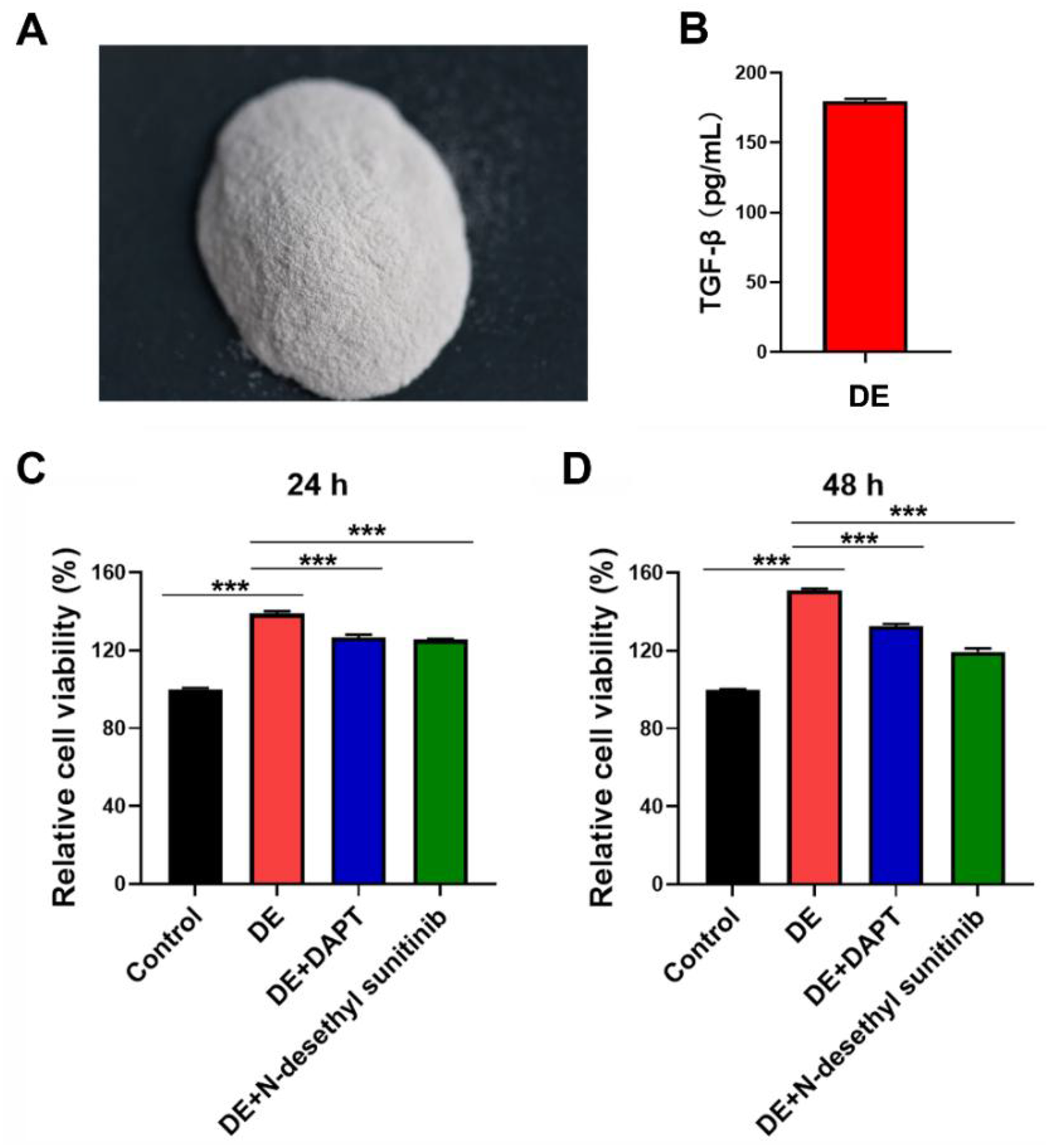
The proliferative effect of DE on HUVECs is inhibited by either notch pathway inhibitor DAPT or tyrosine kinase inhibitor N-desethyl sunitinib. (A) The photograph of dentin powder after mechanically crushed dentin without demineralization. (B) The concentration of TGF-β from DE as determined by ELISA assay. (C-D) The cell viability of HUVECs after treatment of DE, Notch pathway inhibitor DAPT, and/or tyrosine kinase inhibitor N-Desethyl Sunitinib for 24 h (C) and (D) 48 h as assessed by CCK-8 assay. DE, dentin extract. TGF-β, transforming growth factor beta. Data are presented as mean ± SD. Statistical analysis was performed using one-way ANOVA followed by Tukey-Kramer's post hoc test. ***P < 0.001.
DE without demineralization enhances the migration ability of HUVECs
Previous studies have demonstrated that dentine can promote the migration ability of HUVECs.17,19 We further investigated the effects of DE without demineralization on the migration of HUVECs. The scratch assay revealed that DE significantly promoted wound healing, with the wound closure rate increasing from 0.27 ± 0.05 to 0.60 ± 0.06 following DE treatment (Figure 2), consistent with findings from previous studies. However, treatment with either the Notch pathway inhibitor DAPT or the tyrosine kinase inhibitor N-Desethyl Sunitinib significantly reduced the wound closure rates to 0.37 ± 0.02 and 0.33 ± 0.06, respectively, following DE treatment (Figure 2). These results suggest that DE without demineralization improves the migration potential of HUVECs, which is primarily mediated through NOTCH and VEGF signaling pathways.
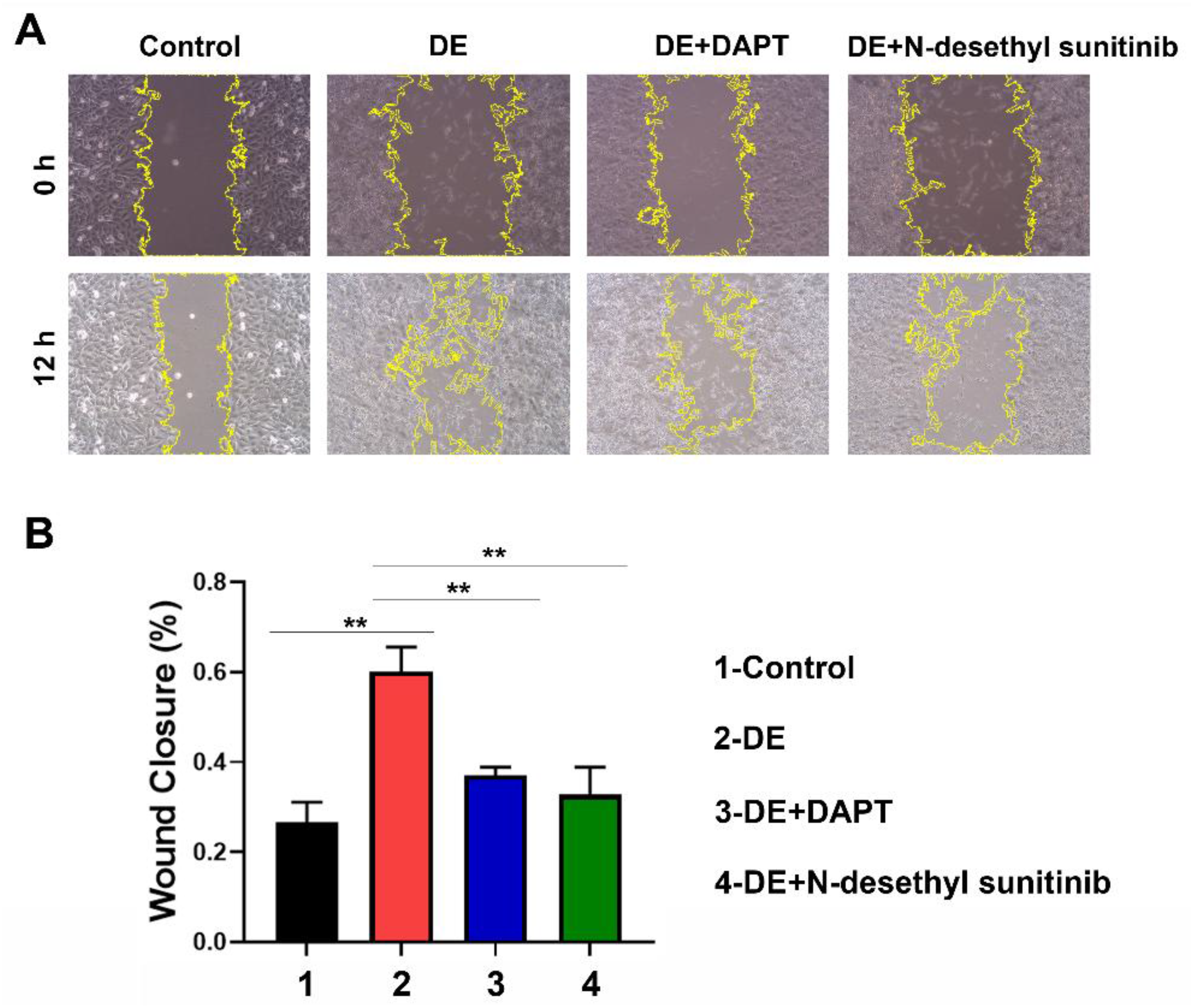
The migration of HUVECs after treatment of DE, notch pathway inhibitor DAPT, and/or tyrosine kinase inhibitor N-desethyl sunitinib. (A) Representative images of HUVEC migration at 0 h and 12 h post-scratching after different treatment conditions. (B) Quantitative analysis of the percentage of wound closure at 12 h post-scratching. DE, dentin extract. Data are presented as mean ± SD. Statistical significance was analyzed using one-way ANOVA followed by Tukey-Kramer's post hoc test. **P < 0.01.
DE without demineralization induces angiogenesis of the HUVECs
We next evaluated the effects of DE without demineralization on the angiogenic potential of HUVECs. Angiogenesis capacity was assessed by the tube formation assay. The number of capillary-like structures, characterized by the number of circles and the length of branches, was significantly increased in the DE-treated group compared to the control group at 4, 8, 12 h post-treatment (Figure 3), indicating a positive effect of DE on tube formation in HUVECs. Consistently, treatment with the Notch pathway inhibitor DAPT or the tyrosine kinase inhibitor N-Desethyl Sunitinib significantly suppressed DE-induced tube formation. Collectively, these findings suggested that DE without demineralization enhances tube formation in HUVECs, primarily through NOTCH signaling and VEGF signaling.
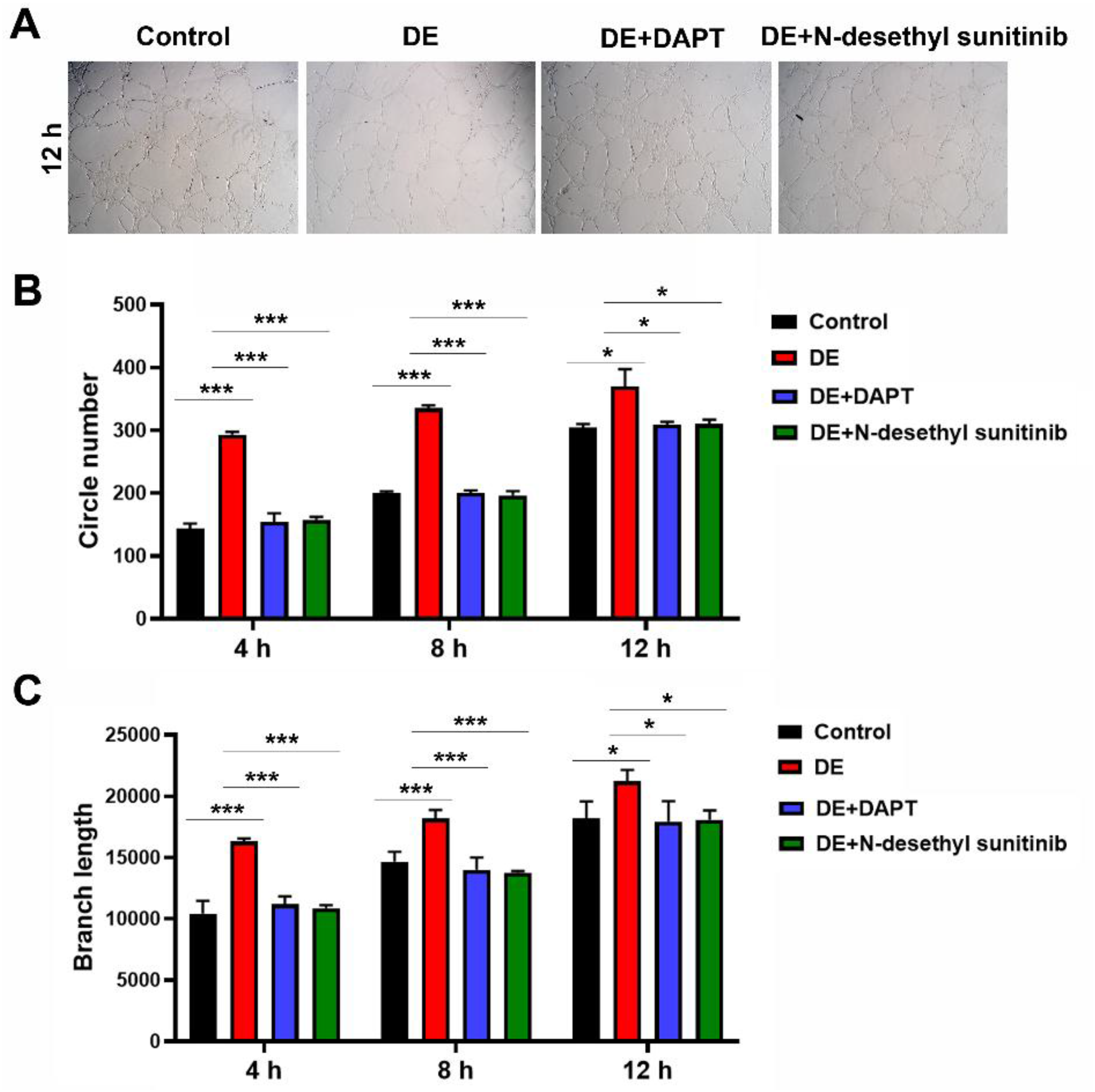
Angiogenesis capacity of HUVECs after treatment of DE, notch pathway inhibitor DAPT, and/or tyrosine kinase inhibitor N-desethyl sunitinib. (A) Representative images of tube formation by HUVECs with different treatment conditions for 12 h. (B) Quantitative analysis of the circle number formed by HUVECs with different treatment conditions for 4, 8, and 12 h. (C) Quantitative analysis of the length of branches formed by HUVECs with different treatment conditions for 4, 8, and 12 h. DE, dentin extract. Data are presented as mean ± SD. Statistical analysis was performed using one-way ANOVA followed by Tukey-Kramer's post hoc test. *P < 0.05, ***P < 0.001.
DE without demineralization activates the notch signaling and VEGF signaling pathway to promote angiogenesis in HUVECs
The Notch signaling pathway regulates cell proliferation, differentiation, and apoptosis and comprises four receptors (Notch1-4) and five ligands (JAG1-2, DLL1-4), which play critical roles in vascular development and differentiation. 20 Endothelial cell migration and tube formation are primarily driven by VEGF signaling. VEGF binding to VEGFR2 activates intracellular signaling pathways that promote cell survival, vascular permeability, migration, and proliferation. 21 Key downstream targets involved in these processes include PDGF, FGF, SMAD2, and SMAD3. To evaluate whether the enhanced tube formation and migration induced by DE are linked to modulation of angiogenic signaling pathways, the mRNA and protein expression levels of angiogenesis-related components were analyzed using RT-qPCR and Western blot in HUVECs treated with DE for 24 h. The results showed that Notch pathway inhibitor DAPT or the tyrosine kinase inhibitor N-Desethyl Sunitinib inactivated the genes related to cell migration and angiogenesis, including Notch1, DLL4, JAG1, HEY1, HEY2, TGFβR I, TGFβR II, PDGF, FGF, SMAD2, SMAD3, VEGFA, VEGFR1/2/3, both at the mRNA (Figure 4) and protein (Figure 5) levels. These changes were statistically significant when compared with the corresponding control groups at the same time point. However, treatment with DE reversed these effects induced by DAPT and N-Desethyl Sunitinib. Taken together, these findings suggest that DE without demineralization activates angiogenesis-regulating signaling pathways, thereby promoting tube formation in HUVECs.
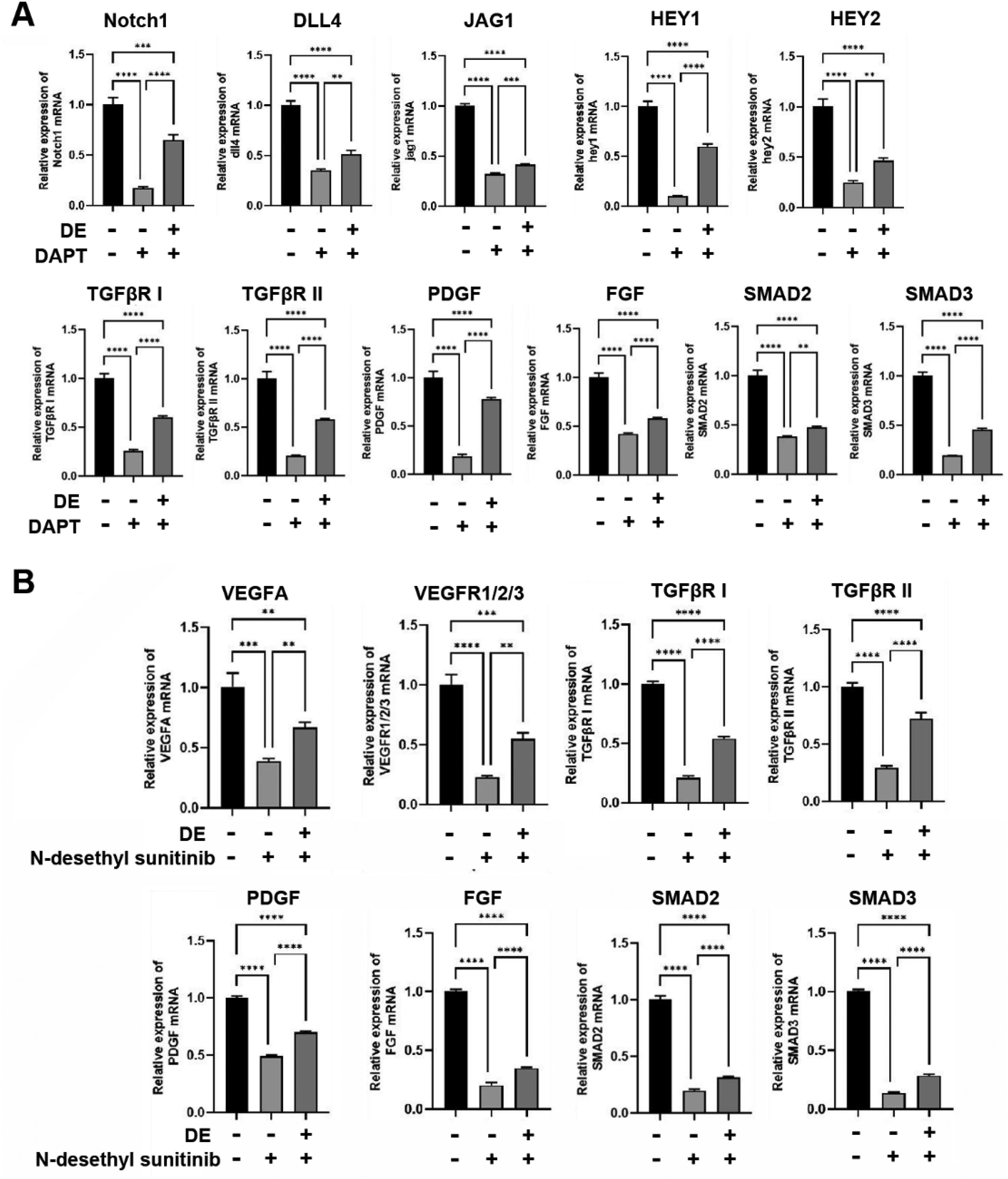
Indicated gene expression of HUVECs after treatment of DE, notch pathway inhibitor DAPT, and/or tyrosine kinase inhibitor N-desethyl sunitinib. (A) RT-qPCR assessment of the mRNA expression of Notch1, DLL4, JAG1, HEY1, HEY2, TGFβR I, TGFβR II, PDGF, FGF, SMAD2, and SMAD3 in HUVECs treated with DE and/or DAPT. (B) RT-qPCR assessment of the mRNA expression of VEGFA, VEGFR1/2/3, TGFβR I, TGFβR II, PDGF, FGF, SMAD2, and SMAD3 in HUVECs treated with DE and/or N-desethyl sunitinib. DE, dentin extract. Data are presented as mean ± SD. Statistical significance was analyzed using the Mann-Whitney U test. **P < 0.01, ***P < 0.001, **** P < 0.0001.
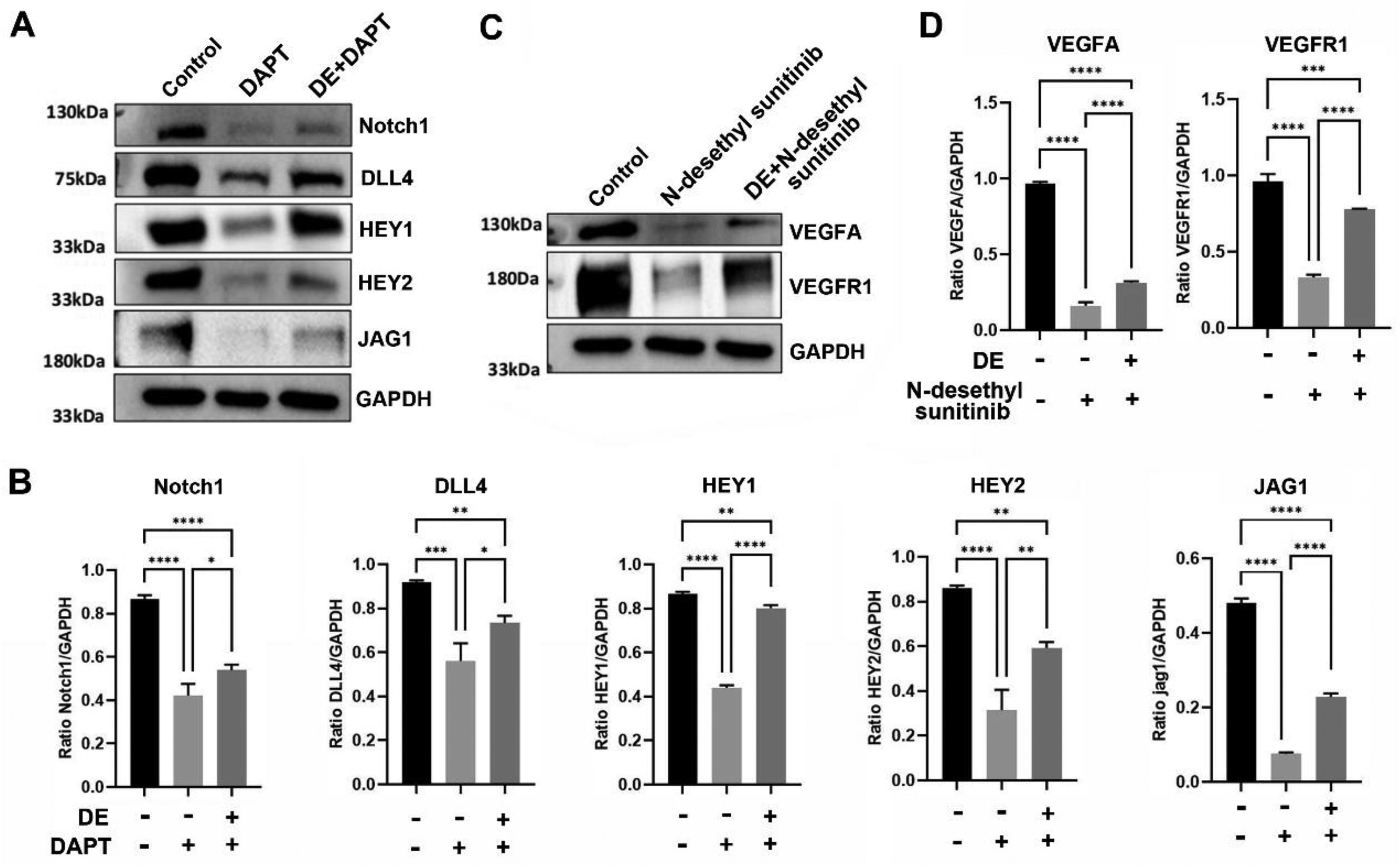
Indicated protein expression of HUVECs after treatment of DE, notch pathway inhibitor DAPT, and/or tyrosine kinase inhibitor N-desethyl sunitinib. (A) Representative western blotting images of the protein expression of Notch1, DLL4, HEY1, HEY2, and JAG1 in HUVECs treated with DE and/or DAPT. (B) Quantitative analysis of indicated protein expression level in HUVECs treated with DE and/or DAPT. (C) Representative western blotting images of the protein expression of VEGFA and VEGFR1 in HUVECs treated with DE and/or N-desethyl sunitinib. (D) Quantitative analysis of indicated protein expression level in HUVECs treated with DE and/or N-desethyl sunitinib. DE, dentin extract. Data are presented as mean ± SD. Statistical significance was analyzed using the Mann-Whitney U test. *P < 0.05, **P < 0.01, ***P < 0.001, **** P < 0.0001.
Discussion
The dentin is a rich source of growth factors and shares significant structural and compositional similarities with bone. To activate its osteogenic potential of the dentin, inductive proteins and factors must be released to stimulate implanted cells. 22 The preparation method and particle size of dentin significantly impact its osteoinductive and osteoconductive properties.13,23 This study evaluated the biological properties of dentin extract (DE) without demineralization. The results revealed that undemineralized DE contained high levels of TGF-β, significantly enhancing HUVEC proliferation, migration, and tube formation. These effects were reduced by the Notch pathway inhibitor DAPT and the tyrosine kinase inhibitor N-Desethyl Sunitinib. DE also reversed the inhibitors’ effects on mRNA and protein expression related to cell proliferation and angiogenesis, highlighting its role in activating the Notch and VEGF/VEGFR2 signaling pathways.
Dentin, the primary hard tissue of teeth, contains various glycoproteins, proteoglycans, and growth factors.12,24 Upon damage, biomolecules released from the dentin matrix play key roles in pulp repair.25–26 Previous studies have demonstrated that demineralized dentin induces mesenchymal stem cell differentiation into chondrocytes, supporting bone and cartilage repair.27,28 Dentin matrix-extracted proteins are rich in growth factors, including TGF-β, bFGF, BMP-2, PDGF, IGF-1, and VEGF,29,30 which contribute to chondro-induction. Among these, TGF-β plays a central role in chondrocyte differentiation by regulating cell aggregation, proliferation, and terminal differentiation. 31 Traditionally, dentin demineralization involves a neutral 10% ethylenediaminetetraacetic acid buffer solution, which facilitates protein extraction to enhance cell adhesion, proliferation, and osteogenic differentiation of various cell types, including dental pulp cells, periodontal ligament cells, and bone marrow mesenchymal stem cells.27,32 In this study, mechanically crushed dentin extract without demineralization retained high levels of TGF-β, promoting cell proliferation, migration, and angiogenesis in HUVECs. These findings suggest that undemineralized dentin retains its biological properties and requires less processing, offering a promising alternative to traditional bone graft materials.
From a clinical perspective, these findings provide important insights into the potential application of DE in bone and dental tissue regeneration. Given its ability to release biologically active growth factors without demineralization, DE may serve as a bioactive additive or conditioning agent to enhance angiogenesis and tissue repair in localized bone defects, such as alveolar ridge preservation, periodontal defects, and guided bone regeneration.15,33 Our preliminary clinical study suggested autogenous undemineralized dentin effectively preserved the three-dimensional contour of extraction sites, and subsequent histological analysis confirmed their osteoinductive potential. Clinically, DE could be prepared from mechanically processed dentin and incorporated into graft materials, scaffolds, or biomaterial carriers, or applied locally to defect sites to enhance vascularization and regenerative outcomes.34,35 Importantly, the simplified preparation process not only preserves biological activity but also reduces processing complexity, highlighting DE as a clinically feasible and translational alternative to conventional demineralized dentin-derived products. Nevertheless, further in vivo and clinical studies are warranted to validate the long-term efficacy and safety of DE in different defect models.
Notch is a conserved regulatory factor that controls cell fate and orchestrates tissue morphogenesis through the juxtacrine Notch signaling pathway.36,37 Its pivotal role in angiogenesis makes it a potential target for promoting de novo tissue vascularization. JAG1, a Notch ligand, is crucial for angiogenesis as it interacts antagonistically with DLL4, guiding vascular branching and propagation, often in collaboration with the VEGFR signaling. 38 VEGF secretion and its receptors as well as bFGF/FGFR signaling are critical for angiogenesis during development.16,39 VEGF/VEGFR2 axis promoted the migration of human dental pulp stem cells via the FAK/PI3 K/Akt and p38 MAPK signaling pathways. 40 In this study, undemineralized DE promoted angiogenesis in HUVECs, mediated through the activation of Notch and VEGF/VEGFR2 signaling pathways, as demonstrated by the inhibitory effects of the Notch pathway inhibitor DAPT and the tyrosine kinase inhibitor N-Desethyl Sunitinib.
Some limitations are shown in this study. First of all, a direct comparison of the biological properties of DE with and without demineralization was not conducted in the current research, and further investigations should address this to better understand their relative biological properties. Secondly, additional research is needed to assess the effects of undemineralized dentin powder on HUVECs, as some organic materials and minerals may remain undissolved in PBS and be discarded during filtering. Lastly, the role of TGF-β should be confirmed using anti-TGF-β antibodies to elucidate its contribution to the observed effects.
In conclusion, the current study demonstrated that dentin extract without demineralization enhances cell proliferation, migration, and angiogenesis in HUVECs via the Notch and VEGF/VEGFR2 signaling pathways. These findings suggest that dentin without demineralization may serve as a potential alternative to traditional bone graft materials, and offer a more streamlined process in clinical applications.
Footnotes
Acknowledgements
The authors have no acknowledgments to report.
Ethics approval and consent to participate
This study doesn’t involve human or animal experiment, thus no ethical approval and patient consent are required.
Author contributions
We declare that all the listed authors have participated actively in the study and all meet the requirements of the authorship. WZ designed the study and wrote the paper. XWZ managed the literature searches. QSJ undertook the data analysis. All authors reviewed the manuscript.
Funding
This research did not receive any specific funding.
Declaration of conflicting interests
The authors declared no potential conflicts of interest with respect to the research, authorship, and/or publication of this article.
Data availability statement
The datasets used or analyzed during the current study are available from the corresponding author on reasonable request.