
Abstract
The origin of tissue culture is commonly dated to 1907 and credited to Ross Harrison at Hopkins Medical School. But an unpublished letter from the 1942 offers a different interpretation and gives priority to Montrose Burrows with important contributions for the development of cell culture by Franklin Mall at Hopkins and Alexis Carrel at the Rockefeller Institute in New York City. The early development of tissue culture is reviewed and its applications in modern biology and medicine are briefly outlined.
Introduction
The standard history of tissue culture traces its origin to two papers with identical text published in 1906 and 1907 by Ross Granville Harrison (1870–1959)1,2 (Figure 1). They described experiments he had performed involving tiny tissue fragments dissected from the neural crest of frog embryos and fixed in a lymph clot onto a glass coverslip. When inverted and placed on top of a glass slide with a concave depression, this was termed ‘a hanging drop preparation.’ There the fragments could be examined microscopically through the thin coverslip. Harrison observed fibers that spread out from the neural cells over several days. In one experiment, a nerve fiber ‘lengthened to almost 20 µ in 25 minutes.’
1
His tissue explants did not grow but merely remained alive for several weeks or so, as evidenced by other morphological changes – epidermal cells exhibiting ciliary action and myotomes differentiating into muscle cells.
Ross Granville Harrison (1870–1959).
Harrison completed the above experiments in 1906 while working in Department of Anatomy at the Johns Hopkins Medical School. He had earned a PhD in Zoology from Hopkins in 1894 and an MD from University of Bonn in 1899. He taught histology and embryology at Hopkins from 1899 until 1907, when he became the Professor of Comparative Anatomy at Yale.
Many of the terms in the new field now termed ‘tissue culture’ were adopted from bacteriology, including the name, ‘culture.’ This implies growth through cell replication. Harrison’s experiments did not demonstrate cell growth as defined by cell multiplication. Instead, they were analogous to short-term physiology preparations measuring contractions in isolated muscle preparations or peristalsis in segments of intestine bathed in various salt solutions. His experiments with tissue fragments were assessed using a microscope, while physiology studies on slices of muscle or intestine involved tracings on a revolving smoked drum (a kymograph). Thus, it could be argued that Harrison did not produce the first tissue ‘cultures,’ although his experiments and method paved the way.
Beginning in 1909, the hanging-drop system was used by other investigators for maintaining tissue fragments of warm blooded animals – chick embryos, dogs, and cats. 3 These tiny explants exhibited cell replication and ‘true’ growth. Foremost among the early tissue culturists was Montrose Thomas Burrows (1884–1947), working with Alexis Carrel (1873–1944) at the Rockefeller Institute in New York City.
Decades later in early 1942, Burrows wrote a letter responding to an inquiry about his pioneering efforts in the field of tissue culture. It disclosed details about the origin of tissue culture different from the standard history, which credits Harrison with priority for achieving the first cell cultures. So far as I am aware, this letter is unknown and unavailable to historians of the field.
4
It is reproduced in large part below with some editorial assists enclosed in brackets, [ ]. Small segments of the letter irrelevant to the theme of this essay are omitted here, as indicated by the dot-filled brackets, […]. One sentence is cited only later in the essay. Burrows’ letter and excerpts from it throughout this essay are in italicized type. The events and persons mentioned by him are explained and discussed in the following sections and endnotes.
26 Jan. 1942
[ Sir: ]
[ …] I had collected a large number of insect galls [and] was convinced that if one was to solve the formation of these galls, the growth of cells in cancer and the body, [then] one must find a means to study the function of body cells
I accepted the place at the Rockefeller Institute to help push this work. [There] I was given the job of studying nerve regeneration. [However,] I took the opportunity to study again pieces of graft tissue. In these experiments I found growth taking place from these grafts most rapidly before they hooked up with the body [ – i.e., connect to the edges of the open wound].
I explained this fact to Flexner and MacCallum. MacCallum [at Columbia University] tried to develop the method of tissue culture then by sending Lambert to New Haven. Lambert was not successful. Lambert failed because Harrison [then at Yale] was convinced that it could not be done. Mall [at Hopkins] had the idea that nerves could be grown
Harrison’s work created a sensation, however. Mall was a politician. He wanted [any future work done at] Yale under his control. He used Harrison and gave [devised] these experiments to accomplish his ends. When I showed Mall my plan for growing cells in vitro, devised independently of his work, he would have it no other way than that I do the [preliminary] work in New Haven.
The problem before me was to reproduce the exudate as it occurs in a wound. I must prepare blood plasma first. Then I must add tissue extracts which occur in the wound exudate. Harrison did not uphold my ideas but finally consented to let me repeat his nerve experiments using a warm blood animal. Chickens were used because it was easy to obtain chick embryos. I wanted to use mammals. Chickens have proved satisfactory and are still used. When I showed the growing and dividing cells to Harrison, he was astonished. Flexner was overjoyed and so was Meltzer. The discovery pleased the Rockefellers. Carrel was used for this reason, I believe, to put it over as it has been put over. Consequently, the tissue culture has not changed biology [by 1942] as it should have done, nor has it advanced since those early days. Carrel was a man of culture but hardly a [practicing] surgeon, pathologist, or biologist. He believed in the fantastic, clairvoyance, etc. He knew mass psychology and was the newspaper favorite of the Institute (as I saw it).
Montrose Thomas Burrows (1884–1947)
Burrows was raised in Halstead, Kansas, and earned a BA degree from the University of Kansas in 1905 (Figure 2). As the letter’s second paragraph indicates, while an undergraduate, he worked with a local surgeon, Dr Arthur E Hertzler (1870–1946).
5
Burrows noted that small skin grafts positioned in the middle of an open wound ( = ‘Reverdin’ grafts = ‘pinch grafts’) grew better than the wound’s edges, suggesting to him that growth in the graft relied on nutrients absorbed from the wound exudate.
Montrose Thomas Burrows (1884–1947).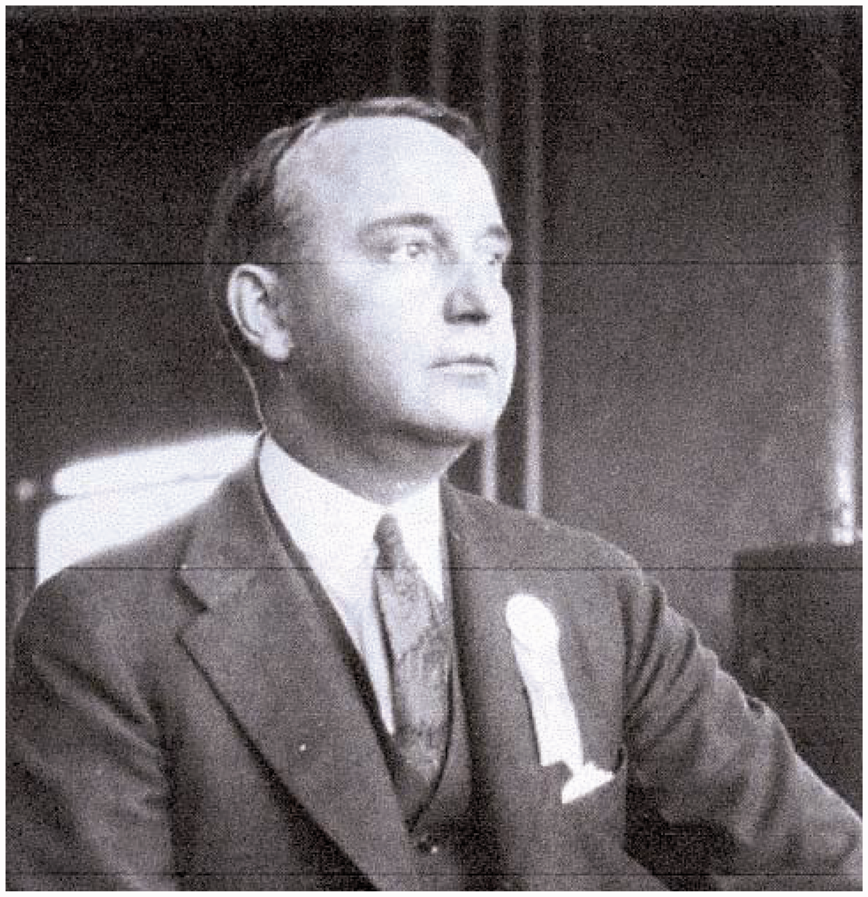
Dr Hertzler was keenly aware of medical research beginning in America. He taught pathology and histology at local medical schools but probably recommended that Burrows obtain a medical degree at the newly opened Johns Hopkins School of Medicine in Baltimore, which was patterned after the research-oriented German schools. Burrows earned an MD degree there in 1909 and immediately joined Alexis Carrel at the Rockefeller Institute in New York as a research fellow. Burrows’ interest in skin grafts may have led him to Carrel, a French surgeon who had been recruited to the Rockefeller in 1905 because of his work in wound healing and experimental surgery.
Early on, Burrows had discussed his interpretation of the rapid growth of Reverdin skin grafts with his new associates – Carrel, Simon Flexner, and William G MacCallum. Simon Flexner (1863–1946) was the director of the Rockefeller Institute for Medical Research from 1902 until 1935.6,7 William G MacCallum (1874–1944) was a professor of pathology at Columbia University (1909–1917) and interacted with members of the nearby Rockefeller Institute. 8 These scientists had read Harrison’s description of frog neural crest fragments surviving in lymph and were interested in testing other animal tissues in the hanging-drop system. Meanwhile, in 1907 Harrison had moved to Yale.
As Burrows’ letter relates, MacCallum sent a young colleague, Robert Archibald Lambert (1883–1960), to New Haven to learn under Harrison’s guidance how to grow nerve cells from higher animals than frogs. Burrows wrote, ‘Lambert failed because Harrison was convinced that it could not be done.’ In 1909, Carrel sent Burrows on the same mission. Carrel may have envisioned tissue culture as a laboratory step to transplanting tissues/organs, while Burrows may have had in mind exploiting this method to maintain strips of skins for later grafting. The letter notes that Burrows’ visit to Yale was encouraged and arranged by Franklin P. Mall.
Franklin P Mall (1862–1917)
Burrows’ letter began with the sentence, ‘I studied medicine because Hertzler convinced me that Mall was the one great man in American medicine at that time’ (Figure 3). Mall was the first Professor of Anatomy at Hopkins (1893–1917), where he established the Department of Embryology of the Carnegie Institute of Washington.
9
There he influenced both Harrison, who was an associate professor of anatomy (1899–1907), and Burrows, who was a medical student there (1905–1909). In his 1942 letter, Burrows wrote that Mall had devised in detail the experiments which he had Harrison carry out, for he (Mall) believed ‘nerves could be grown in vitro.’ The origin of Mall’s interest in this subject came from his two post-graduate training periods in Germany.
10
Franklin P Mall (1862–1917).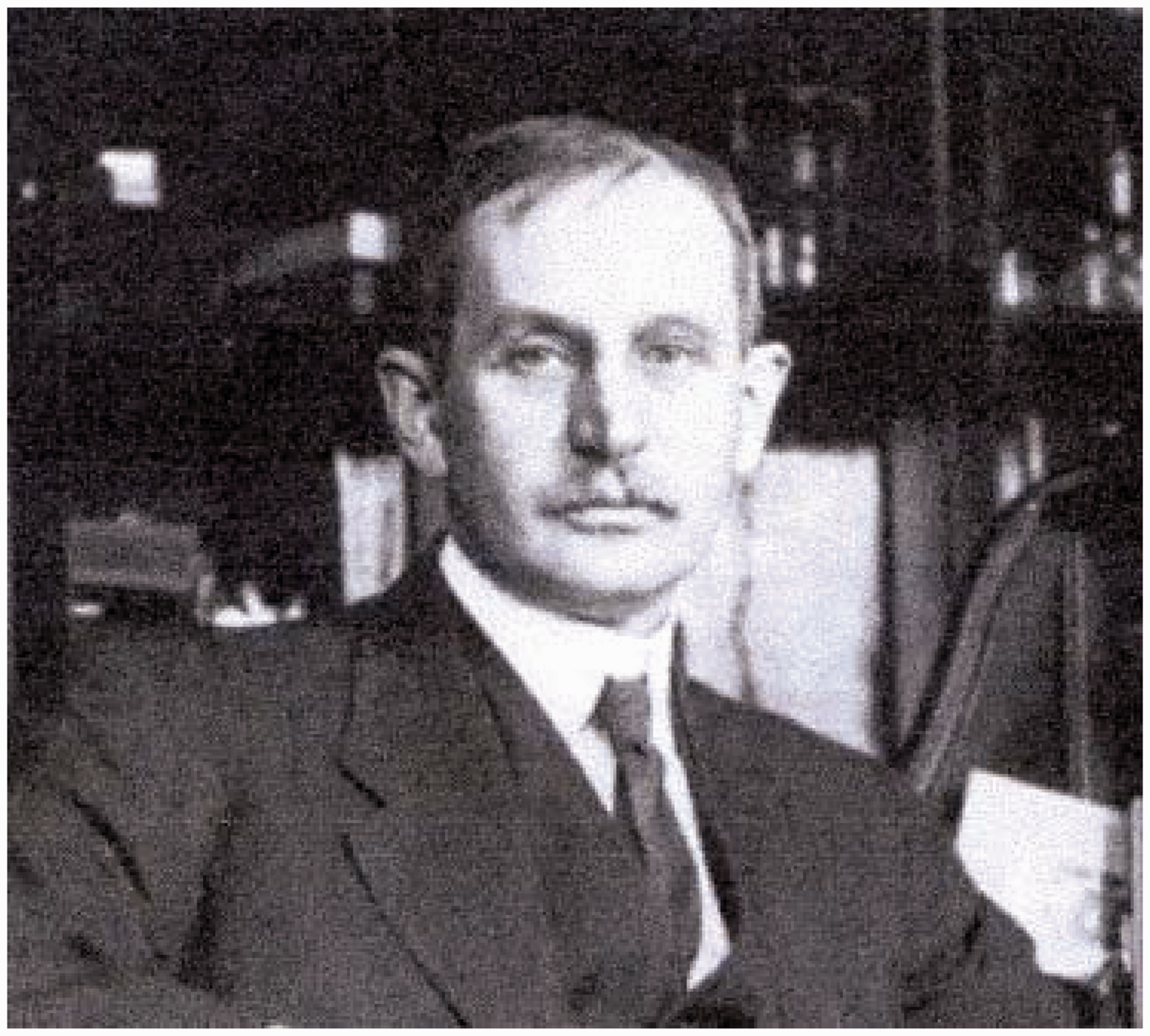
In 1876/7 Mall had studied the nervous system of the eye at Heidelberg University. In 1884 at Leipzig he worked under Wilhelm His (1831–1904), who was known for his researches on the embryological development of the nervous system, and Carl Ludwig (1816–1895), whose research included most of human physiology – notably, the circulation and intestinal tract. One of Mall’s most important discoveries made then, but only published later in 1890, was that of the vasomotor nerves for the portal veins. Mall was doubtlessly familiar with the other German literature in biology. For example, LP Rubin has listed 12 papers prior to 1907 which described the ‘maintenance in vitro or the attempted culture of cells or tissue.’ 11 Leo Loeb (1869–1959), a pathologist at various American universities, had written in 1897 that ‘epithelium from connective tissue cells’ may grow ‘in blood clots or coagulated lymph,’ but it is now believed that he merely achieved ‘survival’ of tissue. 11
As Burrows wrote, Mall’s experiments with frog tissue ‘succeeded as he planned but the cells would not grow and divide in these cultures.’ None-the-less, ‘Harrison’s work created a sensation. … Mall was a politician. He wanted Yale under his control. He used Harrison … to accomplish his ends.’ These three sentences imply that although Harrison was then in New Haven, Mall expected any subsequent work done there in this field to be recognized for his sponsorship from Baltimore.
Meanwhile, Burrows had shown Mall his plan for growing cells in vitro, ‘devised independently of his work.’ Mall directed that he carry out experiments at Yale with Harrison because of the latter’s experience. There, employing Harrison’s hanging-drop system, Burrows placed tissue from chicken embryos in chicken plasma with chick embryo extract – expecting the extract to duplicate nutritive components in wound exudate. His experiments were successful immediately. When Burrows showed ‘the growing dividing cells’ to Harrison, he was ‘astonished.’ Upon learning of this success, Simon Flexner was ‘overjoyed’ and a physiologist at the Rockefeller, Dr Samuel J Meltzer (1851–1920), was likewise duly impressed. 12 ‘The discovery pleased the Rockefellers. Carrel was used for this reason, I believe, to put it over as it has been put over. … He knew mass psychology and was the newspaper favorite of the Institute (as I saw it).’ Burrows seemed to infer here that the trustees (‘the Rockefellers’) sought to publicize the new Institute by having this discovery announced in the press by Carrel, then well-known to the American public. 13
Alexis Carrel (1873–1944)
Many health-conscious Americans once associated Alexis Carrel with human longevity because a tissue culture he prepared from a chick embryo heart was reported to have continued dividing for over 30 years. This is considerably longer than the 7- to 8-year life span of an average chicken. Theoretically, if all the cells grown were collected in one place, ‘their mass would be very much larger than the sun’ – a discredited calculation because nutritional demands of the growing mass could never be satisfied.14,15 It is now generally believed that unknown to Carrel, his fibroblast cultures were occasionally re-seeded with cells from chick embryo extract by a well-meaning or a malicious technician. 14 Thus, his popular renown rested in part on a fibroblast culture whose long life was fraudulently maintained but which was reported upon annually by the newspapers like a national birthday event.
Carrel’s career fell into four periods – the first three involved surgical and scientific areas, while the final fourth concerned social and philosophical issues.
16
Between1896 and 1910, Carrel’s research focused primarily on vascular surgery. Not having received academic recognition in France for his surgical innovations, he visited Canada in 1904 and soon moved to the United States. In Chicago he found interest and support for surgical studies. Between 1906 and 1939, he worked at the Rockefeller Institute, where his lab was called the Division of Experimental Surgery (Figure 4). Carrel was awarded the Nobel Prize in Medicine in 1912 for having perfected a suturing method that repaired severed blood vessels. His careful surgical technique reduced the incidence of thrombosis and infection at suture sites. Connecting blood vessels together then allowed him to transplant organs in dogs – e.g. thyroid, spleen, hind legs, etc. This single technical advance revolutionized vascular surgery and was essential for human organ transplantations he envisioned in the future. His attention to aseptic measures in operations was also critical for long-term tissue cultures.
17
Alexis Carrel (1873–1944).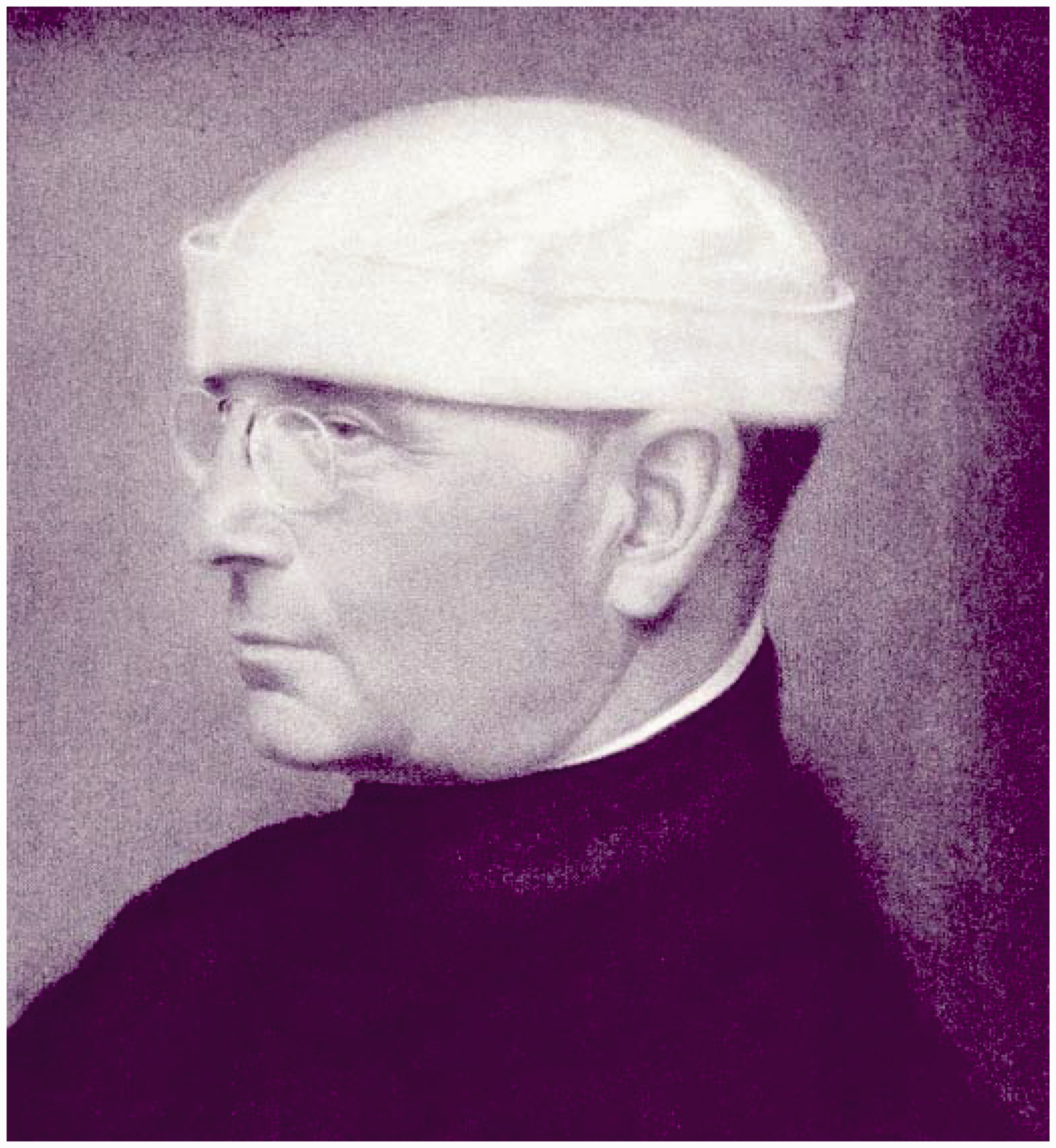
Carrel’s work in this area began in 1909 with Burrows’ return from New Haven and report on his successful experiments. They co-authored a score of papers on tissue culture through 1912, when Burrows left to become an instructor in anatomy at Cornell Clinic. 18 He subsequently published little in the field except for a speculative paper in 1922 on function and growth. 19 With the advent of World War I, Carrel served in the French Army Hospital Corps and focused on surgical matters. He introduced the Carrel-Dakin solution of hypochlorite for irrigating war wounds, which greatly reduced infections from tetanus and gas gangrene. 20 Around 1922 he resumed tissue culture studies in collaboration with Albert H Ebeling (1883–1965), Lillian E Baker (1910–1997), and Raymond Parker (1903–1974).21–23
During the early 1930s, Carrel supported the efforts of Charles A Lindbergh (1902–1974) in developing a glass pump for perfusing isolated small organs with blood and sustaining their function for several weeks without infection – the latter always being a major problem. An ultimate goal envisioned by him was human organ transplantation, but this had to await the new knowledge of histocompatibility immunology. Meanwhile, an immediate possibility was recovering hormones from perfused organs – e.g. insulin. He co-authored several papers with Lindbergh, plus a book entitled The Culture of Organs, 1938. 24 During his final years at the Rockefeller in the mid-1930s, Carrel wrote popular magazine articles on health issues and a ponderous book on social-philosophical themes, much lauded at the time. 25
Throughout his professional career, Carrel published more than 320 scientific papers on surgery, wound infections, and cell cultures. 26 He co-authored 24 articles in three languages with Burrows between 1910 and 1912, and later around 42 with other colleagues, including AH Ebeling, LE Baker, and Ragnvald Ingebrigtsen (1882–1975). 27 These articles and over a hundred papers as the sole author challenge the aspersions on his scientific reputation and productivity made by many critics after World War II.
Later tissue culturists
Besides Carrel’s early associates at Rockefeller noted above, the first generation of tissue culturists included two notable couples at the Hopkins Medical School – Warren and Margaret Lewis (1870–1964; 1881–1970) and a decade later George and Margaret Gey (1899–1970; 1900–1975. 28 The Lewises pursued the cultural conditions essential for tissue growth (media composition, pH, etc.) and studied the behavior of cells – the various forms of mononuclear cells, their movements, pinocytosis, etc. Geo Gey’s significant contributions were the roller drum culture system (used especially for virus studies) and the HeLa cell line (the first ‘permanent’ human cell line). Countless later American cell culturists devised salt solutions and various complex media now identified by their authors’ names – Dulbecco, Eagle, Earle, Gey, Hanks, Parker, Puck, Waymouth, White, and others.29,30
In Great Britain, tissue culture studies began around 1919 at the Cambridge Research Hospital, where Thomas Strangeways (1866–1926), a pathologist, was its director. 31 He acted on the belief that advances in pathology and medicine would come from fundamental medical research at the cellular level. His early technician, ‘a Mr Sheldrick, had previously worked at Carrel’s Rockefeller department.’ 32 Located in London hospitals, both Davis Thomson in 1913 and Albert J Walton had received ‘practical training from Alexis Carrel’ at the Rockefeller Institute. 32
Honor B Fell (1900–1986) began studies on tissue culture at the Cambridge Research Hospital in 1923 and was its director from 1928 to 1970. She helped popularize this new field but rejected efforts to promote it as a replacement for animal experimentation. She urged collaboration among scientists and encouraged women scientists at the renamed Strangeways Research Laboratory. During the 1920s and 30 s it was the ‘only’ British institution that was ‘devoted to cell biology’ and which pursued this new field. 33 The impact of tissue culture in Britain is delightfully discussed in Duncan Wilson’s ‘Tissue Culture in Science and Society.’ 32
Recapitulation
The 1942 letter by Montrose Burrows suggests that both he and Franklin Mall conceived of tissue culture independently. Mall envisioned nerve cells being grown in vitro, based perhaps on early related reports he had read in the German literature. Burrows was intrigued by his observation that pinch grafts of skin in surgical patients appeared to be nourished by tissue juices and did not depend on direct capillary supply. He envisioned skin fragments being maintained in vitro while being bathed in serum and tissue extracts. Mall designed Harrison’s experiments with embryonic frog tissue and later directed Burrows to seek Harrison’s advice for culturing warm-blooded embryonic tissue.
Harrison’s original 1906/7 paper never employed the expressions ‘cell culture, replication, or multiplication’ but used instead the word ‘growth’ or ‘outgrowth’ in the context of a ‘fiber’ or ‘nerve.’ However, the former terms were used in later reports about Harrison’s paper, leading to the inference that he had achieved the ‘cultivation’ of animal cells. The early dates of Harrison’s duplicate papers and these later reports (secondary sources) ‘convinced the world’ of Harrison’s priority/credit for originating tissue culture. However, Burrows’ letter indicates that Harrison did not believe that tissues from higher animals could grow in vitro, and thus he would not likely have developed tissue culture into the major tool of biological research it has become.
Conclusion – priority and credit
Credit and priority of discovery are sensitive issues in science. Sir William Osler (1849–1919), an historian of medicine, once considered the rival claimants for introducing anesthesia (Morton, Long, and Wells) and generalized that ‘in science the credit goes to the man who convinces the world, not to the man to whom the idea first occurs.’ 34 The issue here is whether to give credit to an idea or a technique. The Nobel Prize was awarded to Theodore Svedberg for developing the ultracentrifuge (1926) and to Arne Tiselius for his research on electrophoresis (1948), but no early innovator in tissue culture has been so recognized – perhaps because its potential to yield practical benefits to medicine was slow to be realized and recognized.
A typed copy of Burrows’ letter has been in my files for over 60 years and not previously circulated. It suggests that Montrose Burrows – not Harrison – should receive the primary credit for being the first to establish and maintain true tissue cultures. However, Harrison should be recognized for introducing the hanging-drop preparation employing plasma. Mall may be acknowledged for directing the early experiments in this field by both Harrison and Burrows. Carrel’s scientific reputation should be affirmed not only for his advances in vascular surgery but also for promoting and developing the field of tissue culture with various associates through the mid-1930s.
In his 1942 letter, Burrows seemed disappointed ‘that tissue culture has not changed biology as it should have done.’ However, within a few years the practical impact of this new field on medicine would become evident with virus isolation, viral vaccine production, drug testing, cancer cell therapy, in vitro fertilization, clonal antibody production, and new genetic insights derived from hybrid cells in culture.
Footnotes
Acknowledgement
The author gratefully acknowledges the invaluable assistance of Mrs Amanda Williams, Medical Center Library, University of Kentucky, and also the continued support of IS Tray II.
Declaration of Conflicting Interests
The author(s) declared no potential conflicts of interest with respect to the research, authorship, and/or publication of this article.
Funding
The author(s) received no financial support for the research, authorship, and/or publication of this article.