




Abstract
Objectives
The objective of this study was to determine the effects of Taraxacum officinale extracts on mitochondrial unfolded protein response (UPRmt) and mitophagy in the hippocampi of type 2 diabetes mellitus (T2DM) rats.
Materials and Methods
The T2DM model was established by a high-fat high-sucrose diet (HFSD) for 6 weeks and streptozotocin (STZ) administration. The rats were randomly divided into control, model, metformin (100 mg/kg/day), and T. officinale extracts (1,500 mg/kg/day) treatment groups. Fasting plasma glucose (FPG) levels, 2-h postprandial plasma glucose (2hPPG) levels, and sucrose preference rates (SPR) were ascertained. Enzyme-linked immunosorbent assay (ELISA) was used to quantify the levels of superoxide dismutase (SOD) and malonaldehyde (MDA) in the serum. Finally, the expression levels of UPRmt and mitophagy-related proteins were determined via Western blot analysis.
Results
Rats in the model group exhibited significantly higher levels of FPG and 2hPPG but lower SPR than those in the control group. Rats in the Taraxacum group had lower FPG and 2hPPG levels and higher SPR than those of the model group. Moreover, clpP, HSP60, PINK-1, Parkin, P62, Beclin-1, and LC3-II/I proteins were significantly upregulated in the rats of the model group, relative to the control group. UPRmt and mitophagy-related proteins were markedly downregulated in rats of the Taraxacum group, relative to the model group.
Conclusion
T2DM rats exhibited certain levels of anhedonia and depression, which were effectively alleviated by T. officinale extracts through the downregulation of UPRmt and mitophagy, and also by conferring protection to mitochondrial function.
Keywords
Introduction
Diabetes mellitus (DM) is a metabolic disease typically characterized by chronic hyperglycemia. More than 95% of all patients diagnosed with DM suffer from type 2 diabetes mellitus (T2DM). The prevalence of DM in China is as high as 10.9%, which has become a public health problem. Depression is a mood disorder characterized by a marked and persistent feeling of sadness and loss of interest as the primary symptoms. The global prevalence rate of depression is 2.36 times higher in diabetic patients compared with non-diabetic people (Owusu et al., 2018). DM rats always show depression-like behaviors such as anhedonia and behavioral despair, which are manifested by the decline of sucrose preference rate and the prolongation of forced swimming immobility (Hassan et al., 2019). The underlying factors of diabetes-induced depression mainly include hypothalamic–pituitary–adrenal (HPA) axis dysfunction, 5-hydroxytryptamine (5-HT) dysfunction, inflammatory activation, abnormal neurotrophic factors, and hippocampal damage, among others. Mitochondrial dysfunction is a crucial pathogenesis of DM and associated depression. Mitochondrial unfolded protein response (UPRmt) and mitophagy are the main mechanisms involved in mitochondria-induced self-repair and are activated to restore mitochondrial function when mitochondria are damaged.
Taraxacum officinale is a perennial herb of Asteraceae. It is rich in proteins, fatty acids, polysaccharides, amino acids, vitamins, trace elements, flavonoids, phenolic acids, sterols, and other such compounds (Cai et al., 2023). Extracts from T. officinale demonstrate pharmacological effects, including relieving oxidative stress, regulating blood glucose levels, and improving immunity (Choi et al., 2022). In this study, we explored the efficacy of extracts from T. officinale in alleviating anhedonia and depression in T2DM rats, with a focus on the regulation of UPRmt and mitophagy in the hippocampus.
Materials and Methods
Animals Used
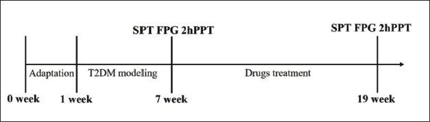
Specific-pathogen free (SPF), Sprague–Dawley (SD) rats, 36 in number (♂, average weight: 200 ± 20 g), were purchased from Beijing HFK Biotechnology Co., Ltd., [Production license number: SCXK (Beijing) 2020-0004]. The Laboratory Animal Ethics Committee of the Medical Department of North China University of Science and Technology scrutinized and approved this experiment. The experimental design is shown in Figure 1.
Reagents Used and Protocols of Drug Preparation
Metformin hydrochloride tablets (0.5 g/tablet) were purchased from Sino-American Shanghai Squibb Pharmaceutical Co., Ltd., Shanghai, China (Batch number: ABS9901). Streptozotocin (STZ) was purchased from Beijing Solarbio Science & Technology Co., Ltd., Beijing, China (CAS: 18883-66-4). Whole herbs of T. officinale were provided by the Institute of Agriculture, Hebei Academy of Agriculture and Forestry Sciences, Hebei, China. To obtain the water extracts of T. officinale, the herbs were crushed with a pulverizer, sifted through a 60-mesh sieve, and soaked within six times of water for 2 h, decocted twice (1½ h each time), and then concentrated in a hot water bath at 60°C. The drug content of the water extracts was ~300 mg/mL. Determination of chlorogenic acid (240 µg/g) and caffeic acid (50 µg/g) in water extracts by high-performance liquid chromatography (HPLC).
Superoxide dismutase (SOD) assay and malonaldehyde (MDA) detection kits were provided by Nanjing Jiancheng Bioengineering Institute, Jiangsu, China (Batch numbers: 20210422 and 20210421, respectively). Anti-glyceraldehyde-3-phosphate dehydrogenase (anti-GAPDH), anti-HSP60, anti-clpP, anti-PINK-1, anti-Parkin, anti-P62, anti-Beclin-1, and anti-LC3 antibodies were purchased from ProteinTech Group Inc. (Cat Nos.: 10494-1-AP, 15282-1-AP, 15698-1-AP, 23274-1-AP, 14060-1-AP, 18420-1-AP, 11306-1-AP, and 14600-1-AP, respectively).
M200PR0 microplate reader (Tecan Group Ltd., Männedorf, Switzerland), desktop refrigerated centrifuge (Thermo Fisher Scientific Inc., Massachusetts, US), microscope (Olympus Shinjuku City, Tokyo, Japan), SDS-PAGE unit, Western blot unit, and SDS-PAGE gel imaging system (Bio-Rad, California, US) were used in this study.
Establishment of a T2DM Rat Model and Protocol for Drug Administration
The rats were raised in an SPF room for a week to adapt to the conditions. Water and food were provided ad libitum. Six rats were randomly selected for the control group (CG), in which routine feeding was continued. The remaining 30 rats were fed with a high-fat high-sucrose diet (HFSD), which had a nutritional composition of 60% fat, 20% protein, and 20% carbohydrate—20 g/day/rat, for 6 weeks. After 6 weeks, the rats were allowed to fast for 12 h. Then streptozotocin (STZ), 28 mg/kg of body weight was injected intraperitoneally to induce T2DM (Zhang et al., 2019). Rats with fasting plasma glucose (FPG) ≥11.1 mmol/L (Tian et al., 2018) and reduced sucrose preference rate (SPR) were randomly divided into three groups: model group (MG) [n = 6], metformin treatment group (MTG) [n = 6], and T. officinale extract-treated group (TETG) [n = 6]. Rats included in the MTG and TETG were intragastrically administered metformin (100 mg/kg/day) and T. officinale water extract (1500 mg/kg/day), respectively (State Pharmacopoeia Commission 2020). Those included in the CG and MG were administered physiological saline (10 mL/kg/day), intragastrically for 12 weeks. Blood was collected from the tail vein of each rat, pre- and post-treatment, and then used to ascertain fasting plasma glucose (FPG) and 2-h postprandial plasma glucose (2hPPG) levels.
Sucrose Preference Test (SPT)
The SPR in rats of each group was estimated pre- and post-treatment through the SPT. The rats were deprived of food and water for 24 h before SPT. They were then allowed to feed on 100 mL of water and 100 mL of 1% sucrose solution for 1 h. When the SPT was carried out for half an hour, the bottle position of the water and sucrose solution was changed to avoid the interference of the bottle position on the drinking water of rats. The SPR was calculated according to the formula: SPR = weight of sucrose solution intake/weight of total intake.
Enzyme-Linked Immunosorbent Assay (ELISA)
Blood was collected from the abdominal aorta and serum was separated. The levels of SOD protein were determined using an ELISA kit, and MDA levels were ascertained using a detection kit, according to the manufacturer’s instructions.
Western Blot Assay
Hippocampal tissue (100 mg) was collected from each rat, placed in a centrifuge tube, and homogenized in 1 mL of Radio Immunoprecipitation Assay (RIPA) lysis buffer. The homogenate was then incubated on ice for 30 min and centrifuged at 4°C for 15 min at 12,000g to obtain the protein contents in the supernatant. Protein concentrations were determined using a BCA assay kit. The proteins were separated by SDS‑PAGE and then transferred to polyvinylidene fluoride membranes using a western blot unit. The membranes were blocked with Tris-Buffered Saline Tween-20 (TBST) containing 5% skimmed milk for 2 h at room temperature and then incubated with primary antibodies (anti-GAPDH, anti-HSP60, anti-clpP, anti-PINK-1, anti-Parkin, anti-P62, anti-Beclin-1, and anti-LC3) for 10 h at 4°C. The membranes were washed four times with TBST and then incubated with an anti‑rabbit secondary antibody for 2 h at room temperature. The membranes were washed four times with TBST. The bands were detected with an enhanced chemiluminescence (ECL) detection system, and their intensities were determined by the ImageJ software.
Statistical Analysis
The SPSS22.0 software was used for data statistics. Data were presented as mean ± SEM deviation and analyzed by one-way analysis of variance. If the variance is homogeneous, the Bonferroni method is used for the post-test. If the variance is not uniform, the Tamhane T2 method is used for the post-test. Statistical differences between the test groups were considered to be significant when the probability factor p was <0.05.
Results
T. officinale Extracts Suppressed FPG Levels in T2DM Rats
Rats of MG exhibited significantly higher FPG levels than those of the CG (p < 0.01). No significant changes in FPG levels among rats in the MFG and the TETG compared with those of MG were observed (p > 0.05). After STZ treatment, rats in the MG exhibited significantly higher FPG levels than those in CG (p < 0.01), while those in the MFG and the TETG had significantly lower FPG levels compared with those in the MG (p < 0.01) (Figure 2A and B).

T. officinale Extracts Suppressed 2hPPG Levels in T2DM Rats
Rats in the other three groups exhibited higher 2hPPG levels than those in the CG (p < 0.01). Notably, 2hPPG levels in rats of the MTG and the TETG were not significantly different from those in rats of the MG (p > 0.05). After STZ treatment, rats of the MG exhibited substantially higher 2hPPG levels than those in the CG (p < 0.01). On the contrary, 2hPPG levels were significantly lower in rats of the MTG and the TETG, relative to those in the CG (p < 0.01) (Figure 3A and B).

T. officinale Extracts Elevated SPR in T2DM Rats
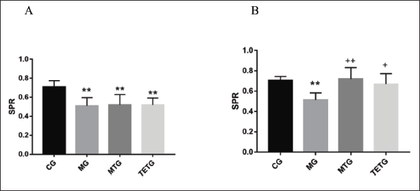
Rats in the other three groups exhibited lower SPR compared with those in the CG (p < 0.01). On the contrary, SPR in rats of the MFG (p < 0.01) and the TETG (p < 0.05) were greater than those in the MG after STZ treatment (Figure 4A and B).
T. officinale Extracts Modulated Serum SOD and MDA Levels in T2DM Rats
Rats of the MG exhibited significantly lower SOD and higher MDA levels, while those of the TETG demonstrated elevated SOD but suppressed MDA levels when compared with those from MG (p < 0.01) (Figure 5A and B).

T. officinale Extracts Downregulated Expression of UPRmt-related Proteins in the Hippocampi of T2DM Rats
UPRmt-related proteins, clpP, and HSP60 were markedly upregulated in the hippocampi of rats from the CG, compared with those from the MG (p < 0.01), while they were downregulated in rats of the TETG (p < 0.01) (Figure 6A and B).

T. officinale Extracts Suppressed the Levels of Mitophagy-related Proteins in the Hippocampi of T2DM Rats
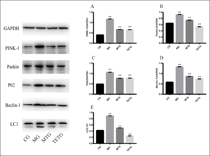
Mitophagy-related proteins: PINK-1, Parkin, P62, Beclin-1, and LC3 I/II were significantly upregulated in the hippocampi of rats from the MG compared with those in the CG (p < 0.01). However, the expression of all mitophagy-related proteins was significantly downregulated in rats from the TETG (p < 0.01) (Figure 7A–E).
Discussion
Current research trends on the pathogenesis of DM have begun to focus on the role of mitochondrial dysfunction. This model of DM pathogenesis is characterized by damage to mitochondrial morphology and mitochondrial (mt) DNA, as well as inhibition of respiratory chain functions (Rowley et al., 2017). The pathogenesis of depression involves hippocampal neurons exhibiting varying stages of impairment in mitochondrial function, reduced mitochondrial numbers, morphological and/or oxidative damage in mitochondria, variations in mtDNA sequence, lowered energy metabolism, and respiratory dysfunction (Klinedinst & Regenold, 2015; Allen et al., 2018). Therefore, improving the mitochondrial quantity, quality, and function may be a potential therapy method for managing diabetes-induced depression.
UPRmt and mitophagy are the main cellular mechanisms involved in the repair of mitochondrial damage. Specifically, UPRmt is a stress response in which transcriptional activation of mitochondrial heat shock proteins and proteases, as well as other nuclear-encoded genes is initiated to maintain mitochondrial protein homeostasis (Jovaisaite et al., 2014). Notably, abnormal protein structure resulting from mitochondrial dysfunction causes activation of UPRmt. HSP60, a molecular chaperone, plays a crucial role in facilitating proper protein assembly and correct protein folding in mitochondria (Richards et al., 2023). clpP assists in protein degradation. HSP60 and clpP were upregulated in the mitochondria of skeletal muscles indicating that UPRmt was activated (Wang et al., 2022).
Mitophagy is target-specific autophagy in which cells selectively degrade damaged or dysfunctional mitochondria. It is mediated by the PINK-1/Parkin signaling pathway, which is a classic mechanism for studying mitophagy in mammalian cells. PTEN-induced kinase 1 (PINK-1) is a mitochondrial serine/threonine-protein kinase. Under normal conditions, PINK-1 is located in the inner mitochondrial membrane. Upon mitochondrial damage, with the help of translocase of the outer mitochondrial membrane (TOMM) and translocase of the inner mitochondrial membrane (TIMM), it enters the cytoplasm, where it is degraded to minuscule levels. PINK-1 identifies damaged mitochondria by constantly checking for the oxidative state of mitochondria and sends signals for the recruitment and activation of Parkin (Nguyen et al., 2016). Parkin, an E3 ubiquitin ligase, promotes the ubiquitination of proteins located on the mitochondrial membrane of damaged mitochondria. Autophagic receptor proteins, such as P62, OPTN, and NDP52 interact with the ubiquitinated proteins on the mitochondrial membrane. The microtubule-associated protein 1 light chain 3 (LC3), binds with the autophagic receptor protein located on the damaged mitochondria (Eiyama & Okamoto, 2015). These mitochondria are then coated with the autophagosome membrane, a phenomenon that activates the autophagic lysosome system, which degrades these mitochondria (Tan et al., 2018).
The potential use of T. officinale in the treatment of T2DM and managing its complications have been proved through several studies (Murtaza et al., 2022). T. officinale can reduce blood glucose levels and significantly improve insulin resistance (Sangeethan et al., 2013; Zhao et al., 2018). It can also reverse mitochondrial oxidative stress caused by alcohol-induced liver injury and thus protect mitochondrial function (Choi et al., 2022). Taraxasterol alleviated mitochondrial damage caused by the toxin nigericin (Yang et al., 2021). However, it is not known whether T. officinale exerts a protective effect on mitochondria in rats with diabetes-induced depression. In this study, we established a T2DM rat model using HFSD and STZ and then estimated SPR. Metformin was used as the control drug as it can improve the symptoms of depression in T2DM patients (Chen et al., 2019). Rats were treated with T. officinale water extracts, and then the expression patterns of UPRmt and mitophagy-related proteins were analyzed. Our experiments revealed that T2DM rats from the MG exhibited varying degrees of anhedonia, manifested as a decrease in SPR, while T. officinale extracts could reverse this state. Moreover, rats in the MG exhibited elevated expression of HSP60 and clpP proteins in their hippocampi. Similarly, rats of the MG exhibited upregulation of mitophagy-related proteins, namely, PINK-1, Parkin, P62, Beclin-1, and LC3II/I in the hippocampi. These results suggested that UPRmt and mitophagy in the hippocampi of T2DM rats were enhanced, which may be a protective mechanism triggered by mitochondrial damage. Interestingly, T. officinale extracts downregulated the expression of HSP60, clpP, and mitophagy-related proteins in the hippocampi of T2DM rats. These results indicate the potential of T. officinale extracts in the treatment of DM-induced depression by protecting mitochondrial function, inhibiting mitochondrial stress response, downregulating the expression of UPRmt and mitophagy-related proteins, and improving the symptoms of depression in T2DM rats. This preliminary experiment tried to explore the effects and underlying mechanisms of T. officinale extracts on depression-like behavior in T2DM rats. In a follow-up study, we intend to further clarify the effects and mechanisms of T. officinale extracts on behavioral changes in T2DM rats. Different doses (concentrations) of T. officinale water extracts will be used for intervention studies, and pathways related to mitochondrial function will be identified.
Conclusion
T. officinale extracts could improve the anhedonia in diabetes rats and had a clear antidepressant effect. T. officinale extracts alleviate classic symptoms of depression in T2DM rats by inhibiting UPRmt and mitophagy in rat hippocampi. Our study laid a research foundation for the clinical application of T. officinale in the treatment of T2DM complicated by depression.
Summary
In this experiment, we studied the effect and mechanism of T. officinale extracts on depression-like behavior in T2DM rats. HFSD and STZ methods were used to model T2DM rats, and depression-like behaviors of T2DM rats were observed. T. officinale extracts were used to treat T2DM rats. The results showed that T2DM rats exhibited certain levels of anhedonia and depression, which were effectively alleviated by T. officinale extracts through the downregulation of UPRmt and mitophagy, and also by conferring protection to mitochondrial function.
Abbreviations
CG: Control group; 2hPPG: 2-hour postprandial plasma glucose; 5-HT: 5-hydroxytryptamine; DM: Diabetes mellitus; ECL: Enhanced chemiluminescence; ELISA: Enzyme-linked immunosorbent assay; FPG: Fasting plasma glucose; HFSD: High-fat high-sucrose diet; HPLC: High-performance liquid chromatography; HPA: Hypothalamic–pituitary–adrenal; MDA: Malonaldehyde; MTG: Metformin treatment group; LC3: Microtubule-associated protein 1 light chain 3; UPRmt: Mitochondrial unfolded protein response; MG: Model group; PINK-1: PTEN-induced kinase 1; RIPA: Radio immunoprecipitation assay; STZ: Streptozotocin; SPT: Sucrose preference test; SPR: Sucrose preference rates; SOD: Superoxide dismutase: SPF: Specific-pathogen free; SD: Sprague–Dawley; T. officinale: Taraxacum officinale; TETG: T. officinale extract treated group; TOMM: Translocase of the outer mitochondrial membrane; TIMM: Translocase of the inner mitochondrial membrane; TBST: Tris-Buffered saline tween-20; T2DM: Type 2 diabetes mellitus.
Authors’ Contributions
All authors have agreed to the submission of this manuscript for publication. JL and XW designed the experimental scheme. MW performed the experiments, analyzed the data, and interpreted the results. ZG and LM performed the animal experiments and behavior tests. ELISA and Western blot were performed by MW and ZL. The figures and manuscript were written by MW and ZG.
Footnotes
Acknowledgments
The authors thank the Diabetes Integrated Traditional Chinese and Western Medicine Prevention and Treatment Team of Tangshan City.
Declaration of Conflicting Interests
The authors indicated no conflicts of interest with respect to the research, authorship, and/or publication of this article.
Funding
This work was supported by the National Natural Science Foundation of China (82205066), the Ministry of Science and Technology Assistance Project for Developing Countries (KY201904005), and the Research Program Project of Hebei Provincial Administration of TCM (2022139).
