
Abstract
Background
Citral is a bioactive compound found in lemongrass oil. It functions as an anti-oxidant and possesses anti-inflammatory qualities.
Objective
The purpose of this study was to assess the anti-arthritic benefits of citral using a rat model with an adjuvant arthritis paradigm.
Experimental Procedures
In Swiss Albino Wistar rats, arthritis was produced by intradermal injection of Freund’s adjuvant (0.1 mL) into their hind paw. Arthritic animals were given citral orally for a duration of 8 days, beginning on the 11th day and lasting until the 18th day after the adjuvant injection. Effect of citral was assessed by measuring liver indicators [aspartate aminotransferase (AST), alanine aminotransferase (ALT), and alkaline phosphatase (ALP)], lipid peroxidation markers [thiobarbituric acid reactive substances (TBARS) and lipid hydroperoxide (LOOH)], anti-oxidant status [superoxide dismutase (SOD), catalase (CAT), glutathione peroxidase (GPx), and glutathione (GSH)], spleen enzyme activity, blood cytokines [tumor necrosis factor-alpha (TNF-α), interleukin-6 (IL-6), and nuclear factor kappa B (NF-κB)], and NF-κB/transforming growth factor-beta (TGF-β) messenger ribonucleic acid (mRNA) expression in control and experimental animals.
Results
Animals with arthritis produced by adjuvant injection exhibited a large rise in hepatic markers, lipid peroxidative markers, lysosomal enzymes, and inflammatory cytokines, while anti-oxidant enzymes showed a significant drop compared to control rats. Administering citral orally resulted in a dramatic modification of the identified biochemical abnormalities in arthritic animals, bringing them closer to normal circumstances and restoring them to normalcy. Moreover, there was an elevation in the levels of inflammatory and fibrotic markers such as NF-κB/TGF-β in rats with induced arthritis. Rats with arthritis showed a significant reduction in NF-κB and TGF-β expression after receiving citral.
Conclusion
This study indicates that citral demonstrates significant anti-arthritic, inflammation-suppressing, and anti-fibrotic properties in experimental arthritis models.
Introduction
The chronic inflammatory disorder known as rheumatoid arthritis (RA) causes joint swelling and discomfort, cartilage and bone deterioration, and possible bone loss. It affects a number of joints, starting with the smaller ones (diarthrodial joints) and progressing to the bigger ones (and even extra-articular tissues) later on. Extreme discomfort, immobility, and deformity (especially in the ankles and knees) are symptoms of the condition’s advanced phases. The pain, edema, and stiffness are clinical characteristics of RA (Scheel et al., 2006; Zhang et al., 2018).
Research has shown a connection between oxidative/nitrosative stress and RA. (Veselinovic et al., 2014). Free radicals play a role, either directly or indirectly, in causing damage to the joints. They play a role in breaking down joint cartilage by targeting its proteoglycan and hindering its synthesis (Quiñonez-Flores et al., 2016). According to Afonso et al. (2007), the damage to the cartilage matrix is intensified by the increased presence of redox-active chemicals, which causes a redox imbalance. Therefore, the dysregulation between proinflammatory and anti-inflammatory states has led to inflammation and damage in the joint’s synovial membrane. Earlier studies have proposed that reducing inflammation and mitigating oxidative stress might be advantageous in preventing damage to articular cartilage (Huang et al., 2018; Lin et al., 2014; Wu et al., 2018). Overexpression of proinflammatory transcription factors [tumor necrosis factor-alpha (TNF-α), interleukin-6 (IL-6), and nuclear factor kappa B (NF-κB)] plays a paramount role in the progression of arthritis. IL-6 promotes the development of blood vessels, which increases inflammation. On the other hand, TNF-α promotes inflammation by activating synovial fibroblasts that express cellular adhesion molecules, leading to an increase in the migration of leukocytes into the joints, ultimately resulting in damage. In the context of RA, there is a significant elevation in NF-κB levels (Miller et al., 2010).
There is a diverse range of medications accessible for the management of RA, namely, anti-rheumatic drugs, steroid hormones, immunosuppressants, biological agents, and inflammation-modifying remedies (Atzeni et al., 2013; Lima-Garcia et al., 2011; Momohara et al., 2011; Pincus & Cutolo, 2015). While arthritis-modifying drugs offer numerous powerful advantages, their clinical use is restricted due to factors such as elevated cost, negative effects, and limited benefit toward defined target areas (Ekambaram et al., 2010). Compounds with potential benefits against RA are examined through diverse animal models to identify their therapeutic advantages, drawing on similarities with the disease in humans (Ghosh et al., 2010). Hence, an effective therapeutic strategy for arthritis requires cost-effective drugs that have a prolonged duration of action and minimal or fewer side effects. Additionally, these drugs should prevent damage to the joints by suppressing inflammation and reducing the production of proinflammatory cytokines.
Amidst diverse natural elements, citral is the active compound found in citrus fruits, plants, and herbs. This monoterpene aldehyde is well-known for its aromatic properties, making it a popular choice in the cosmetic and food industries due to its lemon fragrance and flavor (Souza et al., 2020). The primary components of citral make up approximately 65%–85% of lemongrass leaf oil (Cymbopogon citratus), and it is well-known in tropical and subtropical regions for its various biological activities (Saddiq & Khayyat, 2010). Moreover, citral is recognized for its pharmacological activities, such as hypoglycemic, hypolipidemic (Adeneye & Agbaje, 2007), anti-fungal (Boukhatem et al., 2014), anti-bacterial (Onawunmi et al., 1984; Tofiño-Rivera et al., 2016), immunomodulatory (Bao et al., 2015), anti-tumor (Chaouki et al., 2009), and anti-inflammatory (Meena Priya & Priya, 2017). Additionally, citral has demonstrated inhibition of pathogenic virulence effects against C. sakazakii in vitro (Shi et al., 2020). Notably, citral can restrain the stimulation of macrophage activity and NF-κB, as well as the formation of proinflammatory signaling molecules, including IL-1β (Bachiega & Sforcin, 2011), and it exhibits potent anti-oxidant activity (Barroso et al., 2011; Cheel et al., 2005). Yang et al. (2013) illustrated that the kidney protective effect of citral is due to the early stage of adriamycin-induced focal segmental glomerulosclerosis in mice, which involves the activation of the Nrf2 anti-oxidant pathway and the suppression of NF-κB activation. This study was conducted to investigate the anti-arthritic activity of citral against complete Freund’s adjuvant (CFA)-induced rodents, in light of the global impact of arthritis and the necessity for innovative preventive approaches.
Materials and Methods
Animals
To assess the anti-arthritic impact of citral, experiments were conducted using Swiss Albino rats weighing between 120 and 150 g. The rats were maintained in suitable conditions, receiving a standard diet with unrestricted access to drinking water. The research adhered to approved guidelines, and all parameters were scrutinized in compliance with the Institutional Animal Ethics Committee’s recommendations.
Experimental Induction of Arthritis
The usual way to make the animals develop arthritis was to inject 100 µL of CFA intradermally into the posterior paw of the animals.
Study Design
Citral was diluted with corn oil, and CFA was diluted with liquid paraffin. Four groups were established for the experiment, each comprising six rats (n = 6).
Group I: The rats used as a baseline were given regular animal chow.
Group II: A dosage of 200 mg/kg body weight of citral (Yang et al., 2013) was administered via an intragastric tube over 8 days, spanning from the 11th to the 18th day.
Group III: On the 1st day, Freund’s adjuvant mixture was intradermally injected into the posterior paw.
Group IV: Citral (200 mg/kg of body weight) was administered orally over a period of 8 days, commencing on the 11th day and ending on the 18th day, following the intradermal injection of CFA.
On the 19th day, the rats were euthanized, and both blood and spleen tissue were collected. Plasma and serum were separated from the blood samples, which were then employed for biochemical assays. Spleen tissue was homogenized for biochemical analysis, and ribonucleic acid (RNA) extracted from the spleen tissue was utilized for quantitative reverse transcription polymerase chain reaction (qRT-PCR) analysis.
Assessment of Hepatic Marker Enzymes
We used test kits from Nanjing’s Jiancheng Bioengineering Institute to detect the activity of serum hepatic markers such as aspartate aminotransferase (AST) and alanine aminotransferase (ALT). For AST, initially, 5 µL of serum was mixed well with 5 µL of buffer solution, 20 µL of substrate solution, and 2 µL of 2 mmol/L sodium pyruvate solution. After that, the mixture was incubated for 30 min at 37°C. 20 µL of chromogenic agent was added after incubation, stirred for 10 s, and then incubated for a further 20 min at 37°C. After that, 200 µL of alkali reagent was added, stirred well, then set aside to rest for 15 min at room temperature. Lastly, a microplate reader was used to measure each well’s optical density (OD) at 510 nm.
Alkaline phosphatase (ALP) was conducted utilizing a diagnostic kit procured from the Jiancheng Bioengineering Institute of Nanjing. A mixture of 1.0 mL buffered substrate, 3.1 mL deionized water, and 0.1 mL serum was incubated at 37°C for 15 min. Following incubation, 2.0 mL of color reagent was added to each tube. In control tubes, the enzyme was introduced after the addition of the color reagent. Additionally, 0.1 mL of the standard solution and 0.1 mL of distilled water (blank) were processed under the same conditions. The intensity of the developed color was then measured at 510 nm.
Analysis of Lipid Peroxidative Markers
Thiobarbituric acid reactive substances (TBARS) were measured using standard procedures to evaluate lipid peroxidation (Thangaiyan et al., 2020). First, the material was thoroughly mixed after being diluted with 0.5 mL of double-distilled water. The mixture was incubated in a boiling water immersion for 15 min after 2.0 mL of thiobarbituric acid (TBA)–trichloroacetic acid (TCA)–hydrochloric acid (HCl) reagent was added. The tubes were centrifuged for 10 min at 1,000 rpm after cooling. The supernatant was then collected and measured at 535 nm relative to the reagent blank. The method used to estimate lipid hydroperoxide (LOOH) was as follows: Li et al. (2021) conducted the study. 0.9 mL Fox reagent was mixed with 90 mL methanol and 10 mL 250 mM H2SO4. Next, 0.1 mL of sample was added to the fox reagent mixture. The sample contained mixture was incubated at room temperature for 30 min and read at 560 nm.
Assessment of Anti-oxidant Status
Superoxide dismutase (SOD) and other enzymatic anti-oxidants were studied for their activity (Radhiga et al., 2012), catalase (CAT) (Indirapriyadarshini et al., 2023), and glutathione peroxidase (GPx) (Rotruck et al., 1973) using established methods.
SOD Activity
A 0.5 mL tissue homogenate sample was diluted to 1.0 mL using water. After agitating the mixture for 90 s at 4°C, 1.5 mL of cold chloroform and 2.5 mL of ethanol were added. The mixture was then centrifuged. The enzyme formulation was combined with 1.2 mL of sodium pyrophosphate buffer, 0.1 mL of phenazine methosulfate, and 0.3 mL of nitroblue tetrazolium for the test. The reaction was initiated by adding 0.2 mL of nicotinamide adenine dinucleotide (NADH), and the mixture was subsequently incubated at 30°C for 90 s. The process was halted by the application of 1 mL of glacial acetic acid. The solution was centrifuged and allowed to settle for 10 min after 4 mL of n-butanol was added. The absorbance of the n-butanol layer was assessed at 510 nm.
CAT Activity
0.4 mL of hydrogen peroxide (H2O2) was mixed with 0.1 mL of tissue homogenate. After adding 2.0 mL of a dichromate-acetic acid mixture and boiling for 10 min, the reaction was stopped after 15, 30, 45, and 60 s. The produced color was measured at 620 nm after cooling.
GPx Activity
After mixing 0.2 mL tris buffer and glutathione (GSH), 0.1 mL H2O2 was added. A control with all reagents except tissue homogenate was incubated with the reaction mixture at 37°C for 10 min. Add 0.5 mL of 10% TCA and centrifuge to stop the reaction. GSH was measured in the supernatant using the Ellman technique (1959).
GSH Measurement
Reduced GSH was measured using the Ellman technique (1959). 2.0 mL of 5% TCA precipitated a 0.5 mL tissue homogenate. After centrifugation, 2.0 mL of the supernatant was mixed with 1.0 mL Ellman’s reagent and 4.0 mL 0.3M DHP. The yellow color was detected at 412 nm by a Spectronic 20.
Evaluating Lysosomal Enzyme Activities
King’s method measured acid phosphatase activity on disodium phenyl phosphate. Enzyme activity was assessed by phenol release, moles per minute per milligram of protein. The activity of β-glucuronidase was analyzed following the method outlined by Kawai and Anno (1971), using p-nitrophenyl β-
Measurement of Inflammatory Cytokines by Enzyme-linked Immunosorbent Assay (ELISA) Method
Serum levels of TNF-α, IL-6, and NF-κB were assessed using sandwich ELISA kits from Cell Signaling Technology, USA, in accordance with the manufacturer’s guidelines. 50 µL of the sample and 50 µL of an antibody cocktail were added to the designated wells, and the mixture was incubated at 37°C for 1 h. The wells were rinsed and treated with 100 µL of 3,3′,5,5′-tetramethylbenzidine (TMB) substrate for a 10-min incubation after incubation. To terminate the reaction, incorporate 100 µL of stop solution and measure absorbance at 450 nm using an ELISA reader.
RNA Extraction and Quantitative Real-time Reverse Transcription Polymerase Chain Reaction (RT-PCR) Analysis
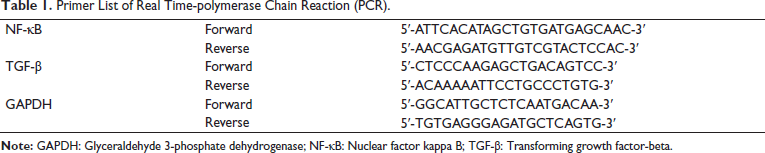
The BioPhotometer (Eppendorf) was used to quantify the total RNA isolated from spleen tissue using TRIzol reagent by measuring absorbance at 260 and 280 nm. The synchronized bacterial reporter (SYBR) Green master mix (Thermo Fisher Scientific, USA) was employed to quantify the messenger ribonucleic acid (mRNA) expression levels of NF-κB, transforming growth factor-beta (TGF-β), and glyceraldehyde 3-phosphate dehydrogenase (GAPDH). The primer sequences are listed in Table 1. PCR settings for complementary DNA (cDNA) synthesis were as follows: 25°C for 10 min, 42°C for 50 min, and 75°C for 15 min. The primer manufacturer’s specifications were followed for the cDNA amplification conditions: 95°C for 2 s, 55°C for 15 s, and 68°C for 20 s. The relative mRNA expression levels were normalized to GAPDH, and the transcript expression levels were adjusted to GAPDH. The 2–∇∇Ct method was employed to calculate the relative mRNA expression. Each reaction was conducted in triplicate.
Primer List of Real Time-polymerase Chain Reaction (PCR).
Statistical Analysis
The data were expressed using the mean values and their corresponding standard deviation (SD). Duncan’s multiple range test was implemented for post hoc analysis, while one-way analysis of variance (ANOVA) was implemented for statistical comparisons. The statistical package for the social sciences (SPSS) software was employed to conduct all analyses, and the threshold for statistical significance was set at p < .05.
Results
Effect of Citral on Hepatic Marker Enzymes in Serum
Figure 1 illustrates the influence of citral treatment on the concentrations of hepatic enzymes (AST, ALP, and ALT). The serum from both control and CFA-induced arthritis rats displayed a substantial rise in hepatic marker levels. Particularly, CFA-induced arthritis rats exhibited a significant surge in AST, ALP, and ALT activities compared to the control group. Conversely, the introduction of citral demonstrated a marked safeguard against hepatic damage, manifesting in a noteworthy reduction in AST, ALP, and ALT activities in citral-treated rats compared to their CFA-induced rats.

Effect of Citral on Lipid Peroxidative Markers in Spleen Tissue
Lipid peroxidation is recognized as a significant contributor to the oxidative stress triggered by the adjuvant arthritis model. In the current examination, the induction of CFA resulted in heightened levels of TBARS and LOOH in spleen tissue. Conversely, the administration of citral demonstrated a marked protective effect, significantly mitigating CFA-induced lipid peroxidation in arthritis-afflicted rats relative to the reference group, as shown in Table 2.
The Effect of Citral on Thiobarbituric Acid Reactive Substances (TBARS), Lipid Hydroperoxide (LOOH) of the Spleen in Complete Freund’s Adjuvant (CFA)-induced Arthritic Rats.

Values not sharing a common marking (a–c) differ significantly at p < .05 [Duncan’s multiple range test (DMRT)].
Significantly different from the control group (p < .05).
Significantly different from the CFA group (p < .05).
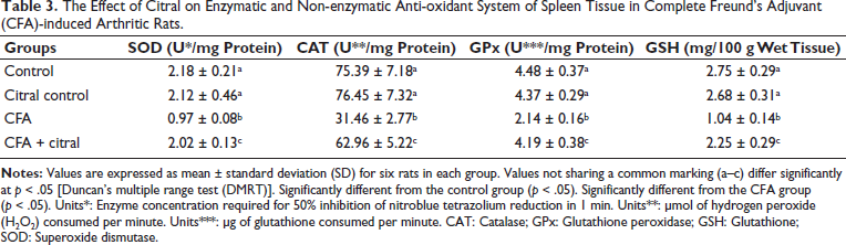
Effect of Citral on Anti-oxidant Status in Spleen Tissue
Recognizing the crucial role of the anti-oxidant defense system in restraining the advancement of arthritis, our investigation involved an analysis of anti-oxidant levels, including SOD, CAT, GPX, and GSH, in the adjuvant arthritis model. The results indicated a notable decline in SOD, CAT, GPX, and GSH levels in arthritis-afflicted rats. However, the administration of citral resulted in a significant enhancement of anti-oxidant activities in rats compared to the control group, as depicted in Table 3.
The Effect of Citral on Enzymatic and Non-enzymatic Anti-oxidant System of Spleen Tissue in Complete Freund’s Adjuvant (CFA)-induced Arthritic Rats.
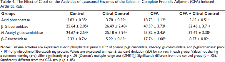
Effect of Citral on Lysosomal Enzyme Activities in Spleen Tissue
Table 4 displays the levels of lysosomal enzyme activity in spleen tissue from the treated and untreated groups. Adjuvant arthritis rats demonstrated considerably greater activity of acid phosphatase, β-glucuronidase, N-acetyl glucosaminidase, and β-galactosidase compared to the control group. However, these elevated activities were successfully regulated in arthritic rats by inducing citral.
The Effect of Citral on the Activities of Lysosomal Enzymes of the Spleen in Complete Freund’s Adjuvant (CFA)-induced Arthritic Rats.
Effect of Citral on Inflammatory Markers in Serum
Activation of oxidative stress triggers the stimulation of transcription factors (NF-κB) and inflammatory markers (TNF-α and IL-6) in Figure 2. Through ELISA analysis, we assessed the increased levels of TNF-α, NF-κB, and IL-6 in CFA-induced arthritis rats. However, the administration of citral demonstrated significantly decreased levels against the increased expression of TNF-α, NF-κB, and IL-6 induced by CFA in rats.

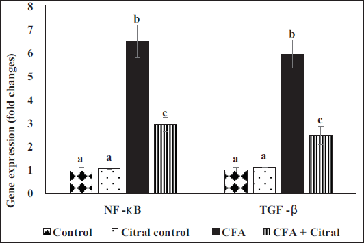
Impact of Citral on Real-time PCR of NF-κB and TGF-β Expression
The expression levels of NF-κB and TGF-β in the spleen tissue of rats with control and CFA-induced arthritis are shown in Figure 3. When compared to control rats, rats given CFA showed a significant rise in NF-κB and TGF-β expression. Citral significantly reduced spleen expression in rats with CFA-induced arthritis when compared to controls.
Discussion
RA is a pervasive global health issue, impacting billions of individuals and standing as a leading cause of disability. The existing treatments for this condition often come with potential side effects, underscoring the urgent need to explore more effective therapies that can benefit a larger segment of patients. In recent years, herbal products have garnered increasing attention for their potential in preventing and treating various diseases. The current work provides evidence of the anti-inflammatory and anti-arthritic properties of citral in experimental rats with CFA-induced arthritis.
The CFA-induced arthritis model in rats is similar to RA in people, making it appropriate for research (Tag et al., 2014). Intradermal CFA injection into rat paws induces both local and chronic systemic inflammation, with a significant sensitivity to heat shock proteins and cartilage proteoglycans. Local inflammation resolves in 3–4 days, but the chronic phase might extend for 2 weeks to several months (Akira et al., 2006). Cytokines, prostaglandins, and enzymes are all inflammatory mediators that are secreted. Body weight alterations in CFA-induced mice are critical for determining medication responsiveness and inflammation duration (Naik & Wala, 2014).
Since liver failure is also a feature of adjuvant arthritis, tissue damage was assessed by measuring enzyme activity in both the serum and the relevant organ. When cells in the damaged organ release aminotransferases, the effect is an increase in aminotransferases. Studies suggest that hepatic markers such as AST, ALT, and ALP are associated with the progression of arthritis (Uttra et al., 2019; Xia et al., 2014). The present study correlates with this finding; CFA-induced animals showed increased ALT, AST, and ALP, whereas oral administration of citral attenuated all the hepatic markers. ALT, AST, and ALP exhibited significant reductions in arthritic mice following the application of citral. This diminishing impact could be associated with their anti-inflammatory properties (Chen et al., 2020).
Numerous studies have extensively documented the involvement of free radicals in the progression of arthritis, highlighting their role in causing damage to joint tissues (Cui et al., 2019). Substantial evidence from diverse investigations supports the notion that oxidative stress and the presence of redox-active molecules play a significant role in inflammation (Comar et al., 2013; Lin et al., 2020). In line with these established findings, our study also revealed an augmentation of oxidative stress in animals induced with the adjuvant arthritis model, while the oral administration of citral effectively suppressed oxidative stress in these CFA-induced animals. The generation of oxidative stress leads to increased production of reactive oxygen species (ROS) and nitric oxide (NO) in the environment, as evidenced by the measurement of TBARS and LOOH levels in the plasma of CFA-induced and control animals. Ka et al. (2015) demonstrated that in a mouse model induced with lipopolysaccharide (LPS), citral relieved lupus nephritis by suppressing the activation of NLRP3 inflammasome and lowering the levels of oxidative stress molecules.
To counteract the harm inflicted by free radicals, a protective system known as the defense system (comprising anti-oxidants) is present in both humans and animals. Anti-oxidants mitigate lipid radical formation by interrupting the chain reaction of free radicals and peroxidation. The results of our ongoing research revealed a decline in the activities of SOD, CAT, and GPx in subjects administered with CFA due to increased oxidative stress. However, the increased lipid peroxidation observed in CFA-induced animals was counteracted and balanced by citral, thanks to its capacity to scavenge free radicals and augment anti-oxidant systems. In accordance with our findings, a study conducted by Kremer et al. (2019) demonstrated that the administration of citral prevented ultraviolet B (UVB)-induced cutaneous carcinogenesis in hairless mice. The research also emphasized the direct anti-oxidant capacity of citral to regulate reactive species levels in the epidermis during UVB-induced carcinogenesis. The depletion of GSH results in significant metabolic alterations, which in turn induces an increase in the sensitivity of cells (Limón-Pacheco et al., 2007). The protective effect of citral as an anti-oxidant against arthritis is demonstrated by the significant increase in GSH levels following citral treatment.
In mouse models of adjuvant-triggered arthritis, lysosomal enzyme extracellular outflow is a hallmark of inflammation progression. In adjuvant-induced arthritis, glycohydrolases released by invading neutrophils, macrophages, and other cell types (such as chondrocytes and synoviocytes) start the production of inflammatory mediators, which are crucial in the development of rheumatic processes. The decline in the efflux of lysosomal enzymes is indicative of potential benefits, indirectly affirming the defensive influence of the drug. Citral infusion diminishes the liberation of lysosomal enzymes in adjuvant-provoked arthritis in rodent subjects, underscoring its anti-inflammatory efficacy. Citral mitigated the inflammatory responses induced by Cronobacter sakazakii in newborn mice, thereby exhibiting its protective effects against intestine inflammation. Furthermore, it is also reported that citral treatment significantly increased the body weight of C. sakazakii-infected mice, thus providing evidence that citral reverses the body weight and inhibits inflammation (Shi et al., 2020).
Extensive documentation supports the understanding that activated macrophages play a role in releasing cytokines within the inflammatory environment, a process linked to the advancement of arthritis (Kshirsagar et al., 2014). Specifically, IL-6, TNF-α, and NF-κB are recognized for their tendency to overexpress and instigate inflammatory cascades at sites of inflammation (Alavala et al., 2020; Alzarea et al., 2022). The results of the present investigation corroborate these findings, showing that the injection of citral in mice with CFA-induced arthritis decreases transcription factors such as NF-κB, IL-6, and TNF-α. Furthermore, citral has exhibited anti-inflammatory responses in various studies, including UVB-induced skin carcinogenesis, where it inhibited cytokine levels such as interleukins and TNF-α (Kremer et al., 2019). The results of this work are in line with data showing that while citral treatment has anti-inflammatory effects, as shown by the downregulation of TNF-α, IL-6, and NF-κB, CFA induction increases proinflammatory cytokines in rats (Chen et al., 2020; Cui et al., 2019; Kumar et al., 2020).
TGF-β is a superfamily of cytokines that play a role in a variety of responses, including apoptosis, proliferation, differentiation, and extracellular matrix synthesis (Wahl, 2007). TGF-β is crucial for the development of RA-related fibrosis (Pohlers et al., 2009). Al-Naqqash et al. (2020) found that TGF-β expression was elevated in RA. Numerous studies have found TGF-β in RA patients’ synovial fluids and tissues, leading to the notion that it plays a role in the disease’s pathogenesis (Bira et al., 2005; Cho et al., 2006; Sakuma et al., 2007). Treatment with citral lowered TGF-β expression levels. Citral may reduce inflammation by preventing the synthesis of the proinflammatory cytokine TGF-β.
Conclusion
Overall, this study’s results show that citral is effective against arthritis caused by total Freund’s enhancer. The results show that citral reduced lysosomal enzymes and reduced paw edema in CFA-induced arthritis. In addition, citral helps animals with arthritis by lowering free radical formation and increasing anti-oxidant levels, which in turn inhibits oxidative stress. Furthermore, it demonstrated action against CFA-induced arthritis via modulating cytokines such as TNF-α, IL-6, and NF-κB. The current inquiry into the mechanisms of action of citral is under progress, but additional molecular-level pharmacological data are necessary to fully understand this natural substance and its potential benefits as an arthritis treatment.
Footnotes
Abbreviations
ALP: Alkaline phosphatase; ALT: Alanine aminotransferase; AST: Aspartate aminotransferase; CAT: Catalase; CFA: Complete Freund’s adjuvant; GPx: Glutathione peroxidase; GSH: Glutathione; IL-6: Interleukin-6; LOOH: Lipid hydroperoxide; NF-κB: Nuclear factor kappa B; RA: Rheumatoid arthritis; SOD: Superoxide dismutase; TBARS: Thiobarbituric acid reactive substances; TGF-β: Transforming growth factor-beta; TNF-α: Tumor necrosis factor-alpha.
Authors’ Contribution
FK: Drafting the manuscript, data acquisition, analysis, and interpretation of data.
JL: Conception and design, review key revision, and approval publishing.
Declaration of Conflicting Interests
The authors declared no potential conflicts of interest with respect to the research, authorship, and/or publication of this article.
Ethical Approval and Informed Consent
The animal ethical approved by People’s Hospital of Dongxihu, Wuhan, China.
Funding
The authors received no financial support for the research, authorship, and/or publication of this article.