
Abstract
Background
Research has found that diabetes and cardiomyopathy are partially caused by hypoxia-reoxygenation damage to myocardial cells in a high-glucose environment. Resveratrol (RSV) exhibits protective effects on the cardiovascular system, and the neuregulin-1/erythroblastic leukemia viral oncogene homolog (NRG1/ErbB) pathway plays a crucial role in cardiac development and functional regulation.
Purpose
This study aims to explore the effect of RSV on myocardial cell hypoxia-reoxygenation injury caused by high glucose (HG) and its relationship with NRG1/ErbB. The relationship between the ErbB pathway can provide new ideas for preventing and treating diabetic cardiomyopathy.
Materials and Methods
The experimental mice were randomly divided into the negative control (NG) group, the HG group, the HG+H/R group, the 0.5 RSV group, the 1.0 RSV group, the 1.5 RSV group, the 1.5 RSV+NRG1 group, and the 1.5 RSV+NRG1+AG1478 group by a blind selection method. Cell proliferation, invasion, and expression of caspase-3, ErbB4, PI3K, Akt, MAPK, and reactive oxygen species (ROS) were detected.
Results
RSV can effectively reduce the apoptosis of myocardial injury caused by HG and can improve its activity; specific blocking of the NRG1 signaling pathway can reverse the protective effect of RSV on myocardial injury caused by HG to a certain extent, and activating NRG1 pathways can amplify their protective effects.
Conclusion
RSV can reduce the expression of caspase-3 through the NRG1/ErbB signaling pathway, not only reducing cell apoptosis but also reducing the hypoxia-reoxygenation injury of cardiomyocytes caused by HG to a certain extent, further playing a protective role in cardiomyocytes.
Keywords
Introduction
Resveratrol (RSV) can reduce the invasion of lung cancer cells and inhibit tumor growth by downregulating the expression of the MMP-2 protein. It also plays an important role in treating lung cancer and other cancer diseases (Li et al., 2019). RSV has been reported to prevent primary cardiovascular diseases by increasing serum interleukin (IL) levels and reducing high-sensitivity C-reactive protein (hs-CRP), tumor necrosis factor (TNF)-α, and sICAM-1 levels (Hu et al., 2021). Its anti-oxidant, anti-inflammatory, and mitochondrial protective functions can reduce cardiomyocyte damage caused by high glucose (HG) and promote cardiomyocyte repair (Xu et al., 2019). Activation of the neuregulin-1/erythroblastic leukemia viral oncogene homolog (NRG1-ErbB) pathway not only promotes cardiomyocyte repair but also inhibits cell apoptosis. Therefore, it is of great significance to explore the effect of RSV on myocardial cell hypoxia-reoxygenation injury caused by HG and its relationship with the NRG1-ErbB pathway (Cui et al., 2022).
RSV is mainly extracted from grapes, peanuts, and other plants. It can scavenge a large number of free radicals while protecting DNA from free radical damage by inhibiting glutathione (GSH) and ·OH. In addition, RSV also has various pharmacological effects, such as anti-oxidant, anti-inflammatory, and anti-cancer (Lixia et al., 2022). In related studies, RSV can not only promote adipocyte differentiation by activating anti-oxidant pathways such as Nrf2, PPARγ, and HO-1, thereby enhancing the anti-oxidant capacity of cells, but also neutralize reactive nitrogen species (RNS) to eliminate the damage of free radicals to cell (Zhou et al., 2020). In addition, it can also enhance the anti-oxidant capacity of cells by increasing the synthesis of GSH (Ali et al., 2021). Second, RSV reduces the degree of cell damage while reducing the inflammatory response by inhibiting pathways such as mitogen-activated protein kinase (MAPK), Janus kinase/signal transducer and activator of transcription (JAK/STAT), and nuclear factor kappa B (NF-κB) (Duan et al., 2020). In addition, the activation of the PPARγ pathway also plays a role in the anti-inflammatory effect, further improving the inflammatory response caused by gastric cancer and liver cancer, thereby alleviating the condition (Tang et al., 2019). During this process, the regulation of Ca2+, K+, and other channels also protects cardiomyocytes from hypoxia-reoxygenation damage to a certain extent (Lei et al., 2022).
The NRG1/ErbB pathway inhibits cell apoptosis and protects cells from stress damage by regulating the expression of genes such as Bcl-2, cyclin D1, and EGFR, thereby playing a role in the development of cardiovascular diseases such as myocardial ischemia and vasodilation (Yang et al., 2019). Studies have shown that activation of the NRG1/ErbB pathway can not only promote the occurrence of macroautophagy and CMA in cardiomyocytes, allowing damaged proteins to be effectively cleared, thereby maintaining normal cell function (Zhao et al., 2020); it can also regulate Na+, Cl–, H+, and other activities of various ion channels, thereby affecting the excitability of cardiomyocytes and further protecting cardiomyocytes from hypoxia-reoxygenation damage (Li et al., 2020). In addition, the NRG1/ErbB pathway can also reduce hypoxia-reoxygenation damage to cardiomyocytes caused by HG by regulating oxidative stress response and cell cycle progression. It also plays a key role in improving cardiovascular diseases, such as schizophrenia and heart failure, as well as cancer diseases (Zhang et al., 2021). In this process, the participation of NF-κB, Jak/Stat, and Wnt pathways affects cell growth, differentiation, apoptosis, and proliferation to a certain extent (Sun et al., 2020). Research suggests that RSV can activate the downstream PI3K/Akt pathway by upregulating the expression of the NRG1/ErbB pathway, thereby promoting cell survival while inhibiting the oxidative stress response induced by HG, further protecting cardiomyocyte immunity (Scisciola et al., 2022). Therefore, exploring the effect of RSV on myocardial cell hypoxia-reoxygenation injury caused by HG and its relationship with the NRG1/ErbB pathway provides new ideas and methods for clinical medical treatment.
Materials and Methods
Experimental Materials
Experimental Animals
Eighty ACL female rats were purchased from Beijing Merial Viton Experimental Animal Technology Ltd., Feeding environment: temperature 20°C–25°C, relative humidity (55 ± 5)%, light 12 h/dark 12 h alternating, ventilation. The research process follows the “3R” principle and is conducted in accordance with conventional animal care guidelines and animal ethics standards.
Experimental Reagents
RSV (purity: 98%, No: 501-36-0, Manufacturer: Hubei Hongjing Chemical Ltd., mainly grape extract, not only can resist oxidation and free radicals, but also effectively resist tumors and protect the cardiovascular system), 7170s automatic detector (Suzhou Industrial Park Beiqi Instruments Ltd.), flow cytometer (Beijing Beijiamei Biotechnology Ltd.), lysate (Hepeng (Shanghai) Biotechnology Ltd.)), paraformaldehyde (Shandong Luoheng Chemical Products Ltd.), and sodium pentobarbital (Shanghai Haring Biotechnology Ltd.).
Method
The hearts of the mice were removed from the chest under sterile conditions. After washing, the hearts were cut into 1 mm³ pieces. The myocardial tissue was digested with collagenase I at a concentration of 1 mg/mL. The tissue was digested several times until it turned red to white and translucent, and then centrifuged at 800 r/min for 5 min to remove the supernatant and obtain cardiomyocytes. Some primary mouse cardiomyocytes were cultured in a normal medium containing 100 mL/L PBS at 37°C and 50 mL/L CO2 as the negative control group (NG). The remaining cardiomyocytes were cultured in a high-glucose medium (HG) with 33 mmol/L glucose.
Cardiomyocytes cultured in HG medium were randomly divided into seven groups: (a) high glucose negative control (HG) group: cultured in HG medium for 24 h; (b) high-glucose hypoxia/reoxygenation (HG+H/R) group: cultured in HG medium for 24 h and then cultured in hypoxia/reoxygenation (H/R) environment for 12 h; (c) RSV low dose (0.5 RSV) group: After HG+H/R treatment, 0.5 mmol/L RSV was added and co-cultured for 12 h; (d) RSV medium dose (1.0 RSV) group: After HG+H/R treatment, 1.0 mmol/L RSV was added to co-culture for 12 h; (e) high dose of RSV (1.5 RSV) group: After HG+H/R treatment, 1.5 mmol/L RSV was added to co-culture for 12 h; (f) in the NRG1 intervention group (1.5 RSV+NRG1), after HG+H/R treatment, 1.5 mmol/L RSV and 1.0 µL R/L NRG1 were added and co-cultured for 12 h; (g) combined NRG1 and AG1478 intervention group (1.5 RSV+NRG1+AG1478): After HG+H/R treatment, 1.5 mmol/L RSV, 1.0 1L R/L NRG1 and 1.5 µL N/L AG1478 were added and cultured for 12 h.
In cardiomyocytes cultured with HG, H/R models were constructed in all groups except the HG group: Put the sample cells that have absorbed the culture medium into the hypoxia box, control the oxygen pressure below 10 mmHg, and incubate the cells in hypoxia mode. During this process, the input of high-purity ammonia is maintained, and the outlet pores are sealed. Finally, the normal culture medium for reoxygenation is replaced.
Determination of Myocardial Mitochondrial Activity, Membrane Swelling, Mn-Superoxide Dismutase (Mn-SOD) Activity, and Malondialdehyde (MDA) Content
Myocardial mitochondria were extracted by differential centrifugation. First, add 1.5 mL of PMSF solvent to the PMSF crystals to dissolve them, and then store them at −20°C. After keeping the backup reagents, wash with PBS and centrifuge to collect the sample cell pellet, then perform a second centrifugation process, and finally collect. The mitochondria were centrifuged twice, and then they were washed and resuspended. The entire process is controlled within 1 h, and the temperature is maintained at 0°C–4°C. Add 1 mL of separation medium B to the precipitate to prepare a mitochondrial suspension and detect the protein content using the Coomassie Brilliant Blue method. First, add phosphoric acid and distilled water to the Coomassie Brilliant Blue dissolved in ethanol and store it at a certain temperature. Use the purified antibody as a standard to draw a protein sample, fuse the sample to be tested with PBS, and combine it with a concentrated dye diluted in distilled water according to a certain ratio. After removing the precipitate, add the dye to combine with the protein, and finally, measure the absorbance. During the determination, the color reaction needs to be controlled for 30 min.
Mn-SOD activity was detected by the xanthine oxidase method, and MDA content was detected by the thiobarbituric acid method. When measuring Mn-SOD activity, first prepare the sample to be tested into a cell suspension of appropriate concentration with physiological saline or phosphate buffer, then add the cell suspension, xanthine, NADPH, and NBT to the kit in sequence and mix them well, detect and record the absorbance and related data, and then draw a standard curve after adding different concentrations of Mn-SOD to calculate the Mn-SOD activity based on the sample absorbance and standard curve; when detecting MDA activity, first prepare a cell suspension, put it into the kit, and mix well. After heating the reaction system for 30 min, react MDA and TBA to generate a red product, which is cooled and extracted. Then, put it into a microplate reader, measure the absorbance, and draw a standard curve. Further analyze the measured data.
Determination of Cardiomyocyte Apoptosis
When detecting cardiomyocyte apoptosis, the collected cells are first washed multiple times and then dissolved in a buffer. Furthermore, the protease activity of the caspase specificity and the peptide bound to the chromogenic molecule ρNA were measured, respectively. And analyze the measured data with a spectrophotometer.
TUNEL Staining of Cardiomyocytes
In situ cell death detection kit fluorescein (Roche, Germany) was used to evaluate the cell apoptosis rate, and the experimental operations were performed in strict accordance with the instructions provided by the reagent manufacturer. TUNEL images were obtained using FITC and DAPI filters. Semi-quantitative analysis was performed using Image-Pro Plus 6.0 software (Media Cybernetics, USA), and TUNEL-positive cells were counted in three fields of view.
Lactate Dehydrogenase (LDH) Leakage, Reactive Oxygen Species (ROS) Content, and SOD Activity
Determine the amount of LDH leakage by the microplate method: homogenize the appropriate sample cells, centrifuge the supernatant, and store it at a certain temperature, then use the LDH Assay buffer to dilute the pyruvate standard, and then pass it through the microplate reader at 440 nm. Measure the absorbance of each well and calculate the LDH leakage amount and activity based on the sample absorbance and standard absorbance.
Determination of ROS content by chemical fluorescence method: The sample cells are treated with drugs to produce ROS, labeled with a DCFH-DA fluorescent probe so that it can enter the cells and be oxidized by ROS, and then the fluorescence signal is detected by a fluorescent microplate reader, and the signal intensity is measured. And the cell number to calculate ROS content.
Aspirate the cell supernatant using the hydroxylamine method to measure SOD activity: collect the cell supernatant after the cell sample is cultured, add an appropriate amount of buffer to prepare the enzyme solution, and then add hydroxylamine to form a hydroxylamine reaction. By adding N-ethylene glycol to the enzyme solution, amine hydrochloride generates diazo compounds, which then reacts with N- to develop color. The absorbance is then measured, and the SOD activity is calculated based on the photometric value.
Determination of Cardiomyocyte Viability
To measure the viability of cardiomyocytes, you first need to count the beating frequency of the cultured sample cells, then fix them on a cell slide with paraformaldehyde, wash them with PBS, incubate them at a certain temperature for 20 min, and then observe the cell apoptosis under a microscope, and record relevant data for further analysis.
LDH and Creatine Kinase (CK) Activity Determination
When measuring LDH activity, first collect the sample tissue, centrifuge it, and obtain the supernatant. Add NADH to the buffer to start the reaction. Measure the NADH generation rate with a spectrophotometer within a certain time to calculate the LDH activity and CK activity. The assay first adds an anti-coagulant to the collected sample and centrifuges it, then mixes the prepared serum with the CK activity detection reagent and incubates it. After terminating the reaction, the absorbance is measured to calculate the CK activity in the sample.
Caspase-3 Activity Detection
Prepare an appropriate amount of diluent according to the standard, detect the absorbance with a microplate reader, and prepare a standard curve. During the caspase-3 activity detection process, it is necessary to collect the myocardial tissue from the periphery of the rat myocardial infarction and add 500 µL of lysis buffer to homogenize, take the supernatant by centrifugation, and incubate it under certain temperature conditions for 60 min before performing data measurement and analysis.
Caspase-3 Protein Immunofluorescence Detection
When detecting caspase-3 protein immunofluorescence, each group of cells needs to be repeatedly washed with PBS, centrifuged, and discarded. Then the mouse anti-human caspase-3 protein monoclonal antibody is added for incubation, and further washed with PBS for a second time and centrifuged. After discarding the supernatant, 100 µL of goat anti-mouse FITC-IgG was added for incubation. Finally, after removing excess fluorescent antibodies, PBS solution was added, and relevant data were detected after filtration.
Western Blot Detection of Caspase-3, ErbB4, PI3K, Akt, and p-p38 MAPK Protein Expression
Take 50 mg of myocardium from the periphery of the myocardial infarction, add pre-cooled protein extraction reagent to homogenate, and centrifuge with AC (12,000 r/min, 15 min). Take the supernatant and quantify the BCA protein. Then incubate through gel preparation, loading, electrophoresis, and transfer, and add primary antibodies caspase-3 (ab32351, 1:1,000), ErbB4 (ab19391, 1:1,000), PI3K (ab302958, 1:1,000), Akt (ab285140, 1:1,000), p-p38 MAPK (1:1,000) overnight; incubate with horseradish peroxidase goat anti-rabbit secondary antibody (1:10,000). Blots were analyzed using glyceraldehyde-3-phosphate dehydrogenase (GAPDH) as an internal control.
RT-qPCR Experiment
Myocardial total RNA was extracted using the TRIzol kit, and the obtained eDNA product was subjected to PCR, using CAP-DH as the reaction internal control. The caspase-3, ErbB4, PI3K, Akt, and GAPDH primers used for qRT-PCR are listed in Table 1. Relative expression levels were estimated using the 2−∇∇Ct method.
Primer Sequences.
DCFH-DA Probe Method
The content of ROS in the cells of each group was detected. DCFH-DA was diluted without serum culture solution (dilution ratio: 1:1,000), and its concentration was 10 µ/L. Cells were collected and suspended in 1 mL of diluted DCFH-DA with a cell concentration of one dilution and incubated in a cell incubator with 5% CO2 and 37°C for 20 min. After three times of serum-free cell culture, the ROS level was detected by flow cytometry.
Statistical Analysis
The data obtained in each of the above experiments were analyzed using SPSS 21.0 and GraphPad Prism software. The missing data are processed using the multiple imputation method. Specifically, the missing values are estimated using a regression model based on existing data to generate multiple complete datasets, which are then statistically analyzed, and the final result is obtained by combining the analysis results of multiple datasets. This method can effectively reduce the impact of missing data on the results and improve the reliability of statistical analysis. If there are no special requirements, p < .05 is used as the test standard.
Results
The Myocardial Injury Caused by High Sugar Has Been Successfully Modeled, and RSV Has a Certain Positive Effect on It
The activity of myocardial cells was observed. The AI of myocardial cells in the HG group was significantly higher than that in the NG group, and that in the HG+H/R group was significantly higher than in the HG and NG groups (Figure 1A). LDH activity of cardiomyocyte culture medium in the NG group, HG group, and HG+H/R group gradually increased after 3 h of hypoxia (Figure 1B). At 3 h after reoxygenation, LDH activity in the cardiomyocyte culture medium in the NG, HG, and HG+H/R groups gradually increased (Figure 1C). After RSV was added, the cardiomyocyte activity was significantly reversed (compared with the HG+H/R group), and the cardiomyocyte AI, the LDH activity of cardiomyocyte culture medium after hypoxia for 3 h, and the LDH activity of cardiomyocyte culture medium after reoxygenation showed a significant downward trend with the increase of RSV dose. It was most obvious in the 1.5 mmol/L RSV intervention group (Figure 2).
Cardiomyocyte Activity. (A) Myocardial Cell Level; (B) Myocardial Cell Culture Medium Level in Each Group After 3 h Hypoxia; (C) Myocardial Cell Culture Medium Level in Each Group After 3 h of Reoxygenation.
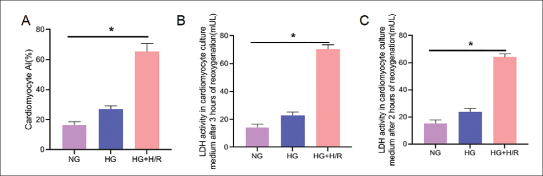
Cardiomyocyte Activity. (A) Myocardial Cell Level; (B) Myocardial Cell Culture Medium Level in Each Group After 3 h of Hypoxia; (C) Myocardial Cell Culture Medium Level in Each Group After 3 h of Reoxygenation.

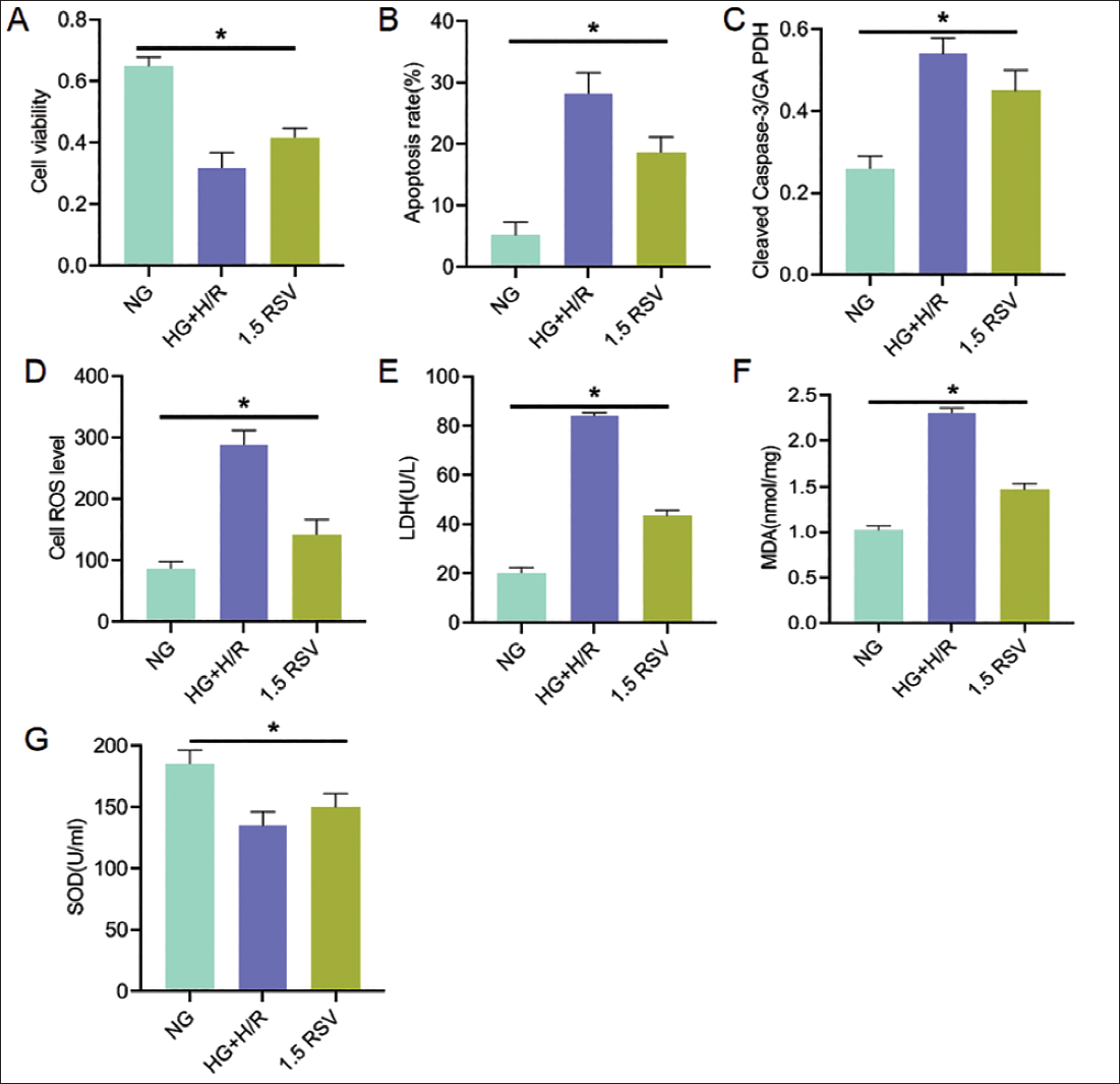
RSV Can Reduce the Apoptosis of Myocardial Injury Caused by High Glucose and Improve its Vitality
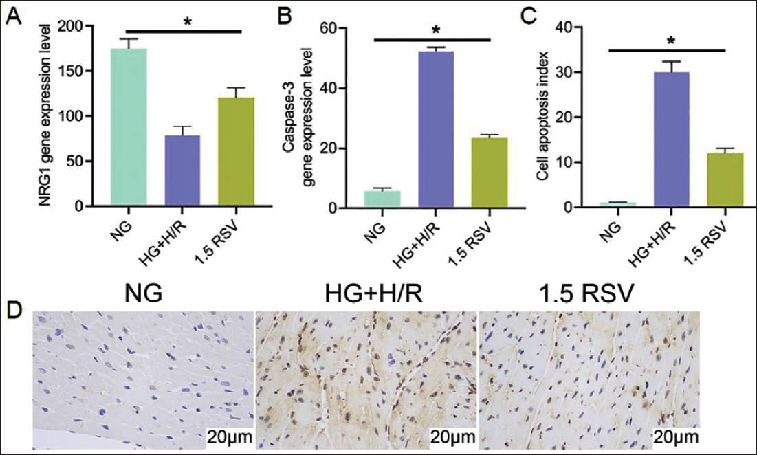
After 1.5 mmol/L RSV intervention, cell viability increased (Figure 3A); at the same time, under the intervention of 1.5 mmol/L RSV, myocardial cell apoptosis rate, caspase-3, ROS, LDH and MDA all showed a downward trend (Figure 3B–F); while in SOD the expression level of RSV group was higher than that of HR group (Figure 3G). In the NRG1 gene expression, by comparing the three administration groups, the 1.5 RSV group was significantly higher than the HG+H/R group (Figure 4A); under further intervention of 1.5 mmol/L RSV, the caspase-3 gene expression was reduced compared with the HG+H/R group (Figure 4B), and overall, the NG group had the lowest apoptosis index, and the 1.5 RSV group had the lowest apoptosis index (vs. HG+H/R group) (Figure 4C), and the apoptosis phenomenon in the 1.5 RSV group was inhibited compared with the HG+H/R group (p < .05) (Figure 4D).
Effects at the Cardiomyocyte Level. (A) Cell Viability Expression; (B) Apoptosis Rate Cell Level; (C) Caspase-3 Cell Level; (D) Reactive Oxygen Species (ROS) Cell Level; (E) Lactate Dehydrogenase (LDH) Cell Level; (F) Malondialdehyde (MDA) Cellular Level; (G) Resveratrol (RSV) Cell Level.
NRG1, Caspase-3 Genes and Apoptosis Index. (A) NRG1 Gene Expression; (B) Caspase-3 Gene Expression Level; (C) Apoptosis Index; (D) Apoptosis Inhibition. Normal Myocardial Cell Nuclei are Blue, and Apoptotic Cell Nuclei are Brown; the Arrows Indicate the Nuclei of Apoptotic Cardiomyocytes.
Specific Blocking of the NRG1 Signaling Pathway Can Reverse the Protective Effect of RSV on Myocardial Damage Caused by High Glucose to a Certain Extent, While Activating the NRG1 Pathway Can Amplify Its Protective Effect
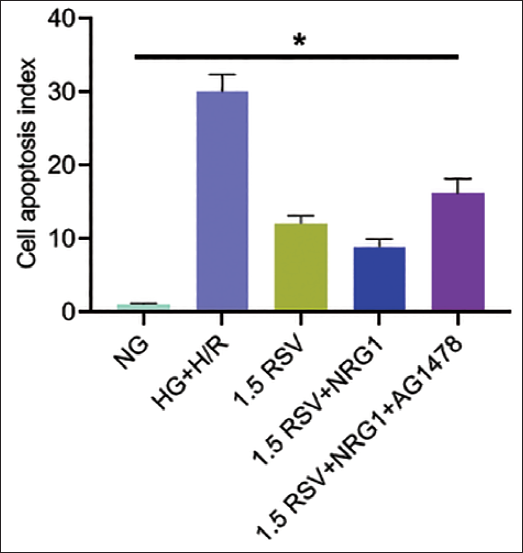
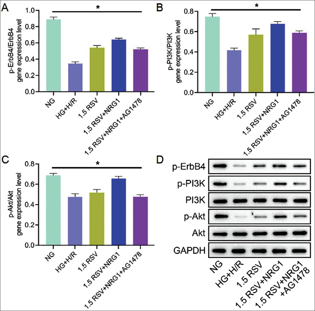
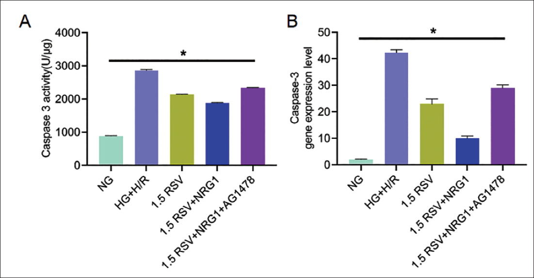
It can be seen from the apoptosis index that the NG group is the lowest and the HG+H/R group is the highest. Among the other three administration groups, the 1.5 RSV+NRG1 group had the lowest apoptosis index (vs. 1.5 RSV group and 1.5 RSV+NRG1+AG1478 group), and the 1.5 RSV+NRG1+AG1478 group had the highest apoptosis (Figure 5). After 1.5 mmol/L RSV intervention, it was found that the degree of myocardial cell membrane swelling increased compared to the HG+H/R group. This promotion phenomenon was further amplified after further use of NRG1, and after the use of AG1478, the increase in membrane swelling was reversed (1.5 RSV+NRG1+AG1478 group vs. 1.5 RSV+NRG1 group, p < .05). The mitochondrial activity of cardiomyocytes also showed the same trend (Figure 6A,B); at the same time, the content of NRG1 in cardiomyocytes was significantly higher than that of the NC group (Figure 6C). In further protein gene detection, under 1.5 mmol/L RSV intervention, the expression level and gene expression level of cardiomyocytes p-ErbB4/ErbB4 also showed an increasing trend (Figure 7A,B). From the Western blot results, it was found that the HG+H/R group compared with the blank group, it showed a decreasing trend (p < .01), while the 1.5 RSV group was significantly higher than the HG+H/R group (p < .01), but this increase was reversed after the addition of AG1478 (vs. 1.5 RSV group). The gene expression levels of p-PI3K/Pl3K and p-Akt/Akt showed the same trend (Figure 7C,D); in addition, the caspase-3 activity of cardiomyocytes decreased. In comparison with the 1.5 RSV group, the caspase-3 activity increased after further use of AG1478 increased (Figure 8A). With further intervention of 1.5 RSV, the expression of the caspase-3 gene was reduced compared with the HG+H/R group, and this inhibitory phenomenon was further amplified after the use of NRG1, while after the use of AG1478, caspase-3 was reversed. The phenomenon of reduced expression of −3 (1.5 RSV+NRG1+AG1478 group vs. 1.5 RSV+NRG1 group, p < .05) (Figure 8B).
Apoptosis Index.
Membrane Swelling, Myocardial Mitochondrial Activity, and NRG1 Content Expression. (A) Membrane Swelling; (B) Myocardial Mitochondrial Activity; (C) NRG1 Content Expression.

Expression Levels of Neuregulin-1/Erythroblastic Leukemia Viral Oncogene Homolog (NRG1/ErbB) Pathway-related Proteins and Genes in Each Group. (A) p-ErbB4/ErbB4 Gene Expression Level; (B) Expression Level; (C) p-PI3K/PI3K Gene Expression Level; (D) p-Akt/Akt Gene Expression Level.
Caspase-3 Activity Level and Gene Expression Level. (A) Caspase-3 Activity Level; (B) Caspase-3 Gene Expression Level.
NRG1 Signaling Pathway Promotes the Protective Effect of RSV on Hyperglycemic Myocardial Injury and Participates in the Regulation of Oxidative Stress
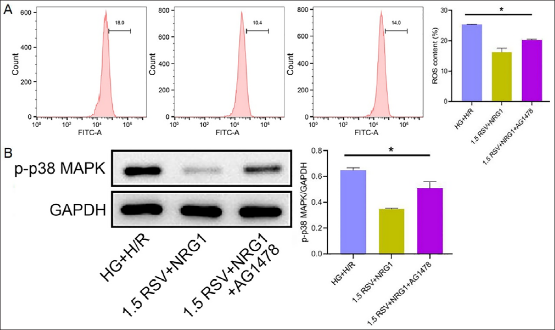
From the point of view of ROS levels in each group, the ROS level in the 1.5 RSV+NRG1 group was significantly lower than that in the HG+H/R group, and after AG1478 treatment, the ROS level was reversed, p < .05 (Figure 9A). At the same time, in the observation of MAPK path-related protein expression, it can be found that the p-p38 MAPK level of the 1.5 RSV+NRG1 group is significantly lower than that of the HG+H/R group. The P-P38 MAPK level in the 1.5 RSV+NRG1+AG1478 group was significantly higher than that in the 1.5 RSV+NRG1 group, p < .05 (Figure 9B).
Reactive Oxygen Species (ROS) Level and MAPK Channel Expression. (A) ROS Level; (B) p-p38 MAPK Expression.
Discussion
Hypoxia-reoxygenation injury to myocardial cells caused by HG is a key contributor to the development of diabetes and heart disease (Cao et al., 2019). Under normal physiological conditions, when myocardial cells are in a hypoxic state, anaerobic metabolic products are produced, leading to myocardial cell toxicity and damage (Mathur & Rani, 2022). During reoxygenation at this time, due to the presence of HG, the production of ROS and RNS in myocardial cells increases. These substances react with proteins, lipids, and so on, in myocardial cells, resulting in myocardial infarction (Aboalgasm et al., 2021). RSV is a polyphenolic compound extracted from plants such as peanuts and grapes (Dugaucquier et al., 2020). Not only can it resist oxidation and free radicals, but it can also play important roles in cardiovascular protection and anti-cancer to a certain extent (Shiraishi et al., 2022). It shows potential to alleviate HG-induced cardiomyocyte damage. Previous studies have shown that RSV can reduce fibrosis associated with diabetic cardiomyopathy by regulating mitochondrial autophagy (Yang et al., 2025). It can alleviate the oxidative stress caused by injury through signaling pathways, reduce the expression levels of inflammatory factors, and so on, to achieve the purpose of alleviating cardiomyocyte injury (Li et al., 2022). However, the specific molecular mechanism of its action is still unclear and requires further research. Some studies have pointed out that RSV can regulate SDF-1/CXCR4 to increase the Bcl-2/Bax value, ultimately inhibiting the apoptosis of neonatal rat neurons induced by ischemia and hypoxia (Dugaucquier et al., 2020). At the same time, some studies have shown that activating NRG1 can activate ErbB to transmit signals to protect cardiomyocytes (Wang, Wei, Zhang, et al., 2022). Clinical studies have shown that the blood sugar level of diabetic patients usually fluctuates between 11 and 33 mmol/L, especially in the case of poor blood sugar control, and the exposure of cardiomyocytes to a high-sugar environment will lead to an increase in oxidative stress, inflammatory response, and apoptosis (Agrawal et al., 2022). Therefore, the glucose concentration of 33 mmol/L selected in this study can not only replicate the clinical hyperglycemia state but also provide a reliable experimental model for studying the pathogenesis of diabetic cardiomyopathy. In this study, RSV can significantly improve myocardial cell damage caused by high sugar, and the protective effect of RSV was reversed with the intervention of AG1478. Therefore, RSV has an ameliorative effect on cardiomyocyte damage caused by HG and may be related to the NRG1/ErbB signaling pathway.
This time, a rat cardiomyocyte injury model was constructed, and different doses of RSV were injected into the model rats. It was found that the AI of cardiomyocytes significantly decreased, and the LDH activity measured after 3 h of hypoxia and 3 h of reoxygenation also significantly decreased. It is suggested that RSV can alleviate cardiomyocyte damage. At the same time, it was also found that the viability of damaged cardiomyocytes increased significantly after RSV intervention. Analysis of the reason may be that RSV can downregulate the expression of the apoptosis protein caspase-3, reduce the oxidative stress and inflammation levels of damaged cells, inhibit cardiomyocyte apoptosis, and alleviate cardiomyocyte damage. Studies have shown that RSV can regulate TLR4 and IKK activity, thereby downregulating IkB phosphorylation and ultimately inhibiting the development of inflammatory responses caused by hypoxia. According to relevant studies, NRG1 can reduce cell apoptosis by inhibiting the expression of caspase-3, thereby playing a protective and therapeutic role (Wang et al., 2021). At the same time, this animal experiment found that under the intervention of RSV, the NRG1 content in myocardial tissue increased significantly compared with the NC group. This demonstrates that RSV’s ability to improve cardiomyocyte hypoxia and reoxygenation caused by HG is related to the NRG1/ErbB signaling pathway.
NRG1 can activate ErbB receptors and is widely involved in biological processes such as cell proliferation, metastasis, and apoptosis (Liu et al., 2021). Studies have found that Tongxinluo can promote the resistance of human cardiac microvascular endothelial cells to H/R injury to a certain extent through the NRG1/ErbB pathway (Ge et al., 2022). In addition, related studies have shown that NRG1/ErbB can regulate PI3K/AKT to upregulate Bcl-2 and downregulate Bax. The reduction of Bcl-2 increases Bax, which in turn causes the opening of the mitochondrial permeability transition pore and promotes the synthesis of pro-apoptotic factors. is released (Lu et al., 2023), activates the cleaving enzyme caspase-3, and ultimately initiates cell apoptosis (Qiu et al., 2019). Therefore, effectively inhibiting the expression of various apoptosis proteins and activating the NRG1/ErbB signaling pathway is crucial for the protection of cardiomyocytes (Wang, Chen, Cao, et al., 2022).
In this animal experiment, we added NRG1 on the basis of RSV alleviating the hypoxia-reoxygenation injury of cardiomyocytes caused by high sugar. While the apoptosis index and caspase-3 were significantly reduced, the mitochondria of cardiomyocytes also decreased. The vitality increased significantly, which enhanced the improvement effect of RSV on myocardial cell damage. In order to further explore the role of NRG1/ErbB in alleviating hypoxia-reoxygenation injury in cardiomyocytes, we added AG1478 in addition to NRG1. We found that AG1478 reversed the improvement effect of RSV on cardiomyocyte injury, causing apoptosis. The death index increased significantly. At the same time, in order to ensure the rigor of the experiment, we also added AG1478 to the RSV group for comparison. We found that the addition of AG1478 also reversed the improvement effect of RSV on myocardial cell hypoxia-reoxygenation injury. It is suggested that the NRG1/ErbB signaling pathway plays an important role in the process of RSV alleviating cardiomyocyte injury. In addition, we also observed that the expression levels of PI3K/AKT-related genes also increased and decreased with the increase of NRG1/ErbB. This shows that RSV can improve cardiomyocyte hypoxia-reoxygenation damage caused by HG through the NRG1/ErbB signaling pathway.
This study also found that ROS levels in cardiomyocytes induced by high sugar were effectively reduced by RSV and NRG1. These results indicate that RSV regulates oxidative stress in the process of myocardial protection through the NRG1/ErbB pathway. Previous studies have shown that the MAPK pathway can reduce myocardial damage by regulating oxidative stress and inflammatory response (Wang et al., 2025). In this study, it was found that the relative expression of p38 MAPK protein in the 1.5 RSV+NRG1 group was significantly lower than that in the HG+H/R group, and the phosphorylation of p38 in cardiomyocytes was effectively inhibited, suggesting that the myocardial protective mechanism of RSV is closely related to the regulation of the MAPK pathway. p38 MAPK activity is a necessary condition for cardiomyocytes to promote angiogenesis. However, after phosphorylation of p38 MAPK, cardiomyocytes further undergo dependent apoptosis, resulting in the inhibition of p38-dependent apoptosis, resulting in resulting in e time, ROS upregulation can further stimulate p38 MAPK activation and cause mitochondrial dysfunction in cardiomyocytes, thus affecting myocardial function (Moris et al., 2017). However, there are complex interactions between these pathways and NRG1/ErbB pathways, which may synergistically regulate cardiomyocyte survival and apoptosis through downstream effector molecules such as PI3K/AKT. Therefore, future studies should further explore the cross-regulatory mechanisms between these pathways to understand the myocardial protective effects of RSV.
Conclusion
In summary, RSV reduces caspase-3 expression and cell apoptosis through the NRG1/ErbB signaling pathway, effectively improving hypoxia-reoxygenation injury in cardiomyocytes caused by HG and exerting a protective effect. This also provides a new method for clinical treatment of myocardial cell hypoxia-reoxygenation injury caused by HG. It can also help us better understand the impact of RSV on the human body, to maximize its effect. However, this study was only observed at the animal level and has not yet been used in clinical studies, and further research is needed to understand the potential difference in the effects of RSV in animals and humans.
Abbreviations
ErbB: Erythroblastic leukemia viral oncogene homolog; HG: High glucose; H/R: hypoxia/reoxygenation; LDH: Lactate dehydrogenase; MDA: Malondialdehyde; NRG1: Neuregulin-1; RSV: Resveratrol; ROS: Reactive oxygen species; SOD: Superoxide dismutase.
Footnotes
Acknowledgments
The authors gratefully acknowledge the Tangshan Central Hospital for providing the necessary equipment for this study.
Declaration of Conflicting Interests
The authors declared no potential conflicts of interest with respect to the research, authorship, and/or publication of this article.
Ethical Approval and Informed Consent
This study was approved by the Ethics Committee of Tangshan Central Hospital.
Funding
The authors received no financial support for the research, authorship, and/or publication of this article.