



Abstract
Background
Repurposing of a hydroxylmethylglutaryl coenzyme A reductase inhibitor like simvastatin has gained significant interest in oncologic treatment owing to the pleiotropic effects. However, the role of simvastatin in inhibiting epithelial-mesenchymal transition (EMT), a critical process in cancer metastasis, remains underexplored.
Purpose
To investigate the anti-EMT potential of simvastatin in human breast cancer MCF-7 cells.
Materials and Methods
MCF-7 cells were treated with simvastatin, and the anti-proliferative and pro-apoptotic effect were evaluated by 3-(4,5-dimethylthiazol-2-yl)-2,5-diphenyltetrazolium bromide assay and fluorescence double staining method, respectively. Cell migration and cell aggregation assays were carried out to determine EMT-associated activities. A key regulator of EMT, β-catenin activity was measured using an indirect enzyme-linked immunosorbent assay, and the gene expression in response to simvastatin treatment was analyzed by reverse transcriptase polymerase chain reaction.
Results
A dose-dependent cytotoxic activity was induced by simvastatin in MCF-7 cells, associated with morphological alterations indicative of apoptosis. Enhanced apoptotic cell death on simvastatin administration was validated by fluorescence nuclear staining. A marked reduction in migratory capacity and cell aggregation was observed in simvastatin-treated cells. Activity of β-catenin and the messenger ribonucleic acid expression were found to be downregulated.
Conclusion
Simvastatin effectively suppressed EMT-related processes in MCF-7 breast cancer cells by inhibiting cell proliferation, migration, aggregation, and β-catenin expression. These findings highlight the potential of simvastatin as a therapeutic agent for preventing breast cancer metastasis through EMT inhibition.
Introduction
The World Health Organization Global Breast Cancer Initiative intends to reduce the mortality due to breast cancer by 2.5% per year, which can ward off nearly 25% of breast cancer deaths by 2030 and 40% by 2040 among women under 70 years of age. 1 Management of breast cancer is regarded as one of the major pillars in the management of the disease; factors like drug resistance and epithelial-mesenchymal transition (EMT) pose major challenges. 2 EMT is a dynamic process where epithelial cells are capable of transforming to mesenchymal stem cells, facilitating rapid adaptation to stress and the development of treatment-resistant phenotypes. 3 Inhibiting EMT is emerging as a novel strategy to address the issue. Drug repurposing is an innovative methodology for identifying a novel therapeutic application of an existing drug to treat a different medical condition. Simvastatin, an established hydroxylmethylglutaryl coenzyme A (HMG-CoA) reductase inhibitor, has been studied as an anti-cancer medication, mainly for EMT inhibition. 4 Simvastatin can inhibit EMT in bladder cancer, causing upregulation of E-cadherin and downregulation of the mesenchymal markers vimentin and β-catenin. 5 For the treatment of hepatocellular carcinoma, Yu et al. reported an immune-based therapeutic mechanism for modifying the immunosuppressive tumor microenvironment. 6 The dual preventive role of simvastatin, as a lipid-lowering drug for cardiovascular disease and as an anti-cancer agent in many cases, has been reported. 7
Cell proliferation, migration, differentiation, and cell death are modulated by the highly conserved Wnt/-β-catenin pathway. The pathway is highly altered in breast cancer and is a target for potential chemotherapy. 8 Hence, a thorough investigation of pathway changes and therapeutic drugs that can regulate Wnt/-β-catenin signaling in breast cancer is warranted.
The EMT is crucial for tumor growth, metastasis, and the creation of tumor cells with stem cell features, which are crucial for resistance to cancer treatment. The purpose of this study was to examine how the hypolipidemic medication simvastatin inhibits EMT in human breast cancer cells MCF-7 and the molecular mechanism behind the process.
Materials and Methods
Materials
Simvastatin was procured from Tocris Biosciences (CAS No: 79902-63-9). Breast cancer MCF-7 cells were obtained from the National Centre for Cell Science, Pune, India, and maintained in Dulbecco’s Modified Eagle Medium (DMEM) (Sigma–Aldrich, USA). The cell line was grown in DMEM supplemented with 10% fetal bovine serum, L-glutamine, sodium bicarbonate (Merck, Germany), and an antibiotic solution comprising penicillin (100 U/mL), streptomycin (100 µg/mL), and amphotericin B (2.5 µg/mL). Cell lines were kept at 37°C in a humidified 5% CO2 incubator (NBS Eppendorf, Germany).
Methods
In Vitro Anti-proliferative Effect of Simvastatin on Cultured MCF-7 Cells
Cells were trypsinized from a confluent monolayer, suspended in 10% growth media, and plated at 5 × 10⁴ cells per well in a 96-well tissue culture plate in a humidified 5% CO2 incubator at 37°C. Simvastatin was prepared (1 mg/mL) in dimethyl sulfoxide (DMSO), syringe-filtered, added in different concentrations ranging from 6.25 to 100 µg/mL, and incubated at 37°C in a humidified 5% CO2 incubator along with an untreated control.
Determination of In Vitro Anti-proliferative Effect of Simvastatin on Cultured MCF-7 Cell Lines Using 3-(4,5-Dimethylthiazol-2-yl)-2,5-diphenyltetrazolium Bromide Assay
Cell viability was studied by a 3-(4,5-dimethylthiazol-2-yl)-2,5-diphenyltetrazolium bromide (MTT) assay. 9 After washing the cell culture suspension with 1× phosphate-buffered saline (PBS), 30 µL of MTT (5 mg/mL in PBS) was added and incubated for 3 h at 37°C. MTT was removed with 1× PBS, and 200 µL of DMSO was added. Following a 30 min incubation at room temperature, the cells were lysed, and a color was obtained. The solution was centrifuged for 2 min to precipitate cell debris. At 540 nm, optical density was measured with DMSO as the blank. The percentage of growth inhibition was calculated.
Determination of Apoptosis by Acridine Orange and Ethidium Bromide Double Staining Method
Different groups comprising untreated control cells and cells treated with simvastatin was incubated for 24 h and proceeded with fluorescent staining as per the methods described by Lekshmy et al. 10 After incubation, the cells were washed with cold PBS and then stained with acridine orange (AO) (100 µg/mL) and ethidium bromide (Et-Br) (100 µg/mL) at room temperature for 10 min. After two washes in 1× PBS, the stained cells were analyzed using an Olympus CKX41 fluorescent microscope with a blue filter. Living cells (green nuclei), early apoptotic (bright green nuclei with condensed or fragmented chromatin), late apoptotic (orange-stained nuclei with condensed or fragmented chromatin), and necrotic cells (orange nuclei) were detected.
Migration Assay
After 24 h of incubation, trypsinized cells were plated at a density of 2 × 10⁵ cells per well in a 12-well plate. A sterile 1 mL pipette tip was used to scratch a premarked line. After removal of the resulting debris from five linear scratches, the cell monolayer was washed three times with PBS and then incubated with simvastatin for 0, 24, 48, and 72 h. After incubation, the effect of the sample on wound closure was assessed microscopically (4× magnification, Olympus CKX41) by photographing the wound areas directly above the intersections between the scratched wound areas and premarked lines. Using the Montpellier Ressources Imagerie-ImageJ analysis software, the effect of the sample on wound closure was assessed in terms of the area. 11
MCF-7 cells were incubated with simvastatin for 24 h. After incubation, 100 µL of each supernatant was put into a 96-well plate and incubated overnight at 37°C. The next day, the supernatant was analyzed for protein by Bradford method, and the wells were drained and washed twice with PBS. 200 µL of freshly prepared blocking buffer in PBS was added and incubated for 1 h at room temperature, then washed twice with PBS-TWEEN. 100 µL of β-catenin primary antibody was added and left at room temperature for 2 h. PBS-TWEEN was used for washing after incubation. After 1 h at room temperature, 100 µL of secondary antibody (horseradish peroxidase (HRP) conjugate, Santa Cruz, USA) was added. The wells were then washed twice with PBS-TWEEN. 200 µL of o-dianisidine hydrochloride was added and left at room temperature for 30 min. 50 µL of 5N HCl was used to stop the reaction. In an enzyme-linked immunosorbent assay (ELISA) reader, the absorbance was measured at 415 nm. Activity was expressed in units per mg protein.
Cell Aggregation Assay
Cell aggregation on agar is an indication of cell-matrix adhesion. 12 Cells were treated as described previously and 100 µL of the cell suspension and 100 µL of the test sample medium were applied to the agar wells and incubated at 37°C in 5% CO2 for 24 h. The aggregation of cells was observed under an inverted microscope and was observed at 24 h intervals for 72 h. Micrographs were taken after the cells had been fixed in 3.7% paraformaldehyde and stained with 0.1% crystal violet.
Determination of Caspase-7 Activity by Indirect Enzyme-linked Immunosorbent Assay
Cultured MCF-7 cells were treated with simvastatin and incubated for 24 h. 100 µL of supernatant was added to each 96-well plate and incubated at 37°C overnight. On the following day, protein was estimated in the supernatant. Washed the wells with PBS, added 200 µL of freshly prepared blocking buffer, and incubated them for 1 h at room temperature. Washed with PBS-TWEEN, added 100 µL of primary antibodies, and left them for 2 h at room temperature. Washed with PBS-TWEEN and added 100 µL of secondary antibody (HRP conjugate, Santa Cruz, USA), and left for 1 h at room temperature. Washed and added 200 µL of o-dianisidine hydrochloride (Sigma–Aldrich, USA) and left for 30 min at room temperature. Stop the reaction by adding 50 µL 5N HCl and read the absorbance at 415 nm in an ELISA reader. Activity is expressed as absorbance/mg protein.
Relative expression study of the β-catenin gene in simvastatin-treated MCF-7 cell lines using reverse transcriptase polymerase chain reaction (PCR).
The effect of simvastatin on the MCF-7 β-catenin gene was examined using reverse transcriptase PCR (RT-PCR). In Roche primer creating software, β-catenin primer pairs were designed with a melting temperature (Tm) of 60°C, a primer length of 20–23 nucleotides (optimum number: 20), and a guanine–cytosine content of 50%–55% (Table 1).
Primer Details.

Ribonucleic Acid Isolation and Complementary Deoxyribonucleic Acid Synthesis
To isolate ribonucleic acid (RNA), an isolation kit was used in accordance with the manufacturer’s instructions (Invitrogen product code 10296010). The dried RNA pellet was suspended in the Tris–EDTA buffer. To create the complementary DNA (cDNA), we used the Thermo Scientific Verso cDNA Synthesis Kit (AB-1453/A). Synthesis of cDNA was programmed into the thermal cycler (Eppendorf Mastercycler). Temperature cycles of 5 min at 25°C, 30 min at 42°C, and 2 min at 95°C were used.
Polymerase Chain Reaction Amplification
The Thermo Scientific amplification kit was employed for the process. The first denaturation lasted 3 min at 95°C, followed by 30 s of denaturation at 95°C, 30 s of annealing, and 1 min of extension at 72°C. This cycle was repeated 30 times, and the final extension was for 15 min at 72°C. The amplified PCR product was separated by electrophoresis in an agarose gel.
Agarose Gel Electrophoresis
The samples were loaded into the gel and ran at 50 V for 30 min. Gel documentation software was used to visualize images of the stained gel (E-Gel Imager, Invitrogen).
Data Analysis
All experiments were done in triplicate, and the values are represented as mean ± standard deviation (SD). One-way analysis of variance (ANOVA) was employed to determine statistical significance using GraphPad Prism version 5.01.
Results
In Vitro Anti-proliferative Effect of Simvastatin on Cultured MCF-7 Cell Lines
Simvastatin produced a dose-dependent decrease in cell viability, along with concomitant changes in cell morphology similar to apoptotic cell death. Cell viability was reduced by 31.33% when cells were treated with simvastatin at a dosage of 100 µg/mL. The morphological alterations exhibited similarities to apoptotic bodies and nuclear condensation. The LC50 value of 55.35 µg/mL, determined using ED50 plus V 1.0 software, can be considered statistically significant (Figure 1).

The morphological changes resembled apoptotic bodies, and nuclear condensation is shown in Figure 2.
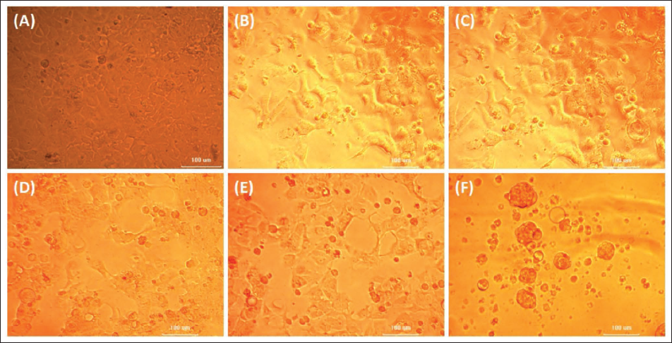
Phase Contrast Images of MCF-7 Cells Exposed to Varied Concentrations of Simvastatin. (A) Untreated MCF-7 Cells and Cells Treated with Simvastatin of (B) 6.25, (C) 12.5, (D) 25, (E) 50, and (F) 100 µg/mL.
Cellular Apoptosis Detection Using Acridine Orange and Ethidium Bromide Double Staining
Differential uptake of fluorescent dyes allowed the observation and identification of viable and non-viable cells. The qualitative analysis showed that the untreated control group showed no signs of apoptosis. In contrast, simvastatin treatment resulted in a dramatic rise in the proportion of apoptosis in MCF-7 cell line. Empirical evidence verified the suppression of cell growth by simvastatin in MCF-7 breast cancer cells (Figure 3).

Determination of Apoptosis by the Double Staining Method. (A) Untreated MCF-7 Cells. (B) MCF-7 Cells Exposed to Simvastatin.
Migration Assay
In the scratch wound-healing experiment, imagery was captured using an inverted microscope at both the 0- and 24-h time points. At 24 h, cells in the untreated set migrated into the cell-free area and completely covered the scratch middle line. Following 24 h of simvastatin treatment, the migration of tumor cells was significantly reduced compared to the untreated group, as indicated in Figures 4 and 5.

Phase Contrast Analysis of Migration Assay: (A) Untreated Control at 0th h. (B) Cells Exposed to Simvastatin at 0th h. (A) 24 Untreated Control at 24th h. (B) 24 Cells Exposed to Simvastatin at 24th h.

Cell Aggregation Assay
The fact that functional integrity of the E-cadherin/catenin complex is essential for cell-cell adhesion among epithelial cells, measuring cell aggregation in vitro is a valuable method for studying distinctions between invasive and non-invasive cell types. From the observed results, it can be confirmed that tumor cells formed a greater number of cell aggregates after 48 h incubation, while in the case of the simvastatin-exposed cells, the aggregation of tumor cells is significantly less than that of the untreated control cells (Figure 6).

Phase Contrast Analysis of Cell Aggregation Assay: (A) Control 0th h. (B) Test 0th h. (A) 24 Control 24th h. (B) 24 Test 24th h. (A)48 Control 48th h. (B)48 Test 48th h. (A)72 Control 72nd h. (B)72 Test 72nd h.
Determination of Caspase-7 Activity and β-catenin Activity by Indirect Enzyme-linked Immunosorbent Assay
Cancer and inflammatory diseases may be treated therapeutically by interfering with caspase-7 activation. The activity of caspase-7 was measured using MCF-7 cells exposed to simvastatin compared to the untreated control by indirect ELISA (Figure 7). The acquired findings showed a substantial increase in the production of caspase-7 in simvastatin-treated groups when compared to the control.

The acquired data indicate a significant downregulation in β-catenin activity in cells treated with simvastatin (0.1863 units/mg protein) compared to cells that were not treated and served as the control (0.2020 units/mg protein). Confirmation of the suppression of β-catenin in simvastatin-treated groups as compared to untreated control cells was achieved using indirect ELISA with anti-β-catenin antibody (Figure 8).

Messenger Ribonucleic Acid Expression Analysis of β-catenin Gene on Simvastatin Exposure on MCF-7 Cell Lines
Based on the agarose gel electrophoretogram and subsequent ImageJ analysis, it is evident that the relative gene expression of β-catenin is reduced in groups treated with simvastatin compared to untreated control cells. The decrease in gene expression can be attributed to the inhibitory potential of simvastatin towards β-catenin expression, which is normally activated in response to TGF-β (Figure 9A and 9B).

(A) Comparative Study of β-catenin in Untreated Control Cells and Cells Treated with Simvastatin Using Agarose Gel Electrophoresis of Amplified Products Obtained from Reverse Transcriptase Polymerase Chain Reaction (PCR) Analysis, Glyceraldehyde-3-phosphate Dehydrogenase is the Reference Standard. (B) Relative Gene Expression of β-catenin as Determined by ImageJ Analysis Software. Along the Y Axis Expression Change in Terms of Arbitrary Units and Along the X Axis Untreated Control.
Discussion
Simvastatin, a competitive inhibitor of 3-hydoxy-3-methylglutaryl coenzyme A reductase, is a well-tolerated hypocholesterolemic drug. A catalytic process aided by 3-hydroxy-3-methylglutaryl coenzyme A reductase limits mevalonate synthesis, which is necessary for cholesterol and other non-steroidal isoprenoid derivatives needed for cell proliferation, differentiation, and survival. 13 Statins may exert their pleiotropic effects by inhibiting the isoprenylated small guanosine triphosphate-binding proteins, Rho, Ras, and Rac. Preclinical studies employing cancer cell lines and animal models showed statins’ anti-proliferative, proapoptotic, and anti-invasive effects. Statins preferentially affect cancer cells with comparatively lower toxicity toward healthy ones.14, 15 Although several reports have shown the beneficial impacts of statins on malignancies, including breast cancer, the absence of scientific validation and understanding of their mechanisms of action requires further study in this domain. The objective of this investigation is to evaluate the anti-metastasis effect of simvastatin on human breast cancer cells.
Simvastatin elicited a robust anti-proliferative effect in MCF-7 breast cancer cells, characterized by a dose-dependent reduction in viability and pronounced apoptotic morphology. The cell viability was declined by more than 30%, along with considerable morphological alterations like nuclear condensation, membrane blebbing, cell shrinkage, and apoptotic body formation at concentrations exceeding 6.25 µg/mL. The LD50 value of 55.35 µg/mL indicates a highly significant cytotoxic impact. These findings indicate the therapeutic potential of simvastatin as a powerful anti-cancer agent, inducing apoptosis in breast cancer cell lines.
Several studies have postulated that statins modulate apoptosis and impact the expression of Bcl-2 family members. By using Et-Br/AO fluorescence staining, the effect of statin supplementation on apoptosis in MCF-7 cells was investigated. Our findings clearly demonstrate an increase in apoptotic cells. The observed apoptotic shift corroborates the anti-proliferative effect. Variations in the expression levels of pro- and anti-apoptotic Bcl-2 family members are a hypothesized mechanism by which statins exert their effects in non-cardiovascular diseases. Multiple investigations have shown that statins modulate apoptosis and cell death by reducing the expression of the anti-apoptotic protein Bcl-2.16, 17
Active migration of tumor cells is required for invasion and metastasis, and several variables influence this movement. Cell migration inhibition directly inhibits metastasis. The scratch assay measured cell migration and antimetastasis activity. The migratory and invasive nature of tumor-derived mesenchymal cells affects the metastatic process. These cells can escape the main tumor and enter the bloodstream after this transformation. This change enables these cells to leave the primary tumor and infiltrate the bloodstream, which is encouraging tumor cells to migrate and invade, a crucial phase in tumor metastasis. 18 The scratch wound healing assay showed that, in simvastatin treatment groups, the wound was not closed even after 72 h of incubation, displaying significantly reduced migratory capacity, whereas the control group efficiently closed the wound gap within 24 h. The decrease in wound migration is attributable to the decreased EMT in the simvastatin-treated groups versus the control groups. Experimental evidence has shown that statins suppress the proliferation of breast cancer cells by reducing the expression of pituitary tumor-transforming gene 1, a crucial gene linked to the invasion and spread of breast cancer. 19 Simvastatin effectively suppressed the growth of bladder cancer cells and induced arrest in the cell cycle in the G1/G0 phase via activating the peroxisome proliferator activated receptor gamma signal transduction pathway. 20 Dosage- and time-dependent cell death was induced in MCF-7 and MDA-MB-231 cells by the administration of simvastatin. Simvastatin’s anti-neoplastic activity on breast cancer cells was consistently linked to apoptosis and synergistic efficacy when used with doxorubicin. 21 As reported by Kan et al., simvastatin induces apoptosis and slows the development of osteosarcoma cells predominantly through inhibiting prenylation. Elevated levels of c-Jun activation seem to be essential in the development of osteosarcoma. 22 By inducing apoptosis, simvastatin suppresses the proliferation, migration, and invasion of gastric cancer cells. Mechanistic investigations indicated that simvastatin inhibits β-catenin, YAP, and their downstream targets in gastric cancer cells. 23
Recent scientific interest in producing EMT inhibitors can be attributed to the expansion of knowledge on the processes of EMT in breast cancer. Even though simvastatin is reported to have apoptotic activity in breast cancer cells, little is understood about its influence on EMT. Hence, our study was centered on Wnt signaling, a pivotal factor in the proliferation and development of breast cancer cells. E-cadherin/catenin complex downregulation is a characteristic feature of invasive carcinomas.24, 25 Therefore, in vitro measurement of cell aggregation is an effective technique for investigating the differences between invasive and non-invasive cell types, as the functional integrity of the complex is a requirement for cell-cell adhesion between epithelial cells.18, 26 In our study, tumor cells developed a greater number of cell aggregates after 48 h of incubation; however, tumor cell aggregation is much lower in simvastatin treated cells than in untreated control cells.
Caspases, a group of endoproteases that connect cell regulatory networks governing inflammation and cell death, are intricately regulated by their production as inactive zymogens. Once signaling events occur, these zymogens acquire catalytic activity and then aggregate into dimers or macromolecular complexes. 27 Apoptotic caspases activate a signaling cascade that controls cell destruction. Inflammatory caspases activate proinflammatory cytokines and promote innate immune responses to internal and external insults. Interfering specifically with caspase-7 activation has therapeutic promise for cancer and inflammatory diseases. 28 β-catenin was first identified as a cell-cell adhesion protein. β-catenin-mediated signaling may mediate the development of inflammation and malignancies. β-catenin is crucial for intercellular junctions and the Wnt signaling pathway, which is a downstream signaling molecule induced by canonical Wnt signaling. β-catenin plays two roles in EMT: mediating cell-cell adhesion with cadherin complexes at adhesion junctions and coordinating transcription with T-cell factor/lymphoid enhancer factor family. 29
The Wnt/β-catenin signaling pathway significantly influences the EMT in breast cancer cells by facilitating the accumulation and stability of β-catenin via the degradation of the adenomatous polyposis coli complex and E-cadherin, consequently activating genes that promote EMT. 30 The TGF-β and Wnt/β-catenin signaling pathways play a crucial role in the onset of EMT, and the inhibition of β-catenin production might result in enhanced protein degradation, subsequently leading to the suppression of the Wnt pathway. In line with this, our results suggest that simvastatin treatment has led to an appreciable decrease in messenger RNA (mRNA) expression of β-catenin, suggesting inhibition of mRNA transcription, which can lead to decreased protein turnover and leading to inhibition of EMT.
Findings of this investigation position simvastatin as a potential chemotherapeutic agent with significant translational relevance.
Conclusion
The present study elucidates the promising role of simvastatin, a widely used HMG-CoA reductase inhibitor, in modulating EMT processes in human breast cancer MCF-7 cells. While the anti-cancer properties of simvastatin have been acknowledged due to its pleiotropic effects, its direct impact on EMT, a critical mechanism driving cancer progression and metastasis, has remained underexplored. Our comprehensive in vitro analyses revealed that simvastatin exerts significant anti-proliferative and proapoptotic effects in a dose-dependent manner, as evidenced by the MTT assay and fluorescence double staining. Furthermore, simvastatin markedly suppressed cellular migration and aggregation, two fundamental hallmarks of EMT, highlighting its potential to hinder metastatic dissemination. Mechanistically, simvastatin treatment led to the downregulation of β-catenin activity, a central mediator of EMT, as confirmed by indirect ELISA and RT-PCR-based mRNA expression analysis. The observed reduction in β-catenin expression further supports simvastatin’s role in attenuating EMT signaling pathways, which are often aberrantly activated in breast cancer metastasis. Morphological alterations accompanying these molecular changes substantiate the phenotypic reversion of cells away from a mesenchymal, migratory state toward an epithelial phenotype. Collectively, these findings underscore simvastatin’s ability to disrupt EMT progression, suggesting a novel therapeutic avenue wherein this statin could be repurposed as an adjunctive agent in breast cancer treatment strategies aimed at impeding metastasis. The study paves the way for further in-depth molecular investigations and translational studies to validate simvastatin’s efficacy in in vivo models and clinical settings. Given its established clinical safety profile, simvastatin holds substantial potential to be repositioned as an anti-metastatic therapeutic in breast cancer, thereby contributing to more effective management of disease progression and improving patient outcomes.
The investigation was confined to the human breast cancer cell line MCF-7, which may not represent the heterogeneity of various subtypes of breast cancer. Moreover, the study was entirely in vitro, lacking in vivo validation to confirm the therapeutic efficacy of simvastatin in a complex tumor microenvironment. Furthermore, EMT markers like E-cadherin, N-cadherin, and vimentin are to be studied in detail. The molecular mechanism underlying the suppression of simvastatin-mediated is to be deeply explored before going to the translational level.
Footnotes
Abbreviations
Acknowledgments
The authors extend their sincere appreciation to the entire team at the Center for Research on Molecular Biology and Applied Science, Thiruvananthapuram, Kerala, India, for their invaluable support and assistance throughout the execution of this work. We would like to express our sincere gratitude to Dr. Dhanya CR for the technical support for the preparation of the manuscript.
Authors Contribution
RR: Conceptualization, methodology, supervision, writing – original draft, writing – review and editing.
ML: Conceptualization, validation, writing – original draft, writing – review and editing.
NS: Formal analysis, investigation, validation.
RC: Formal analysis, investigation.
SA: Methodology.
SS: Formal analysis, investigation, supervision, writing – review and editing.
SC: Supervision, writing – original draft.
RC: Writing – original draft.
Data Availability Statement
The datasets analyzed during the current study are available from the corresponding author on reasonable request.
Declaration of Conflict of Interests
The authors declared no potential conflicts of interest with respect to the research, authorship, and/or publication of this article.
Ethical Approval
This article does not contain any studies with human participants or animals performed by any of the authors. The study adhered to all relevant institutional and national guidelines for the use of cell lines.
Funding
The authors received no financial support for the research, authorship, and/or publication of this article.
Informed Consent
Human volunteers are not involved in the present study.
